Chinese Journal of Tissue Engineering Research ›› 2026, Vol. 30 ›› Issue (27): 7053-7060.doi: 10.12307/2026.425
Previous Articles Next Articles
Interactive biomechanical effects between medial meniscus anterior horn transverse tears and osteoarthritic degeneration: a finite element simulation analysis
Chen Jiahao1, Zhang Jiahao1, Tian Jiaqing1, Xiang Ruian1, Wang Shuai1, Xu Xuemeng1, 2
- 1Fifth Clinical Medical College of Guangzhou University of Chinese Medicine, Guangzhou 510095, Guangdong Province, China; 2Guangdong Second Traditional Chinese Medicine Hospital, Guangzhou 510095, Guangdong Province, China
-
Received:2025-09-20Accepted:2026-01-05Online:2026-09-28Published:2026-05-14 -
Contact:Xu Xuemeng, Chief physician, Doctoral supervisor, Fifth Clinical Medical College of Guangzhou University of Chinese Medicine, Guangzhou 510095, Guangdong Province, China; Guangdong Second Traditional Chinese Medicine Hospital, Guangzhou 510095, Guangdong Province, China -
About author:Chen Jiahao, MS candidate, Fifth Clinical Medical College of Guangzhou University of Chinese Medicine, Guangzhou 510095, Guangdong Province, China -
Supported by:Key Research & Development Program of Guangzhou Municipal Science and Technology Bureau, No. 202206010048 (to XXM); "Guangdong Special Support Program" Provincial Health Commission Leading Talent Project, No. [2024]54 (to XXM); Guangzhou Regional Traditional Chinese and Western Medicine Collaborative Clinical Major Innovation Technology Construction Project, No. [2023]2318 (to XXM)

CLC Number:
Cite this article
Chen Jiahao, Zhang Jiahao, Tian Jiaqing, Xiang Ruian, Wang Shuai, Xu Xuemeng. Interactive biomechanical effects between medial meniscus anterior horn transverse tears and osteoarthritic degeneration: a finite element simulation analysis[J]. Chinese Journal of Tissue Engineering Research, 2026, 30(27): 7053-7060.
share this article
Add to citation manager EndNote|Reference Manager|ProCite|BibTeX|RefWorks
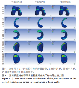
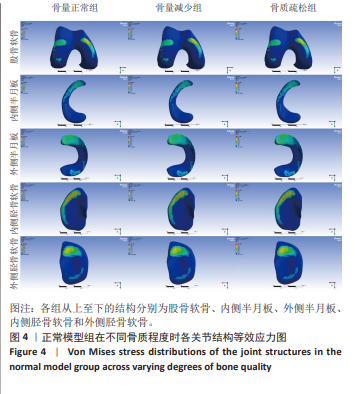
2.1 模型验证结果 根据Ansys软件网格划分的生成结果,正常骨质膝关节有限元模型的节点数812 371,单元数为559 898个。通过进一步细分网格的计算结果表明,当进一步细化网格后,关键应力指标的数值变化趋于平稳,其相对差异已小于2%,显著低于预设5%的判定标准。因此,该应力结果对网格尺寸不再敏感,可认为已实现网格无关化。前抽屉试验的结果显示,胫骨平台中点的位移为4.75 mm,与既往文献结果相近[26-27]。轴向加载试验结果显示内外侧间室分别占比63.90%和36.09%。股骨软骨内外侧等效应力最高值分别为3.54 MPa和2.84 MPa,胫骨内外侧软骨等效应力最高值分别为2.47 MPa和1.87 MPa,内侧半月板与外侧半月板等效应力最高值分别为13.27 MPa和11.08 MPa,均与既往研究结果相近[23,28]。 2.2 内侧半月板正常模型与前角横行撕裂模型中各部位的应力数据 2.2.1 正常模型股骨软骨、半月板以及胫骨软骨的等效应力峰值及应力分布 在正常骨质、骨量减少以及骨质疏松组中,股骨软骨的等效应力峰值分别为2.74,2.97和3.19 MPa;外侧半月板的等效应力峰值为8.05,8.71和9.38 MPa;内侧半月板的等效应力峰值为14.30,15.51和16.72 MPa;胫骨内侧软骨的等效应力峰值为3.03,2.81和2.59 MPa;胫骨外侧软骨的等效应力峰值为2.05,1.91和1.76 MPa。可见内侧半月板的峰值应力大于外侧部分,且随着骨质的减少,半月板应力峰值逐渐增大,而胫骨软骨的应力峰值随着骨质的下降逐渐减少,在3组模型中,尽管皮质骨和松质骨的骨质不一,但表面的应力分布趋势基本一致,详见图4。"
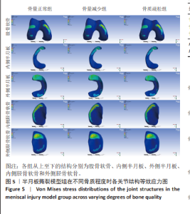
2.2.2 前角横行撕裂模型股骨软骨、半月板以及胫骨软骨的等效应力峰值与应力分布 在前角横行撕裂模型的骨质、骨量减少以及骨质疏松组中,股骨软骨的等效应力峰值分别为3.54,3.78和4.13 MPa;外侧半月板的等效应力峰值为10.31,10.99和12.01 MPa;内侧半月板的等效应力峰值为21.15,22.57和24.70 MPa;胫骨内侧软骨的等效应力峰值为4.01,3.66和3.43 MPa;胫骨外侧软骨的等效应力峰值为2.62,2.40和2.25 MPa。在前角横行撕裂模型中,股骨软骨、外侧半月板及内侧半月板、内外侧胫骨软骨均较正常模型组承受更多的等效应力;且内侧半月板损伤后承受的等效应力相较于正常组提升了47.90%,且应力的分布更加集中在前角损伤部位,损伤的内侧半月板表面的高应力值随着骨质的下降而升高,见图5。"
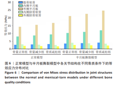
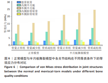
正常模型与半月板撕裂模型中各关节结构在不同骨质条件下的等效应力分布对比见图6。"
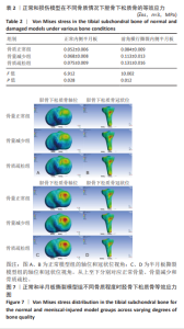
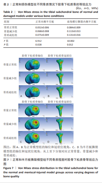
2.2.3 正常组与损伤组胫骨平台下松质骨的应力改变和应力分布 在正常组和损伤组中,模型的应力主要集中在胫骨平台内侧下松质骨的前中部,外侧松质骨上部的类圆形区域内,随着骨质的减少,该类圆形区域的等效应力逐渐增大。其中骨量减少组和骨质疏松组正常内侧半月板的等效应力改变相较于正常骨质组分别增加了30.77%和44.23%;而在损伤组中分别增加了34.52%和55.95%,详见表2。此外,可见在损伤组中,松质骨的应力范围更大,所承受的等效应力更大,详情见图7。"
| 1] WANG S, HASE K, KITA S, et al. Biomechanical effects of medial meniscus radial tears on the knee joint during gait: A concurrent finite element musculoskeletal framework investigation. Front Bioeng Biotechnol. 2022;10:957435. [2] LAU BC, CONWAY D, MULVIHILL J, et al. Biomechanical Consequences of Meniscal Tear, Partial Meniscectomy, and Meniscal Repair in the Knee. JBJS Rev. 2018;6(4):e3. [3] PENG X, WU F, HU Y, et al. Current advances in animal model of meniscal injury: From meniscal injury to osteoarthritis. J Orthop Translat. 2025;50: 388-402. [4] CHEN H, LIU L, ZHANG Y. Finite element analysis of the knee joint stress after partial meniscectomy for meniscus horizontal cleavage tears. BMC Musculoskelet Disord. 2023;24(1):744. [5] D’AMBROSI R, MEENA A, RAJ A, et al. In elite athletes with meniscal injuries, always repair the lateral, think about the medial! A systematic review. Knee Surg Sports Traumatol Arthrosc. 2023;31(6):2500-2510. [6] MAMERI ES, DASARI SP, FORTIER LM, et al. Review of Meniscus Anatomy and Biomechanics. Curr Rev Musculoskelet Med. 2022;15(5):323-335. [7] FOX AJS, WANIVENHAUS F, BURGE AJ, et al. The Human Meniscus: A Review of Anatomy, Function, Injury, and Advances in Treatment. Clin Anat. 2015;28(2):269-387. [8] DASZKIEWICZ K, LUCZKIEWICZ P. Biomechanics of the medial meniscus in the osteoarthritic knee joint. Peer J. 2021;9:e12509. [9] CAMPBELL SL, FRANKLIN SP. Medial Meniscal Tears Increase Risk of Moderate to Severe Cartilage Damage to the Medial Femoral Condyle. Vet Comp Orthop Traumatol. 2025. doi: 10.1055/a-2542-2200. [10] 张程, 吴忠书, 李子祺, 等. 膝骨关节炎与骨质疏松症的相关性研究进展[J]. 中国骨质疏松杂志,2021,27(4):618-624. [11] HUANG K, CAI H. The interplay between osteoarthritis and osteoporosis: Mechanisms, implications, and treatment considerations - A narrative review. Exp Gerontol. 2024;197:112614. [12] TAKAHASHI A, UMEHARA J, KAMIMURA M, et al. Obesity is a risk factor for osteoarthritis progression and spontaneous osteoporosis is a risk for the development of spontaneous osteonecrosis in patients with medial meniscus posterior root tear. J Orthop Sci. 2021;26(5):844-849. [13] DONG Y, HU G, DONG Y, et al. The effect of meniscal tears and resultant partial meniscectomies on the knee contact stresses: a finite element analysis. Comput Methods Biomech Biomed Engin. 2014;17(13):1452-1463. [14] YANG Q, ZHU XY, BAO JY, et al. Medial meniscus posterior root tears and partial meniscectomy significantly increase stress in the knee joint during dynamic gait. Knee Surg Sports Traumatol Arthrosc. 2023;31(6): 2289-2298. [15] ZHANG K, LI L, YANG L, et al. The biomechanical changes of load distribution with longitudinal tears of meniscal horns on knee joint: a finite element analysis. J Orthop Surg Res. 2019;14(1):237. [16] LO G H, NIU J, MCLENNAN CE, et al. Meniscal damage associated with increased local subchondral bone mineral density: a Framingham study. Osteoarthritis Cartilage. 2008;16(2):261-267. [17] 韩龙飞, 侯文渊, 陆舜, 等. 胫骨平台下低密度区骨量及体积对膝关节内软骨及半月板影响的有限元分析[J]. 中国修复重建外科杂志,2025, 39(3):296-306. [18] WELLS ME, SCANALIATO JP, DUNN JC, et al. Meniscal Injuries: Mechanism and Classification. Sports Med Arthrosc. 2021;29(3):154-157. [19] MAO Z, YANG Q, MENG X, et al. Dynamic biomechanical effects of medial meniscus tears on the knee joint: a finite element analysis. J Orthop Surg Res. 2025;20(1):26. [20] 杨骏良, 路坦, 徐彪, 等. 前交叉韧带部分断裂对膝关节应力影响的三维有限元分析[J]. 中国组织工程研究,2024,28(9):1347-1353. [21] REN S, SHI H, LIU Z, et al. Finite Element Analysis and Experimental Validation of the Anterior Cruciate Ligament and Implications for the Injury Mechanism. Bioengineering (Basel). 2022;9(10):590. [22] LUO J, ZOU Z, ZOU Q, et al. Effects of different microfracture drilling parameters on bone quality: a finite element analysis. Front Bioeng Biotechnol. 2025;12:1515136. [23] 刘蒙飞, 马鹏程, 尹灿, 等. 不同骨密度对膝关节单髁置换后关节内各结构影响的三维有限元分析[J]. 中国组织工程研究,2024,28(24): 3801-3806. [24] WANG Z, MA R, CAI Z, et al. Biomechanical Evaluation of Stand-Alone Oblique Lateral Lumbar Interbody Fusion Under 3 Different Bone Mineral Density Conditions: A Finite Element Analysis. World Neurosurg. 2021;155: E285-E293. [25] 李钟鑫, 刘璐, 高丽兰, 等. 人体全膝关节精细有限元模型建立及有效性验证[J]. 生物医学工程与临床,2020,24(5):501-507. [26] 马鹏程, 张思平, 孙荣鑫, 等. 单髁置换股骨假体不同放置位置的生物力学效应[J]. 中国组织工程研究,2022,26(12):1843-1848. [27] SHAO B, XING J, ZHAO B, et al. Role of the proximal tibiofibular joint on the biomechanics of the knee joint: A three-dimensional finite element analysis. Injury. 2022;53(7):2446-2453. [28] 李钟鑫. 膝关节单间室假体置换的生物力学研究[D]. 天津: 天津理工大学,2021. [29] MASS H, KATZ JN. The influence of meniscal pathology in the incidence of knee osteoarthritis: a review. Skelet Radiol. 2023;52(11):2045-2055. [30] YAN M, LIANG T, ZHAO H, et al. Model Properties and Clinical Application in the Finite Element Analysis of Knee Joint: A Review. Orthop Surg. 2024; 16(2):289-302. [31] ZELLMANN P, RIBITSCH I, HANDSCHUH S, et al. Finite Element Modelling Simulated Meniscus Translocation and Deformation during Locomotion of the Equine Stifle. Animals. 2019;9(8):502. [32] PAPALIA R, VASTA S, FRANCESCHI F, et al. Meniscal root tears: from basic science to ultimate surgery. Br Med Bull. 2013;106(1):91-115. [33] WANG JY, QI YS, BAO HRC, et al. The effects of different repair methods for a posterior root tear of the lateral meniscus on the biomechanics of the knee: a finite element analysis. J Orthop Surg Res. 2021;16(1):296. [34] KIM YM, JOO YB, LEE WY, et al. Remodified Mason-Allen suture technique concomitant with high tibial osteotomy for medial meniscus posterior root tears improved the healing of the repaired root and suppressed osteoarthritis progression. Knee Surg Sports Traumatol Arthrosc. 2021;29(4): 1258-1268. [35] XU Z, LI Y, RAO J, et al. Biomechanical assessment of disease outcome in surgical interventions for medial meniscal posterior root tears: a finite element analysis. BMC Musculoskelet Disord. 2022;23(1):1093. [36] SHARMA L, SONG J, FELSON DT, et al. The role of knee alignment in disease progression and functional decline in knee osteoarthritis. JAMA. 2001;286(2):188-195. [37] LI L, YANG L, ZHANG K, et al. Three-dimensional finite-element analysis of aggravating medial meniscus tears on knee osteoarthritis. J Orthop Translat. 2020;20:47-55. [38] ENGLUND M, GUERMAZI A, LOHMANDER SL. The Role of the Meniscus in Knee Osteoarthritis: a Cause or Consequence? Radiol Clin North Am. 2009; 47(4):703-712. [39] GOLDRING MB. Articular cartilage degradation in osteoarthritis. HSS J. 2012;8(1):7-9. [40] 韩龙飞, 林天烨, 何敏聪, 等. 胫骨平台下低密度区骨量及体积对下肢力线影响的有限元分析[J]. 中国修复重建外科杂志,2024,38(6):734-741. [41] ANWAR A, HU Z, ZHANG Y, et al. Multiple Subchondral Bone Cysts Cause Deterioration of Articular Cartilage in Medial OA of Knee: A 3D Simulation Study. Front Bioeng Biotechnol. 2020;8:573938. [42] KISHIRO S, UEHARA K, MINAKAWA N, et al. Subchondral insufficiency fractures of the medial tibial condyle are associated with medial meniscus extrusion: A retrospective observational study. J Orthop Sci. 2025;30(2): 333-338. [43] SHOJI-MATSUNAGA A, ONO T, HAYASHI M, et al. Osteocyte regulation of orthodontic force-mediated tooth movement via RANKL expression. Sci Rep. 2017;7(1):8753. [44] HASHIMOTO T, AKAGI M, TSUKAMOTO I, et al. RANKL-mediated osteoclastic subchondral bone loss at a very early stage precedes subsequent cartilage degeneration and uncoupled bone remodeling in a mouse knee osteoarthritis model. J Orthop Surg Res. 2025;20(1):226. [45] CHEN Y, HU Y, YU YE, et al. Subchondral Trabecular Rod Loss and Plate Thickening in the Development of Osteoarthritis. J Bone Miner Res. 2018;33(2):316-327. [46] GOLDRING SR, GOLDRING MB. Changes in the osteochondral unit during osteoarthritis: structure, function and cartilage-bone crosstalk. Nat Rev Rheumatol. 2016;12(11):632-644. [47] DAY JS, DING M, VAN DER LINDEN JC, et al. A decreased subchondral trabecular bone tissue elastic modulus is associated with pre-arthritic cartilage damage. J Orthop Res. 2001;19(5):914-918. |
| [1] | Chen Huiting, Zeng Weiquan, Zhou Jianhong, Wang Jie, Zhuang Congying, Chen Peiyou, Liang Zeqian, Deng Weiming. Tail anchoring technique of vertebroplasty in treatment of osteoporotic vertebral compression fractures with intravertebral cleft: a finite element analysis [J]. Chinese Journal of Tissue Engineering Research, 2026, 30(9): 2145-2152. |
| [2] | Zeng Xuan, Weng Rui, Ye Shicheng, Tang Jiadong, Mo Ling, Li Wenchao. Two lumbar rotary manipulation techniques in treating lumbar disc herniation: a finite element analysis of biomechanical differences [J]. Chinese Journal of Tissue Engineering Research, 2026, 30(9): 2153-2161. |
| [3] | Cheng Qisheng, Julaiti·Maitirouzi, Xiao Yang, Zhang Chenwei, Paerhati·Rexiti. Finite element analysis of novel variable-diameter screws in modified cortical bone trajectory of lumbar vertebrae [J]. Chinese Journal of Tissue Engineering Research, 2026, 30(9): 2162-2171. |
| [4] | Wu Hongxu, Liu Xuanyu, Wang Taoyu, Wang Shiyao, Cheng Jingyi, Zhang Mingwen, Zhang Yinxia, Liu Zhihua, Wang Xiaojie. Finite element simulation of scoliosis with muscle unit introduction: verification of correction effect under bidirectional load [J]. Chinese Journal of Tissue Engineering Research, 2026, 30(9): 2172-2181. |
| [5] | Liu Jiafu, Ren Ruxia, Liao Zhouwei, Zhou Xiali, Wu Yihong, Zhang Shaoqun. Three-dimensional finite element analysis of cervical spine biomechanical characteristics in a rat model of cervical vertigo [J]. Chinese Journal of Tissue Engineering Research, 2026, 30(9): 2182-2190. |
| [6] | Zhou Daobin, Wang Kehao, Xie Yang, Ning Rende. Biomechanical characteristics of volar locking plate only versus combined dorsal mini-plate fixation of distal radius fractures with dorsal ulnar fragment [J]. Chinese Journal of Tissue Engineering Research, 2026, 30(9): 2255-2261. |
| [7] | Liu Wenlong, Dong Lei, Xiao Zhengzheng, Nie Yu. Finite element analysis of tibial prosthesis loosening after fixed-bearing unicompartmental knee arthroplasty for osteoporosis [J]. Chinese Journal of Tissue Engineering Research, 2026, 30(9): 2191-2198. |
| [8] | Zheng Wangyang, Fei Ji, Yang Di, Zhao Lang, Wang Lingli, Liu Peng, Li Haiyang. Finite element analysis of the force changes of the supraspinatus tendon and glenohumeral joint during the abduction and flexion of the humerus [J]. Chinese Journal of Tissue Engineering Research, 2026, 30(9): 2199-2207. |
| [9] | Cai Qirui, Dai Xiaowei, Zheng Xiaobin, Jian Sili, Lu Shaoping, Liu Texi, Liu Guoke, Lin Yuanfang. Mechanical effects of Long’s traction orthopedic method on cervical functional units: quantitative analysis of biomechanical model of head and neck [J]. Chinese Journal of Tissue Engineering Research, 2026, 30(9): 2208-2216. |
| [10] | Rao Jingcheng, Li Yuwan, Zheng Hongbing, Xu Zhi, Zhu Aixiang, Shi Ce, Wang Bing, Yang Chun, Kong Xiangru, Zhu Dawei. Biomechanical differences between the new proximal femoral stable intramedullary nail and traditional intramedullary nail#br# [J]. Chinese Journal of Tissue Engineering Research, 2026, 30(9): 2217-2225. |
| [11] | Chen Long, Wang Xiaozhen, Xi Jintao, Lu Qilin. Biomechanical performance of short-segment screw fixation combined with expandable polyetheretherketone vertebral body replacement in osteoporotic vertebrae [J]. Chinese Journal of Tissue Engineering Research, 2026, 30(9): 2226-2235. |
| [12] | Yan Xiangning, Chen Lei, Chen Yonghuan, Wang Chao, Li Xiaosheng. Influence of different depths and loads on knee joint mechanics and peripheral muscle force characteristics during squatting [J]. Chinese Journal of Tissue Engineering Research, 2026, 30(9): 2236-2247. |
| [13] | Zhang Nan, Meng Qinghua, Bao Chunyu. Characteristics and clinical application of ankle joint finite element models [J]. Chinese Journal of Tissue Engineering Research, 2026, 30(9): 2343-2349. |
| [14] | Zhang Zizheng, Luo Wang, Liu Changlu. Application value of finite element analysis on unicompartmental knee arthroplasty for medial knee compartmental osteoarthritis [J]. Chinese Journal of Tissue Engineering Research, 2026, 30(9): 2313-2322. |
| [15] | Zhang Xianxu, Ma Zhong, Liu Xin, Huang Lei, Shen Wenxiang, Luo Zhiqiang . Lumbar fusion combined with unilateral fixation for lumbar degenerative diseases: biomechanics, technical evolution, and clinical applications [J]. Chinese Journal of Tissue Engineering Research, 2026, 30(9): 2334-2342. |
| Viewed | ||||||
|
Full text |
|
|||||
|
Abstract |
|
|||||