Chinese Journal of Tissue Engineering Research ›› 2026, Vol. 30 ›› Issue (35): 9231-9238.doi: 10.12307/2026.300
Previous Articles Next Articles
Applications and advances of tissue/organ perfusion
Yuan Yue1, Chang Guoxin1, Lin Dingmei1, Mo Zixuan1, Yu Jingjing2, Zhu Yixia2, Zheng Zhaoguang2, 3, Wang Yan1
- 1Guangdong Pharmaceutical University, Guangzhou 510006, Guangdong Province, China; 2School of Medicine, Foshan University, Foshan 528000, Guangdong Province, China; 3Foshan Newtopcome Pharmaceutical Technology Co., Ltd., Foshan 528000, Guangdong Province, China
-
Received:2025-10-29Revised:2025-12-20Online:2026-12-18Published:2026-04-28 -
Contact:Wang Yan, PhD, Professor, Master’s supervisor, Guangdong Pharmaceutical University, Guangzhou 510006, Guangdong Province, China -
About author:Yuan Yue, MS candidate, Guangdong Pharmaceutical University, Guangzhou 510006, Guangdong Province, China -
Supported by:Guangdong Provincial Key Research and Development Program, No. 2023ZDZX2058 (to ZZG)
CLC Number:
Cite this article
Yuan Yue, Chang Guoxin, Lin Dingmei, Mo Zixuan, Yu Jingjing, Zhu Yixia, Zheng Zhaoguang, Wang Yan. Applications and advances of tissue/organ perfusion[J]. Chinese Journal of Tissue Engineering Research, 2026, 30(35): 9231-9238.
share this article
Add to citation manager EndNote|Reference Manager|ProCite|BibTeX|RefWorks
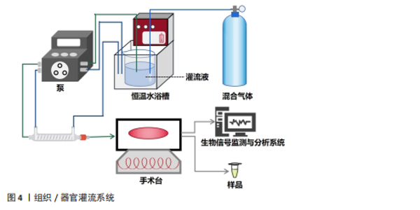
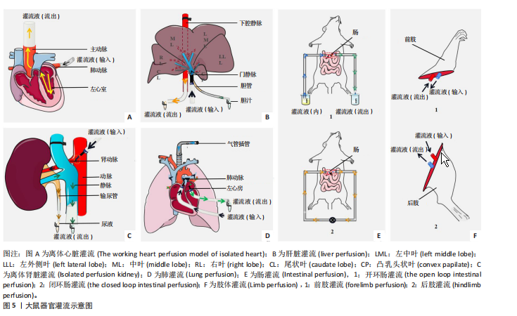
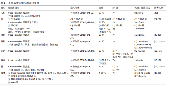
2.1 灌流技术分类 组织/器官灌流是指在组织/器官或组织/器官的血管上插管并持续通入灌流液。整个组织/器官灌流系统主要包括混合器官、恒温水浴、泵、手术台、生物信号检测和分析系统,见图4。通常灌流液会先被混合气体罐中混合气体(O2和CO2)饱和,并在恒温水浴中保持恒温,通过泵来调节灌流液的流速并灌流组织/器官。最后,生物信号检测和分析系统可以获得实时心电图、灌流心脏压力等生物数据,与此同时可以收集流出的灌流液样本。根据灌流过程中是否将组织/器官离体可将灌流分为原位灌流和离体灌流。原位灌流,也称在体灌流,是保持组织/器官留在体内直接进行灌流。原位灌流最能接近动物的真实生理状态,但一般很难排除机体其他器官或组织的影响。离体灌流,也称体外灌流,是组织/器官迅速与动物分离后并置于特定仪器和环境里进行灌流。离体灌流可以更稳定地进行定性或定量分析,但相较于体内真实生理状态势必存在误差。组织/器官灌流可能受到灌流液成分、温度、pH值、流速和混合气体组成的影响。生理盐水,又称平衡盐溶液,因其缓冲能力、等渗性和营养供应特性,可以维持渗透压和pH值的稳定,提供一些简单的营养物质,以满足实验中细胞、组织或器官生存和代谢的基本需要。因此,大多数灌流实验都选择生理盐水作为灌流液。Krebs-Henseleit缓冲液是组织/器官灌流的基本生理盐水(NaCl、KCl、CaCl2、MgSO4、KH2PO4、NaHCO3、Glucose),可根据不同组织/器官的具体生理条件添加合适的牛血清白蛋白、乙二胺四乙酸或肝素等进行改良。此外,也可以对混合气体成分进行细微的修改,以满足不同类型的组织/器官。目前,主要的组织/器官类型包括心脏灌流、脑灌流、肾脏灌流、肠灌流、肺灌流、肝脏灌流、神经灌流、肢体灌流等。主要组织/器官灌流如图5所示,下面将详细介绍它们的特性。 2.1.1 心脏灌流 在1895年,哺乳动物的离体心脏模型被Oscar Langendorff首次建立,由于他的巨大贡献,该模型被命名为Langendorff离体心脏灌流系统。通过生物信号分析软件,Langendorff离体心脏灌流系统可以记录心内压、动脉血压和心电图信号。作为心脏研究中的一种强大技术,Langendorff离体心脏灌流系统促进了医学研究的巨大发展,主要是在药理学和生理学方面[2-3]。动物麻醉(吸入或注射)后,将心脏迅速分离取出,置于4 ℃的KHB中轻按排除血液,然后迅速将灌流设备的针管插入心脏主动脉用手术线结扎(插管不可损伤动脉瓣),接着在恒压或恒流模式下进行灌流并检测信号。也有一些研究者先在体插管结扎再分离心脏。为了使离体心脏模型更接近生理条件,研究人员还使用了工作心脏灌流模型,该模型在肺静脉中添加了一个插管,将灌流液注入左心房。两种模型的结合可以更准确地评估离体心脏[4]。迄今为止,心脏灌流研究侧重于心脏缺血再灌注,缺血模型可通过结扎升主动脉或灌流一段零流量来模拟[5]。心脏手术操作、灌流液成分、温度、流速和压力等因素都会影"

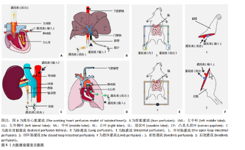
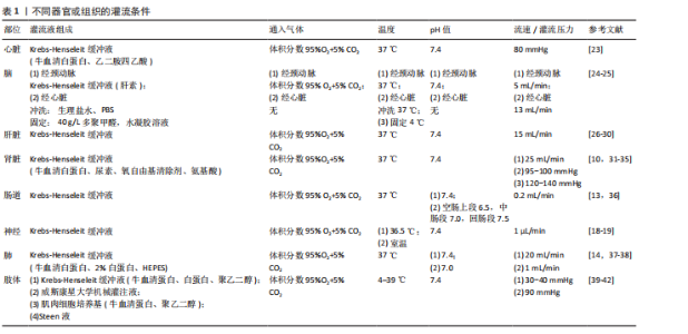
响灌流实验的结果。灌流实验要求操作者熟练掌握手术技巧,并尽可能缩短手术时间来减少缺血性损伤和惊厥的影响。对于离体心脏灌流实验,从打开胸腔到分离心脏并建立灌流系统的整个手术过程应在2 min内完成[6]。 2.1.2 脑灌流 在1984年,SMITH等[7]发展了一套以大鼠为实验对象的右颈外动脉插管逆向原位脑灌流技术,目前主要被广泛运用于研究物质跨血脑屏障转运机制、物质跨血脑屏障转运动力学特征和血脑屏障的性质等方面。颈动脉插管是一项要求高、难度大的手术操作,而通过心脏插管灌流大脑则相对简单易操作,目前多用于固定脑组织。固定液从心脏左心室流入血液循环系统,通过循环系统和毛细血管网到达所有细胞以固定组织。首先将动物麻醉并打开胸腔,将灌流管插入心脏或者升主动脉通入PBS冲洗血管,通常以肝脏和其他组织变白作为清洗结束标准,再将灌流液改为40 g/L多聚甲醛,待灌流结束后将脑组织取出进行切片和染色,使用免疫组化技术可继续研究脑内蛋白表达和药物作用机制。值得注意的是,灌流时间过长会造成器官或组织损伤。为了确保大脑的完整性和活性,灌流时间控制在30 min内可以保障大脑血脑屏障功能正常[8]。灌流固定脑组织的血管和组织完整性还受灌流压的影响,125-150 mmHg是保持脑血管系统和神经元结构的最佳压力范围[9]。 2.1.3 肾脏灌流 离体肾灌流模型是有WEISS等[10]于1959年首次提出的。在哺乳动物中,药物主要是通过含丰富代谢酶的肾脏代谢的。因此,离体肾灌流也是研究药代动力学和药理学的一个非常有用的模型。离体肾脏灌流的操作过程类似于离体心脏灌流。离体肾灌流的手术操作程序[11],一般是先将实验动物麻醉,肾动脉插管以灌流液恒速灌流,再进行肾静脉和输尿管插管,在体灌流平衡一段时间后肾被摘离置于特定的离体装置进行灌流。 2.1.4 肠灌流 口服给药仍然是首选的给药途径,肠灌流作为一种非常重要的药物吸收模型,可以很好地了解药物的肠道吸收。将小肠段在开环或闭环装置里进行原位灌流,开环装置不循环插管段的灌流液;闭环装置中灌流液可以通过插管肠段再循环使用。原位肠灌流模型中,血液和神经系统保持完整,创造的实验条件也比较接近体内情况。LOZOYA-AGULLO等[12]分别用单向肠灌流和闭环肠灌流两种实验方法,发现了药物在空肠不同节段有着显著的区域依赖性渗透性,并且不论是采用单向肠灌流还是闭环肠灌流,所得数据都同样可靠。离体肠灌流也可以实时评估药物对不同节段小肠的影响,但一般用于初步的定性研究。值得注意的是,在肠灌流实验中,在肠段与身体分离之前,用冷的(4 ℃)HEPES缓冲液冲洗腹膜腔可以减少缺血性损伤[13]。 2.1.5 肺灌流 离体肺灌流也是生物制药研究的一个合适模型,不仅可以探索药物在肺部代谢过程,还可以确定吸入给药的药物沉积与吸收过程。与上述灌流插管操作不同的是,离体肺灌流首先需要切开气管并迅速插上通有100%氧气的气管,然后切开肾动脉放血,在肺动脉和左心房插灌流管,灌流液从肺动脉流出左心房。在灌流液中加入药物可以研究药物在肺部代谢情况,倘若研究吸入制剂,可将通气正压变为负压,将药物定期深入吹入。肺灌流过程要持续监测肺部力学数据,在整个操作过程也可以通过目视肺部是否有水肿来检查肺活力[14-15]。 2.1.6 肝脏灌流 肝脏是由多个叶片组成的结构复杂的器官,在肝脏灌流中,通常在门静脉插管使灌流液流入,从腔静脉流出,在胆管插管后可以收集胆汁并检测流量。大部分研究者会控制灌流液温度为37 ℃,但部分研究人员会稍微提高温度至37.8 ℃[16],这可能与肝脏是体内温度最高的器官有关。但是,对于这微弱的温度差异是否会影响实验或影响肝脏活动,目前还没有研究或指南做出明确解释。肝脏作为代谢器官,除了可以很好地研究药物代谢外,还以研究药物对胆汁分泌的影响[17]。 2.1.7 神经灌流 关于神经灌流的研究较少,这可能与手术操作难度高有关。另外,与上述气管灌流不同,神经灌流并不是通过血管插管来进行灌流的。研究人员将微透析探针植入并定位于大鼠纹状体并灌流人工脑脊液,通过测定收集脑透析液中神经递质的含量可以确定药物对神经的影响[18]。将离体脊髓段横向切成厚片(400-500 μm)孵育后,将切片移至自制的全浸式记录浴槽底网上,固定切片后,在室温下持续灌流经混合气体饱和人工脑脊液,再利用微控电极拉制仪记录细胞电生理[19]。 2.1.8 肢体灌流 离体肢体灌流是一种实验技术,用于在离体维持动物肢体存活并进行研究。通常采用猪、兔、鼠的前肢或后肢构建灌流模型。在离体后肢灌流中需分离出股动脉、股静脉及股神经,结扎并离断同侧旋髂动、静脉及阴部动、静脉。插管使灌流液从股动脉流入,从股静脉流出,并精确控制流速、压力和温度[20]。该方法常用于测试药物在肢体组织中的分布、研究肌肉代谢功能、观察血管反应,以及模拟缺血等疾病来评估新疗法。其优势在于能排除生物体全身干扰,实现对局部生理病理过程的精准控制和分析。值得注意的是前肢灌流模型使用较低的流速即可达到与后肢灌流相似的结果[21]。目前尚无在各种温度设置和模型中都能显示出一致结果的最佳灌流液成分。离体肢体灌流目前缺乏标准化方案,其灌流液成分与温度等关键参数需根据具体实验条件进行优化[22]。不同组织/器官灌流的操作实验条件如表1所示。 2.2 组织/器官灌流技术的应用 组织/器官灌流技术目的在于高度可控的离体环境中,精"
"
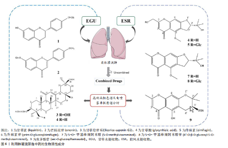
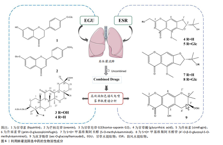
准模拟目标组织/器官的核心生理或病理状态。无论是用于中药多组分、多靶点的活性筛选,还是探究药物的吸收、分布、代谢和排泄(药代动力学),其根本目标都是克服传统二维细胞模型与整体动物实验的局限性,在更好地再现人体组织复杂结构和微环境的同时,实现对特定生物学过程的独立操控与深入解析,从而显著提高研究的预测价值和对临床应用的指导意义。从原理上讲,在离体后尽可能长时间地维持其正常的生理活性和功能完整性,从而允许研究人员进行精确的实时监测和分子干预。基于上述目的和原理,组织/器官灌流技术已用于病理机制、中药活性成分筛选、药效与机制、药物代谢、药物治疗、组织/器官移植等方面的研究。 2.2.1 病理机制研究 缺血再灌注损伤研究是灌流技术应用最多的方面。缺血再灌注是指在血管部分或完全急性阻塞后,血流在一定时间内恢复,从而造成组织损伤并逐渐加重的病理过程。在再灌注过程中会产生大量活性氧,活性氧会破坏细胞膜和细胞结构,线粒体的通透性也会过度增加。心肌缺血在临床上可以通过心肌缺血再灌注治疗,但也可能导致心肌缺血再灌注损伤,临床表现为心律失常、心肌狭窄和微血管阻塞。采用Langendorff 离体心脏灌流系统的研究发现过表达哺乳动物雷帕霉素靶蛋白可抑制心肌缺血再灌注损伤中心肌细胞的坏死,不仅抑制了炎症因子白细胞介素6、白细胞介素1β、肿瘤坏死因子α、单核细胞趋化蛋白1、巨噬细胞炎症蛋白1α的表达,增加了抗炎细胞因子白细胞介素10的表达[23]。在心肌缺血再灌注损伤中,Apelin作为G蛋白偶联APJ受体的内源性配体,增强了超氧化物歧化酶活性和细胞外信号调节激酶1/2、Akt磷酸化[43],除了Bcl-2关联永生基因3,磷酸二酯酶3B也在心脏缺血再灌注损伤中发挥保护作用[44-45],可作为心肌缺血再灌注损伤的治疗靶点。 在离体肾脏灌流中,中性粒细胞加剧了肾脏的缺血再灌注损伤,这主要与O2代谢物有关[46]。此外,离体肾灌流模型可以很好地监测肾小管功能。通过灌流黏蛋白然后计算平均清除率可以发现黏蛋白在肾脏的大量积聚,从而揭示黏蛋白治疗铜绿假单胞菌和其他革兰阴性杆菌引起肾毒性的原因,也能观察黏蛋白在肾脏的分布情况[47]。在肾消融诱导的肾衰竭中,血管紧张素Ⅱ1型受体刺激介导血管紧张素Ⅱ诱导的血管收缩,而血管紧张素Ⅱ2型受体刺激介导血管舒张。离体肾灌流显示肾功能衰竭导致血管舒张增强,因此血管紧张素Ⅱ2型受体依赖性血管舒张与CYP-AA代谢物有关,血管紧张素Ⅱ2型受体mRNA表达在肾功能衰竭中上调[48]。鞘氨醇-1-磷酸诱导鞘氨醇-1-磷酸2受体介导的血管收缩且这种反应在糖尿病大鼠中增强,这也意味着鞘氨醇-1-磷酸2可能是控制糖尿病引起的血管并发症的药物开发靶点之一[49]。蛇毒会导致急性肾损伤,在灌流罗素蛇毒后发现会导致灌流压和肾血管阻力显著降低,随后逐渐升高至正常水平。罗素蛇毒还会导致肾小球滤过率、尿流量和渗透压清除率显著降低。血小板活化因子受体拮抗剂WEB2086对上述变化有不同程度的抑制作用,这表明罗素蛇毒及其成分对肾脏的影响是协同作用,部分由血小板活化因子介导[30]。 其他组织/器官灌流液被应用于病理机制研究。LARUE等[50]用脑灌流模型证明了淀粉样蛋白Aβ在阿尔茨海默病转基因小鼠模型中的积累,在肝脏灌流过程中用N2取代O2引起肝脏缺氧,可以明显看到肝细胞质膜和细胞质通过内皮开口向肝窦腔内囊状突出,中央小叶区域的细胞发生了显著的结构变化[51]。 2.2.2 中药生物活性成分筛选 现代药理学研究表明,药物与细胞或膜上的受体或通道相互作用的能力是决定药物在生物体内行为至关重要的第一步。因此,化合物与细胞膜、细胞或器官的相互作用已被成功用作中药生物活性成分假说的基础[52-53]。2011年首次建立了用于筛选中药生物活性成分的离体组织/器官灌流:首先,离体组织/器官并用KHB洗去血液,然后灌流中药提取物,用PBS冲洗掉未结合的成分,最后通过改变灌流液的pH值来解离结合的成分,并用高效液相色谱及电喷雾串联质谱分析鉴定结合的成分。使用离体肺灌流结合高效液相色谱及电喷雾串联质谱分析,分别从甘草和防风的水提取物中快速筛选活性成分。从离体肺灌流筛选中获得了来自甘草的4种化合物和来自防风的5种化合物(图6)。此外,来自甘草的化合物可以在体外显著抑制肺上皮细胞(A549细胞)的损伤。今后,离体组织/器官有可能发展成为中药生物活性成分强大而可靠的筛选工具[54-55]。"
| [1] LE GALLOIS M. Experiments on the principle of life, and particularly on the principle of the motions of the heart, and on the seat of this principle: including the report made to the first class of the Institute, upon the experiments relative to the mtions of the heart: M.Thomas, 1813:328. [2] LANGENDORFF O. Untersuchungen am überlebenden Säugethierherzen. Archiv für die gesamte Physiologie des Menschen und der Tiere. 1885;26(1):285-295. [3] 李楠,张晨,孙艳君,等.Langendorff心脏灌流技术(针对心肌缺血再灌注)的应用和发展[J].中国实验动物学报,2023,31(8):1070-1077. [4] MUESSIG JM, KAYA S, MOELLHOFF L, et al. A model of blood component-heart interaction in cardiac ischemia-reperfusion injury using a langendorff-based ex vivo assay. J Cardiovasc Pharmacol Ther. 2020;25(2):164-173. [5] ZHANG S, LI H, YANG SJ. Tribulosin protects rat hearts from ischemia/reperfusion injury. Acta Pharmacol Sin. 2010;31(6):671-678. [6] SUTHERLAND FJ, SHATTOCK MJ, BAKER KE, et al. Mouse isolated perfused heart: characteristics and cautions. Clin Exp Pharmacol Physiol. 2003;30(11): 867-878. [7] SMITH QR, TAKASATO Y, SWEENEY DJ, et al. Regional cerebrovascular transport of leucine as measured by the insitu brain perfusion technique. J Cereb Blood Flow Metab. 1985;5(2):300-311. [8] ZLOKOVIC BV, BEGLEY DJ, DJURICIC BM, et al. Measurement of solute transport across the blood-brain barrier in the perfused guinea pig brain: method and application to N-methyl-alpha-aminoisobutyric acid. J Neurochem. 1986;46(5): 1444-1451. [9] SCHWARZMAIER SM, KNARR M, HU S, et al. Perfusion pressure determines vascular integrity and histomorphological quality following perfusion fixation of the brain. J Neurosci Methods. 2022; 372:109493. [10] WEISS C, PASSOW H, ROTHSTEIN A. Autoregulation of flow in isolated rat kidney in the absence of red cells. Am J Physiol. 1959;196(5):1115-1118. [11] GONZALEZ-VIEDMA A, VAN DYCK R, DE BEULE J, et al. Unraveling metabolism during kidney perfusion using tracer studies: a systematic review. Artif Organs. 2022;46(11):2118-2134. [12] LOZOYA-AGULLO I, ZUR M, BEIG A, et al. Segmental-dependent permeability throughout the small intestine following oral drug administration: Single-pass vs. Doluisio approach to in-situ rat perfusion. Int J Pharm. 2016;515(1-2):201-208. [13] FINOTTI M, BARAHONA M, MAINA RM, et al. L-arginine prevents ischemic injury in explanted rat intestinal regions in an ex vivo perfusion model. Transplantation Reports. 2022;7(2): 100096. [14] DONG M, MURDTER TE, PHILIPPI C, et al. Pulmonary delivery and tissue distribution of aerosolized antisense 2’-O-Methyl RNA containing nanoplexes in the isolated perfused and ventilated rat lung. Eur J Pharm Biopharm. 2012;81(3):478-485. [15] WEKSLER B, LENERT J, NG B, et al. Isolated single lung perfusion with doxorubicin is effective in eradicating soft tissue sarcoma lung metastases in a rat model. J Thorac Cardiovasc Surg. 1994; 107(1):50-54. [16] ESMAEILI Z, MOHAMMADI S, NEZAMI A, et al. A disposition kinetic study of Tramadol in bile duct ligated rats in perfused rat liver model. Biomed Pharmacother. 2017;91:251-256. [17] AVNER DL, LEE RG, BERENSON MM. Protoporphyrin-induced cholestasis in the isolated in situ perfused rat liver. J Clin Invest. 1981;67(2): 385-394. [18] 潘凌云, 王雨, 刘新华, 等. UPLC-MS/MS同时测定大鼠脑微透析样品中11个神经递质含量[J].中国中药杂志,2022,47(12):3242-3250. [19] 吴金蓉, 汪萌芽. 多巴胺对脊髓运动神经元下行激活和外周传入突触传递的差异性影响[J].皖南医学院学报,2023,42(1):4-8. [20] ILHAN E, BUYUKAFSAR K, TIFTIK RN, et al. Effects of the Rho/Rho-Kinase Pathway on Perfusion Pressure in the Isolated-Perfused Rat Hind Limb Vascular Bed. Balkan Med J. 2021;38(5):304-309. [21] PENDEXTER CA, HAQUE O, MOJOUDI M, et al. Development of a rat forelimb vascularized composite allograft (VCA) perfusion protocol. PLoS One. 2023;18(1):e266207. [22] DURU C, BINIAZAN F, HADZIMUSTAFIC N, et al. Review of machine perfusion studies in vascularized composite allotransplant preservation. Front Transplant. 2023;2:1323387. [23] AOYAGI T, KUSAKARI Y, XIAO CY, et al. Cardiac mTOR protects the heart against ischemia-reperfusion injury. Am J Physiol Heart Circ Physiol. 2012;303(1):H75-H85. [24] DEMEULE M, CURRIE JC, BERTRAND Y, et al. Involvement of the low-density lipoprotein receptor-related protein in the transcytosis of the brain delivery vector angiopep-2. J Neurochem. 2008;106(4):1534-1544. [25] MURAKAMI H, TAKANAGA H, MATSUO H, et al. Comparison of blood-brain barrier permeability in mice and rats using in situ brain perfusion technique. Am J Physiol Heart Circ Physiol. 2000; 279(3):H1022-H1028. [26] TAKEDA K, IKENAKA Y, TANAKA KD, et al. Investigation of hepatic warfarin metabolism activity in rodenticide-resistant black rats (Rattus rattus) in Tokyo by in situ liver perfusion. Pestic Biochem Physiol. 2018;148:42-49. [27] AHMED I, ATTIA MS, AHMAD N, et al. Use of isolated perfused rat liver model for testing liver preservation solutions. Transplant Proc. 2001; 33(7):3710-3712. [28] WANG HJ, BENET LZ. Protein binding and hepatic clearance: re-examining the discrimination between models of hepatic clearance with diazepam in the isolated perfused rat liver preparation. Drug Metab Dispos. 2019;47(12): 1397-1402. [29] CHEUNG K, HICKMAN PE, POTTER JM, et al. An optimized model for rat liver perfusion studies. J Surg Res. 1996;66(1):81-89. [30] DIBAEI M, HOSSEINI A, LAVASANI H, et al. Assessment of metabolic interaction between curcumin and tramadol using the isolated perfused rat liver. Heliyon. 2024;10(15):e35070. [31] CHAIYABUTR N, CHANHOME L, VASARUCHAPONG T, et al. The pathophysiological effects of Russell’s viper (Daboia siamensis) venom and its fractions in the isolated perfused rabbit kidney model: a potential role for platelet activating factor. Toxicon: X. 2020;7:100046. [32] HILLIARD LM, OSICKA TM, CLAVANT SP, et al. Characterization of the urinary albumin degradation pathway in the isolated perfused rat kidney. J Lab Clin Med. 2006;147(1):36-44. [33] LI J, WANG DH. Increased GFR and renal excretory function by activation of TRPV1 in the isolated perfused kidney. Pharmacol Res. 2008; 57(3):239-246. [34] VILLALTA M, SAMPAIO TL, DE MENEZES R, et al. Nephrotoxicity induced by the venom of Hypnale hypnale from Sri Lanka: studies on isolated perfused rat kidney and renal tubular cell lines. Toxicon. 2019;165:40-46. [35] CZOGALLA J, SCHWEDA F, LOFFING J. The mouse isolated perfused kidney technique. J Vis Exp. 2016;(117):54712. [36] JAPPAR D, WU SP, HU Y, et al. Significance and regional dependency of peptide transporter (PEPT) 1 in the intestinal permeability of glycylsarcosine: in situ single-pass perfusion studies in wild-type and Pept1 knockout mice. Drug Metab Dispos. 2010;38(10):1740-1746. [37] SAKURAI F, NISHIOKA T, YAMASHITA F, et al. Effects of erythrocytes and serum proteins on lung accumulation of lipoplexes containing cholesterol or DOPE as a helper lipid in the single-pass rat lung perfusion system. Eur J Pharm Biopharm. 2001;52(2):165-172. [38] ERIKSSON J, THORN H, LENNERNAS H, et al. Pulmonary drug absorption and systemic exposure in human: Predictions using physiologically based biopharmaceutics modeling. Eur J Pharm Biopharm. 2020;156:191-202. [39] VERAZA RJ, LOPEZ R, PARRY O, et al. Proof of concept study for a closed ex vivo limb perfusion system for 24-hour subnormothermic preservation using acellular perfusate. J Trauma Acute Care Surg. 2022;93(2S Suppl 1): S102-S109. [40] KRUIT AS, BROUWERS K, VAN MIDDEN D, et al. Successful 18-h acellular extracorporeal perfusion and replantation of porcine limbs - Histology versus nerve stimulation. Transpl Int. 2021;34(2): 365-375. [41] BURLAGE LC, LELLOUCH AG, TAVEAU CB, et al. Optimization of Ex Vivo Machine Perfusion and Transplantation of Vascularized Composite Allografts. J Surg Res. 2022;270:151-161. [42] HAUG V, KOLLAR B, ENDO Y, et al. Comparison of Acellular Solutions for Ex-situ Perfusion of Amputated Limbs. Mil Med. 2020;185(11-12): e2004-e2012. [43] ZENG XJ, ZHANG LK, WANG HX, et al. Apelin protects heart against ischemia/reperfusion injury in rat. Peptides. 2009;30(6):1144-1152. [44] SU F, MYERS VD, KNEZEVIC T, et al. Bcl-2-associated athanogene 3 protects the heart from ischemia/reperfusion injury. JCI Insight. 2016; 1(19):e90931. [45] CHUNG YW, LAGRANHA C, CHEN Y, et al. Targeted disruption of PDE3B, but not PDE3A, protects murine heart from ischemia/reperfusion injury. Proc Natl Acad Sci U S A. 2015;112(17): E2253-E2262. [46] LINAS SL, SHANLEY PF, WHITTENBURG D, et al. Neutrophils accentuate ischemia-reperfusion injury in isolated perfused rat kidneys. Am J Physiol. 1988;255(4 Pt 2):F728-F735. [47] MA Z, WANG J, NATION RL, et al. Renal disposition of colistin in the isolated perfused rat kidney. Antimicrob Agents Chemother. 2009;53(7): 2857-2864. [48] BAUTISTA R, SANCHEZ A, HERNANDEZ J, et al. Angiotensin II type AT(2) receptor mRNA expression and renal vasodilatation are increased in renal failure. Hypertension. 2001;38(3 Pt 2): 669-673. [49] BAUTISTA-PEREZ R, ARELLANO A, FRANCO M, et al. Sphingosine-1-phosphate induced vasoconstriction is increased in the isolated perfused kidneys of diabetic rats. Diabetes Res Clin Pract. 2011;94(1):e8-e11. [50] LARUE B, HOGG E, SAGARE A, et al. Method for measurement of the blood-brain barrier permeability in the perfused mouse brain: application to amyloid-beta peptide in wild type and Alzheimer’s Tg2576 mice. J Neurosci Methods. 2004;138(1/2):233-242. [51] LEMASTERS JJ, JI S, THURMAN RG. Centrilobular injury following hypoxia in isolated, perfused rat liver. Science. 1981;213(4508): 661-663. [52] ZHENG ZG, DUAN TT, HE B, et al. Macrophage biospecific extraction and HPLC-ESI-MSn analysis for screening immunological active components in Smilacis Glabrae Rhizoma. J Pharm Biomed Anal. 2013;77:44-48. [53] HU Q, JIA L, ZHANG X, et al. Accurate construction of cell membrane biomimetic graphene nanodecoys via purposeful surface engineering to improve screening efficiency of active components of traditional Chinese medicine.Acta Pharm Sin B. 2022;12(1):394-405. [54] ZHENG ZG, WANG RS, CHENG HQ, et al. Isolated perfused lung extraction and HPLC-ESI-MSn analysis for predicting bioactive components of Saposhnikoviae Radix. J Pharm Biomed Anal. 2011;54(3): 614-618. [55] ZHENG ZG, XU YH, LIU F, et al. Screening bioactive components of Glycyrrhiza uralensis Fisch. with isolated perfused lung extraction and HPLC-ESI-MSn analysis. J Pharm Biomed Anal. 2019;169: 127-132. [56] 邵柯捷,任安经.Salusinβ减轻内皮素-1诱导的大鼠离体心脏功能损伤[J].南昌大学学报(医学版),2022,62(6):1-5. [57] ZHANG Z, CHEN J, SU S, et al. Luteolin ameliorates hypoxic pulmonary vascular remodeling in rat via upregulating K(V)1.5 of pulmonary artery smooth muscle cells. Phytomedicine. 2024;132:155840. [58] AKINMOLADUN AC, OLOWE JA, KOMOLAFE K, et al. Antioxidant activity and protective effects of cocoa and kola nut mistletoe (Globimetula cupulata) against ischemia/reperfusion injury in Langendorff-perfused rat hearts. J Food Drug Anal. 2016;24(2):417-426. [59] 曹瑀莹, 杜丙秀, 李劭恒, 等. 人参皂苷Re对异丙肾上腺素诱导离体灌流大鼠心脏心律失常的调节作用[J]. 中草药,2021,52(20):6234-6244. [60] LOZOYA-AGULLO I, GONZÁLEZ-ÁLVAREZ I, GONZÁLEZ-ÁLVAREZ M, et al. In situ perfusion model in rat colon for drug absorption studies: comparison with small intestine and Caco-2 cell model. J Pharm Sci. 2015;104(9):3136-3145. [61] 程珍珍, 周本宏, 姜珊, 等. 基于原位单向肠灌流模型研究没食子酸的肠吸收特性[J]. 中国药理学通报,2021,37(5):669-673. [62] 华悦, 魏晓峰, 李喆, 等. 盐巴戟天有效成分在体肠灌流研究[J]. 中国中医药信息杂志,2021, 28(9):105-110. [63] ANDLAUER W, KOLB J, STEHLE P, et al. Absorption and metabolism of genistein in isolated rat small intestine. J Nutr. 2000;130(4):843-846. [64] GRADOLATTO A, CANIVENC-LAVIER MC, BASLY JP, et al. Metabolism of apigenin by rat liver phase I and phase ii enzymes and by isolated perfused rat liver. Drug Metab Dispos. 2004; 32(1):58-65. [65] EGEN-SCHWIND C, ECKARD R, KEMPER FH. Metabolism of garlic constituents in the isolated perfused rat liver. Planta Med. 1992; 58(4):301-305. [66] BEN-HARARI RR, PARENT EA, KLEINERMAN J. Metabolism of 5-hydroxytryptophan in the isolated perfused rat lung. Pharmacology. 1990; 41(5):272-279. [67] BRIER ME, LATHON PV, ARONOFF GR, et al. Pharmacodynamics of atrial natriuretic peptide in isolated perfused Dahl rat kidneys. Hypertens Res. 1995;18(3):219-225. [68] ARELLANO M, MALET-MARTINO M, MARTINO R, et al. The anti-cancer drug 5-fluorouracil is metabolized by the isolated perfused rat liver and in rats into highly toxic fluoroacetate. Br J Cancer, 1998;77(1):79-86. [69] NG B, LENERT JT, WEKSLER B, et al. Isolated lung perfusion with FUDR is an effective treatment for colorectal adenocarcinoma lung metastases in rats. Ann Thorac Surg. 1995; 59(1):205-208. [70] HENDRIKS JM, VAN SCHIL PE, VAN OOSTEROM AA, et al. Isolated lung perfusion with melphalan prolongs survival in a rat model of metastatic pulmonary adenocarcinoma. Eur Surg Res. 1999; 31(3):267-271. [71] LINDNÉR P, FJÄLLING M, HAFSTRÖM L, et al. Isolated hepatic perfusion with extracorporeal oxygenation using hyperthermia, tumour necrosis factor alpha and melphalan. Eur J Surg Oncol. 1999;25(2):179-185. [72] VORON T, ZINZINDOHOUÉ F, JOURNOIS D, et al. Hyperthermic isolated liver perfusion with melphalan and bevacizumab. J Visc Surg. 2013; 150(1):60-66. [73] DONDOSSOLA D, LONATI C, BATTISTIN M, et al. Twelve-hour normothermic liver perfusion in a rat model: characterization of the changes in the ex-situ bio-molecular phenotype and metabolism. Sci Rep. 2024;14(1):6040. [74] 李唐波, 宋迪煜, 郝国兵, 等. 兔离断后肢深低温冻存中二甲基亚砜导入效果的定量分析[J]. 中国组织工程研究,2025,29(34):7326-7332. [75] TIAN X, GAO M, LI A, et al. Protocol for Isolation of Viable Adult Rat Cardiomyocytes with High Yield. STAR Protoc. 2020;1(2):100045. [76] BUTOVA XA, MYACHINA TA, KHOKHLOVA AD. A combined Langendorff-injection technique for simultaneous isolation of single cardiomyocytes from atria and ventricles of the rat heart. MethodsX. 2021;8:101189. [77] TIRITICCO V, CODOTTO G, BLARASIN B, et al. Rat Liver Perfusion and Primary Hepatocytes Isolation: An Old Procedure Crucial for Cutting-Edge 3D Organoids Culture. J Vis Exp. 2024;(213). doi: 10.3791/66857. [78] CARDIFF RD, MILLER CH, MUNN RJ. Mouse tissue fixation. Cold Spring Harb Protoc. 2014;2014(5): 461-466. [79] GAGE GJ, KIPKE DR, SHAIN W. Whole animal perfusion fixation for rodents. J Vis Exp. 2012;(65): e3564. [80] HADE AC, PHILIPS MA, PROMET L, et al. A cost-effective and efficient ex vivo, ex situ human whole brain perfusion protocol for immunohistochemistry. J Neurosci Methods. 2024;404:110059. [81] MOLCK AM, POULSEN M, TINDGARD LS, et al. Lack of histological cerebellar changes in Wistar rats given pulegone for 28 days. Comparison of immersion and perfusion tissue fixation. Toxicol Lett. 1998;95(2):117-122. [82] BAY V, IVERSEN NK, SHIADEH SMJ, et al. Tissue processing and optimal visualization of cerebral infarcts following sub-acute focal ischemia in rats. J Chem Neuroanat. 2021;118:102034. [83] 傅婷婷,刘艳.大鼠离体肾脏灌流模型在筛选慢性肾病药物的初步应用[J].实验动物与比较医学,2017,37(5):357-362. [84] HOREJS C. Organ chips, organoids and the animal testing conundrum. Nat Rev Mater. 2021;6(5): 372-373. [85] YOKOI F, DEGUCHI S, TAKAYAMA K. Organ-on-a-chip models for elucidating the cellular biology of infectious diseases. BBA-Mol Cell Res. 2023; 1870(6):119504. [86] MESSELMANI T, LE GOFF A, SONCIN F, et al. Investigation of the metabolomic crosstalk between liver sinusoidal endothelial cells and hepatocytes exposed to paracetamol using organ-on-chip technology. Toxicology. 2023;492:153550 |
| [1] | Liu Yuxiao, Huang Sijing, Geng Longyu, Gao Beiyao, Yang Guang, Ge Ruidong, Gao Qi. The functions and underlying molecular mechanisms of PIEZO channels in nervous system diseases [J]. Chinese Journal of Tissue Engineering Research, 2026, 30(34): 9017-9023. |
| [2] | Yang Yunhong, Guo Lihua, Tang Han, Lin Lvping, Kuang Hongjun, Zhao Hong. Proteomic analysis of the mechanism of moxibustion intervention in a rat model of atopic dermatitis [J]. Chinese Journal of Tissue Engineering Research, 2026, 30(29): 7581-7591. |
| [3] | Bai Ruokun, Mo Jian, Han Jie, Li Kunjian, Nie Xiayu, Chen Shuai. Visualization analysis on research literature about animal models for osteonecrosis of the femoral head [J]. Chinese Journal of Tissue Engineering Research, 2026, 30(25): 6643-6653. |
| [4] | Jiang Chao, Che Yanjun. Biological mechanisms and future research trends of cartilaginous endplate degeneration [J]. Chinese Journal of Tissue Engineering Research, 2026, 30(23): 5915-5924. |
| [5] | Ma Zhennan, Wang Yinfeng, Yao Lijuan, Chen Leqin. Glycocalyx: the new link between exercise and disease [J]. Chinese Journal of Tissue Engineering Research, 2026, 30(23): 5972-5981. |
| [6] | Shi Gaolong, Ge Caijun, Chen Jianpeng, Wang Yuanbin, Fan Zelin, Yan Jun, Wang Qianliang. Mechanism by which the paraventricular nucleus of the hypothalamus is involved in chronic pain and anxiety in mice with lumbar disc herniation [J]. Chinese Journal of Tissue Engineering Research, 2026, 30(22): 5707-5715. |
| [7] | Huang Sijing, Cui Rui, Geng Longyu, Gao Beiyao, Ge Ruidong, Jiang Shan. Application and molecular mechanism of extracorporeal shock wave for anti-fibrosis [J]. Chinese Journal of Tissue Engineering Research, 2026, 30(17): 4417-4429. |
| [8] | Peng Hao, Jiang Yang, Song Yanping, Wu Quan, Yao Na, Chen Qigang, Shen Zhen. H-type angiogenesis and its role in various skeletal disease animal models [J]. Chinese Journal of Tissue Engineering Research, 2026, 30(16): 4154-4165. |
| [9] | Wang Qifei, Du Xingbin, Kong Jianda. Neural physiological basis and exercise-induced mechanism of central fatigue [J]. Chinese Journal of Tissue Engineering Research, 2025, 29(32): 6979-6988. |
| [10] | Liang Zhou, Zhang Chi, Pan Chengzhen, Yang Bo, Pu Zhanglin, Liu Hua, Peng Jinhui, Wen Lichun, Ling Guanhan, Chen Feng. Anti-osteoporotic mechanisms of kaempferol based on gut microbiota and comprehensive targeted metabolomics [J]. Chinese Journal of Tissue Engineering Research, 2025, 29(20): 4190-4204. |
| [11] | Long Chenyan, Cheng Biao, Tian Ju. Cellular and molecular mechanisms of platelet-rich plasma in promoting wound healing [J]. Chinese Journal of Tissue Engineering Research, 2025, 29(13): 2793-2801. |
| [12] | Tang Wenjing, Wu Siyuan, Yang Chen, Tao Xi. Inflammatory responses in post-stroke depression [J]. Chinese Journal of Tissue Engineering Research, 2022, 26(8): 1278-1285. |
| [13] | Chen Yanlin, Xu Lin, Xu Shengjia. Effects of physical activity on hippocampal plasticity and cognition [J]. Chinese Journal of Tissue Engineering Research, 2020, 24(5): 773-779. |
| [14] | Si Qing-zong, An Xiao-li, An Ying-fei, Liu Bin. Medical ultra-high molecular weight polyethylene modification and tribological characteristics [J]. Chinese Journal of Tissue Engineering Research, 2013, 17(3): 496-500. |
| Viewed | ||||||
|
Full text |
|
|||||
|
Abstract |
|
|||||