Chinese Journal of Tissue Engineering Research ›› 2026, Vol. 30 ›› Issue (34): 8852-8859.doi: 10.12307/2026.889
Previous Articles Next Articles
Role of myeloid-derived suppressor cells in osteoclast differentiation in primary osteoporosis
Cheng Xinyi, Chen Yida, Wang Yi, Liu Daihui, Zheng Yi, Shi Qin
- Department of Orthopedics, First Affiliated Hospital of Soochow University, Institute of Orthopedics of Soochow University, Suzhou 215006, Jiangsu Province, China
-
Received:2025-10-18Revised:2026-02-13Online:2026-12-08Published:2026-04-11 -
Contact:Shi Qin, PhD, Professor, Department of Orthopedics, First Affiliated Hospital of Soochow University, Institute of Orthopedics of Soochow University, Suzhou 215006, Jiangsu Province, China -
About author:Cheng Xinyi, MS, Department of Orthopedics, First Affiliated Hospital of Soochow University, Institute of Orthopedics of Soochow University, Suzhou 215006, Jiangsu Province, China -
Supported by:National Natural Science Foundation of China (General Program), No. 82172485 (to SQ)
CLC Number:
Cite this article
Cheng Xinyi, Chen Yida, Wang Yi, Liu Daihui, Zheng Yi, Shi Qin. Role of myeloid-derived suppressor cells in osteoclast differentiation in primary osteoporosis[J]. Chinese Journal of Tissue Engineering Research, 2026, 30(34): 8852-8859.
share this article
Add to citation manager EndNote|Reference Manager|ProCite|BibTeX|RefWorks
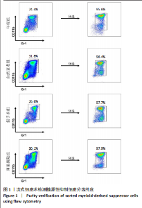
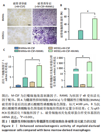
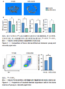
2.1 髓源性抑制细胞的分选纯度鉴定结果 流式细胞术检测结果显示,未分选前,年轻组、自然衰老组、假手术组、卵巢摘除组小鼠骨髓细胞中CD11b+Gr1+髓源性抑制细胞比例分别为24.4%,31.8%,26.6%,30.1%;经磁珠分选后,骨髓细胞中髓源性抑制细胞比例分别为95.6%,96.4%,97.7%,97.9%,见图1,说明分选后的髓源性抑制细胞纯度较高,可以进行后续实验。 2.2 髓源性抑制细胞与骨髓源性巨噬细胞自身破骨活化能力的比较 抗酒石酸酸性磷酸酶染色显示,破骨诱导分化后,髓源性抑制细胞组中多核破骨细胞(≥3个核)数量显著多于骨髓源性巨噬细胞组,并且髓源性抑制细胞组细胞体积更大、抗酒石酸酸性磷酸酶染色阳性区域比例更高,见图2A,B。qRT-PCR检测显示,破骨诱导分化后,髓源性抑制细胞组T细胞核因子1、破骨细胞相关免疫球蛋白样受体基因的mRNA表达高于骨髓源性巨噬细胞组(P < 0.001),见图2C。结果表明,相较于骨髓源性巨噬细胞,髓源性抑制细胞具有更强的活化为成熟破骨细胞的能力。 2.3 年轻和自然衰老雌性小鼠骨微结构的比较 年轻组与自然衰老组小鼠股骨远端骨微结构,如图3A所示,可见自然衰老组小鼠骨质流失严重。定量分析结果显示,与年轻组比较,自然衰老组小鼠骨密度、骨体积分数与骨小梁数量均降低(P < 0.05,P < 0.01),骨小梁分离度增加(P < 0.05),见图3B。 2.4 年轻和自然衰老雌性小鼠骨髓细胞中髓源性抑制细胞比例的比较 流式细胞术检测结果显示,自然衰老组小鼠骨髓细胞中CD11b+Gr1+髓源性抑制细胞比例(33.6±0.3)%高于年轻组(25.4±0.5)%(P < 0.001),见图4。"
"
"

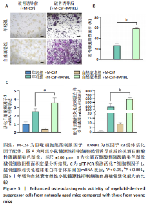
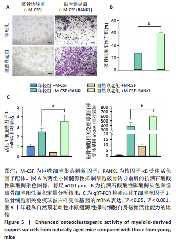
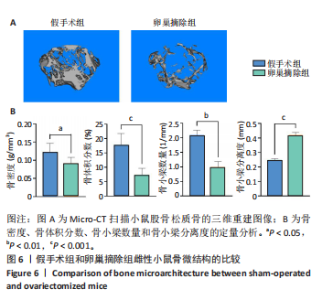
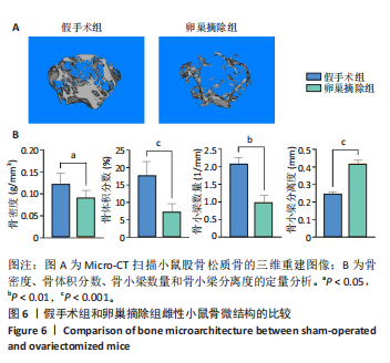
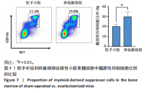
2.5 年轻和自然衰老雌性小鼠髓源性抑制细胞自身破骨活化能力的比较 抗酒石酸酸性磷酸酶染色显示,破骨诱导分化后,自然衰老组髓源性抑制细胞中多核破骨细胞(≥3个核)数量高于年轻组,并且自然衰老组髓源性抑制细胞细胞体积更大、抗酒石酸酸性磷酸酶染色阳性区域比例更高,见图5A,B。qRT-PCR检测显示,破骨诱导分化后,自然衰老组细胞中T细胞核因子1、破骨细胞相关免疫球蛋白样受体基因的mRNA表达高于年轻组(P < 0.05,P < 0.001),见图5C。结果表明,与年轻组髓源性抑制细胞相比,自然衰老组髓源性抑制细胞具有更强的活化为成熟破骨细胞的能力。 2.6 假手术组和卵巢摘除组小鼠骨微结构的比较 假手术组和卵巢摘除组小鼠股骨远端骨微结构,如图6A所示,可见卵巢摘除组小鼠骨质流失严重。定量分析结果显示,与假手术组比较,卵巢摘除组小鼠骨密度、骨体积分数与骨小梁数量均减少(P < 0.05,P < 0.001,P < 0.01),骨小梁分离度增加(P < 0.001),见图6B。 2.7 假手术组和卵巢摘除组小鼠骨髓细胞中髓源性抑制细胞比例的比较 流式细胞术检测结果显示,卵巢摘除组小鼠骨髓细胞中CD11b+Gr1+髓源性抑制细胞比例(29.8±4.7)%高于假手术组(20.1±1.9)%(P < 0.01),如图7所示。"
"
2.8 假手术组与卵巢摘除组小鼠髓源性抑制细胞自身破骨活化能力的比较 抗酒石酸酸性磷酸酶染色显示,破骨诱导分化后,卵巢摘除组髓源性抑制细胞中多核破骨细胞(≥3个核)数量高于假手术组,并且卵巢摘除组髓源性抑制细胞细胞体积更大、抗酒石酸酸性磷酸酶染色阳性区域比例更高,见图8A,B。 qRT-PCR检测显示,破骨诱导分化后,卵巢摘除组细胞中T细胞核因子1、破骨细胞相关免疫球蛋白样受体基因的mRNA表达"

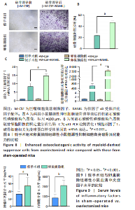
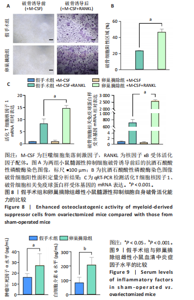
高于假手术组(P < 0.001),见图8C。结果表明,与假手术组髓源性抑制细胞相比,卵巢摘除组髓源性抑制细胞具有更强的活化为成熟破骨细胞的能力。 2.9 假手术组与卵巢摘除组小鼠血清中炎症因子水平的比较 如图9所示,卵巢摘除组小鼠血清中白细胞介素6和肿瘤坏死因子α水平高于较假手术组(P < 0.001,P < 0.05)。"
| [1] FANG H, DENG Z, LIU J, et al. The Mechanism of Bone Remodeling After Bone Aging. Clin Interv Aging. 2022;17:405-415. [2] GENANT HK, COOPER C, POOR G, et al. Interim report and recommendations of the World Health Organization Task-Force for Osteoporosis. Osteoporos Int. 1999;10(4):259-264. [3] PERIS P, MARTÍNEZ-FERRER A, MONEGAL A, et al. Aetiology and clinical characteristics of Male osteoporosis. Have they changed in the last few years? Clin Exp Rheumatol. 2008;26(4):582-588. [4] SØE K, DELAISSE JM, BORGGAARD XG. Osteoclast formation at the bone marrow/bone surface interface: Importance of structural elements, matrix, and intercellular communication. Semin Cell Dev Biol. 2021;112:8-15. [5] SONG S, GUO Y, YANG Y, et al. Advances in pathogenesis and therapeutic strategies for osteoporosis. Pharmacol Ther. 2022;237:108168. [6] UMUR E, BULUT SB, YIĞIT P, et al. Exploring the Role of Hormones and Cytokines in Osteoporosis Development. Biomedicines. 2024;12(8):1830. [7] PAROLINI C. Pathophysiology of bone remodelling cycle: role of immune system and lipids. Biochem Pharmacol. 2025;235:116844. [8] ZHAO Z, DU Y, YAN K, et al. Exercise and osteoimmunology in bone remodeling. FASEB J Off Publ Fed Am Soc Exp Biol. 2024;38(7):e23554. [9] GEUSENS P, LEMS WF. Osteoimmunology and osteoporosis. Arthritis Res Ther. 2011;13(5):242. [10] ZHANG W, GAO R, RONG X, et al. Immunoporosis: role of immune system in the pathophysiology of different types of osteoporosis. Front Endocrinol. 2022;13:965258. [11] PARK Y, KWOK SK. Recent Advances in Cell Therapeutics for Systemic Autoimmune Diseases. Immune Netw. 2022;22(1):e10. [12] FINN OJ. Immuno-oncology: understanding the function and dysfunction of the immune system in cancer. Ann Oncol. 2012;23 Suppl 8(Suppl 8):viii6-9. [13] ZHAO F, GONG W, SONG J, et al. The paradoxical role of MDSCs in inflammatory bowel diseases: From bench to bedside. Front Immunol. 2022;13:1021634. [14] ZOU L, JIANG W, WANG Z, et al. Effect of advanced oxidation protein products (AOPPs) and aging on the osteoclast differentiation of myeloid-derived suppressor cells (MDSCs) and its preliminary mechanism. Biochem Biophys Res Commun. 2022;636(Pt 2):87-96. [15] LI Z, ZHAO Y, CHEN Z, et al. Age-related expansion and increased osteoclastogenic potential of myeloid-derived suppressor cells. Mol Immunol. 2021;137:187-200. [16] ABDELMAGID SM, BARBE MF, SAFADI FF. Role of inflammation in the aging bones. Life Sci. 2015;123:25-34. [17] LI Z, XIA Q, HE Y, et al. MDSCs in bone metastasis: mechanisms and therapeutic potential. Cancer Lett. 2024;592:216906. [18] MONTERAN L, ERSHAID N, SCHARFF Y, et al. Combining TIGIT Blockade with MDSC Inhibition Hinders Breast Cancer Bone Metastasis by Activating Antitumor Immunity. Cancer Discov. 2024;14(7):1252-1275. [19] SAWANT A, DESHANE J, JULES J, et al. Myeloid-derived suppressor cells function as novel osteoclast progenitors enhancing bone loss in breast cancer. Cancer Res. 2013;73(2):672-682. [20] REN F, ZHENG S, LUO H, et al. Fibroblast derived C3 promotes the progression of experimental periodontitis through macrophage M1 polarization and osteoclast differentiation. Int J Oral Sci. 2025;17(1):30. [21] HU L, XIE X, XUE H, et al. MiR-1224-5p modulates osteogenesis by coordinating osteoblast/osteoclast differentiation via the Rap1 signaling target ADCY2. Exp Mol Med. 2022;54(7):961-972. [22] FUJIKAWA Y, SENDO S, DEL PERAL FANJUL A, et al. Myeloid-derived suppressor cell-derived osteoclasts with bone resorption capacity in the joints of arthritic SKG mice. Front Immunol. 2024;15:1168323. [23] SENDO S, SAEGUSA J, MORINOBU A. Myeloid-derived suppressor cells in non-neoplastic inflamed organs. Inflamm Regen. 2018;38:19. [24] KWACK KH, MAGLARAS V, THIYAGARAJAN R, et al. Myeloid-derived suppressor cells in obesity-associated periodontal disease: a conceptual model . Periodontol 2000. 2021;87(1):268-275. [25] FRANCESCHI C, CAMPISI J. Chronic inflammation (inflammaging) and its potential contribution to age-associated diseases. J Gerontol A Biol Sci Med Sci. 2014;69 Suppl 1:S4-9. [26] FRANCESCHI C, GARAGNANI P, PARINI P, et al. Inflammaging: a new immune–metabolic viewpoint for age-related diseases. Nat Rev Endocrinol. 2018;14(10): 576-590. [27] YU K, YU C, JIAO L, et al. The Function and Therapeutic Implications of TNF Signaling in MDSCs. Biomolecules. 2022;12(11):1627. [28] WEBER R, GROTH C, LASSER S, et al. IL-6 as a major regulator of MDSC activity and possible target for cancer immunotherapy. Cell Immunol. 2021;359:104254. [29] NEO SY, TONG L, CHONG J, et al. Tumor-associated NK cells drive MDSC-mediated tumor immune tolerance through the IL-6/STAT3 axis. Sci Transl Med. 2024;16(747):eadi2952. [30] GABRILOVICH DI, NAGARAJ S. Myeloid-derived-suppressor cells as regulators of the immune system. Nat Rev Immunol. 2009;9(3):162-174. [31] TAKAGI R, SAKAMOTO E, KIDO JI, et al. S100A9 Increases IL-6 and RANKL Expressions through MAPKs and STAT3 Signaling Pathways in Osteocyte-Like Cells. Biomed Res Int. 2020;2020:7149408. [32] ZHANG W, FANG X, GAO C, et al. MDSCs in sepsis-induced immunosuppression and its potential therapeutic targets. Cytokine Growth Factor Rev. 2023;69:90-103. [33] YAN L, LIANG M, YANG T, et al. The immunoregulatory role of myeloid-derived suppressor cells in the pathogenesis of rheumatoid arthritis. Front Immunol. 2020;11:568362. [34] REN Y, BÄCKER H, MÜLLER M, et al. The role of myeloid derived suppressor cells in musculoskeletal disorders. Front Immunol. 2023;14:1139683. [35] HEGDE S, LEADER AM, MERAD M. MDSC: Markers, development, states, and unaddressed complexity. Immunity. 2021;54(5):875-884. [36] ZHUANG J, ZHANG J, LWIN ST, et al. Osteoclasts in multiple myeloma are derived from gr-1+CD11b+myeloid-derived suppressor cells. PLoS One. 2012;7(11):e48871. |
| [1] | Zhang Haiwen, Zhang Xian, Xu Taichuan, Li Chao. Bibliometric and visual analysis of the research status and trends of senescence in osteoporosis [J]. Chinese Journal of Tissue Engineering Research, 2026, 30(6): 1580-1591. |
| [2] | Yang Jing, Wang Houmei, Wang Yi, Song Min, Ren Jie, Dai Lujun, Xiao Ziwen. Constructing a rat animal model of pelvic organ prolapse: a comparison of three modeling methods [J]. Chinese Journal of Tissue Engineering Research, 2026, 30(4): 864-872. |
| [3] | Xia Wenyu, Zhang Wei, Li Wenhao, Jiang Kunlong, Wu Zebin, Yang Huilin. Mechanism by which Hernandezine alleviates osteoporosis through macrophage polarization and osteoclast activation [J]. Chinese Journal of Tissue Engineering Research, 2026, 30(34): 8860-8867. |
| [4] | Li Yiwei, Luo Zongming, Rong Yifa, Jiang Kai, Zhang Jiahao, Lu Bowen, Li Gang. Druggable gene and single cell analyses reveal potential therapeutic targets for osteoporosis [J]. Chinese Journal of Tissue Engineering Research, 2026, 30(25): 6654-6660. |
| [5] | Hu Jie, He Hui, Ma Fengyu, Shen Xiaotian, Yuan Zhangqin, Liang Ting, Han Fengxuan. 10-Hydroxy-2-decenoic acid facilitates osteogenic differentiation via the enhancement of autophagy and antioxidant capacity [J]. Chinese Journal of Tissue Engineering Research, 2026, 30(25): 6433-6445. |
| [6] | Wang Yan, Lyu Hao, Hu Zhimu, Zhou Yao, Liu Qiang, Yang Yuxiang, Yi Hairu, Wang Jiuxiang, Jiang Ting. Intervention with Compound Kidney-Invigorating Granules in a mouse model of osteoporosis: role of the TRIB3/beta-catenin axis [J]. Chinese Journal of Tissue Engineering Research, 2026, 30(23): 6142-6149. |
| [7] | Du Xingbin, Jiang Fugao, Kong Jianda. Traditional Chinese sports in the treatment of osteoporosis: potential biological mechanisms and clinical application progress [J]. Chinese Journal of Tissue Engineering Research, 2026, 30(23): 5943-5953. |
| [8] | Wu Lingjie, Zheng Kaiyuan, Wang Guangrong, Yin Chong . Strategies for the application of miRNA-targeted therapy in the treatment of osteoporosis [J]. Chinese Journal of Tissue Engineering Research, 2026, 30(22): 5792-5803. |
| [9] | Wang Siwei, Yao Xiaosheng, Qi Xiaonan, Wang Yu, Cui Haijian, Zhao Jiaxuan. Matrix metalloproteinase 9 mediates mitophagy to regulate osteogenesis and myogenesis [J]. Chinese Journal of Tissue Engineering Research, 2026, 30(18): 4557-4567. |
| [10] | Fu Jingyue, Zhou Qinfeng, Li Muzhe, Ma Yong, Pan Yalan, Sun Jie, Huang Xiangyang, Guo Yang. Preparation and evaluation of an animal model of osteoporosis and osteoarthritis comorbidity in rats [J]. Chinese Journal of Tissue Engineering Research, 2026, 30(17): 4299-4308. |
| [11] | Wang Wentao, Hou Zhenyang, Wang Yijun, Xu Yaozeng. Apelin-13 alleviates systemic inflammatory bone loss by inhibiting macrophage M1 polarization [J]. Chinese Journal of Tissue Engineering Research, 2025, 29(8): 1548-1555. |
| [12] | Yang Peng, Zhang Wei, Li Wenming, Li Wenhao, Wu Zebin, Zhou Jun, Geng Dechun. Linagliptin alleviates wear particle-induced inflammatory osteolysis by regulating macrophage polarization and osteoclast formation [J]. Chinese Journal of Tissue Engineering Research, 2025, 29(12): 2421-2428. |
| [13] | Yang Cheng, Li Yusheng, Jiao Hongzhuo, Shang Man, Liu Qi, Li Linzhen, Fan Fangyang, Zhang Chenglong, Zhang Xiaoyu, Zhang Juntao. Establishment and validation of the Sprague-Dawley rat model of osteoarthritis with kidney deficiency and blood stagnation [J]. Chinese Journal of Tissue Engineering Research, 2024, 28(27): 4273-4280. |
| [14] | Zhang Shudong, Huang Yilin, Yao Qi. Punicalagin treats postmenopausal osteoporosis by promoting osteogenesis [J]. Chinese Journal of Tissue Engineering Research, 2024, 28(26): 4101-4105. |
| [15] | Yang Shanshan, Ouyang Renjun, Tian Jia, Linghu Min, Wang Zhen, Yang Xiaohong. Detection of immune-related cytokines of bone marrow mesenchymal stem cells in postmenopausal osteoporosis mice by antibody chip and analysis of key differential genes [J]. Chinese Journal of Tissue Engineering Research, 2024, 28(25): 3947-3954. |
| Viewed | ||||||
|
Full text |
|
|||||
|
Abstract |
|
|||||