[1] NCD Risk Factor Collaboration (NCD-RisC). Worldwide trends in diabetes prevalence and treatment from 1990 to 2022: a pooled analysis of 1108 population-representative studies with 141 million participants. Lancet. 2024;404(10467):2077-2093.
[2] CHHILLAR A, JAISWAL A. Hyaluronic Acid-Based Self-Healing Hydrogels for Diabetic Wound Healing. Adv Healthc Mater. 2025;14(4):e2404255.
[3] LIU C, LIU K, ZHANG D, et al. Dual-layer microneedles with NO/O2 releasing for diabetic wound healing via neurogenesis, angiogenesis, and immune modulation. Bioact Mater. 2024;46:213-228.
[4] WANG Y, ZHANG Y, YANG YP, et al. Versatile dopamine-functionalized hyaluronic acid-recombinant human collagen hydrogel promoting diabetic wound healing via inflammation control and vascularization tissue regeneration. Bioact Mater. 2024;35:330-345.
[5] DIXON SJ, OLZMANN JA. The cell biology of ferroptosis. Nat Rev Mol Cell Biol. 2024;25(6):424-442.
[6] 俞瑾瑶,孙佳怡,王泽洁,等.细胞器特异性铁死亡调控机制研究进展[J].中华实验外科杂志,2024,41(11):2666-2672.
[7] DAI W, SHU R, YANG F, et al. Engineered Bio-Heterojunction Confers Extra- and Intracellular Bacterial Ferroptosis and Hunger-Triggered Cell Protection for Diabetic Wound Repair. Adv Mater. 2024;36(9): e2305277.
[8] 李敏,周亚茹.铁死亡在糖尿病肾脏病中的研究进展[J].中华糖尿病杂志,2022,14(9):1001-1004.
[9] 张中伟,高鸿,吴学艳,等.低温缺氧-复氧心肌成纤维细胞源性外泌体对心肌细胞损伤的机制研究[J].解放军医学院学报,2024, 45(9):954-959.
[10] 陈宏睿,杨小凡,康禹,等.糖尿病大鼠来源成纤维细胞外泌体对创面愈合影响的研究[J].中国糖尿病杂志,2024,32(11):849-855.
[11] HYUN J, EOM J, IM J, et al. Fibroblast function recovery through rejuvenation effect of nanovesicles extracted from human adipose-derived stem cells irradiated with red light. J Control Release. 2024; 368:453-465.
[12] FAN MH, PI JK, ZOU CY, et al. Hydrogel-exosome system in tissue engineering: A promising therapeutic strategy. Bioact Mater. 2024;38: 1-30.
[13] 李聪聪,孔晨,王宋庆,等.DMSCs衍生外泌体在口腔组织再生工程的应用新进展[J].安徽医学,2025,46(2):251-256.
[14] 赵建伟,李勋胜,吕金朋,等.鹿茸干细胞外泌体复合水凝胶促进烫伤皮肤的修复[J].中国组织工程研究,2025,29(34):7344-7352.
[15] 刘皓珩,郭倩如,田锦,等.负载MXene纳米片的冻融水凝胶的制备及防治术后胰瘘的研究[J].材料导报,2025,39(9):224-232.
[16] 王卓,孙盼盼,程焕芝,等.壳聚糖在口腔软硬组织修复与再生中的应用[J].中国组织工程研究,2026,30(2):459-468.
[17] 伍志鑫,蒋雯雯,詹健辉,等.水凝胶:口腔颌面部组织缺损修复中的作用与问题[J].中国组织工程研究,2025,29(10):2178-2188.
[18] 姚金池,韩忠孝,王雪,等.BMP2复合壳聚糖水凝胶对大鼠胫骨骨折愈合的影响及可能机制[J].解剖科学进展,2024,30(1):63-66.
[19] 曲彦隆,关正瑞,南飞,等.负载桑黄素的碳点紫外光交联壳聚糖水凝胶对大鼠软骨损伤的作用及其机制研究[J].中国修复重建外科杂志,2022,36(12):1524-1533.
[20] TENG L, MAQSOOD M, ZHU M, et al. Exosomes Derived from Human Umbilical Cord Mesenchymal Stem Cells Accelerate Diabetic Wound Healing via Promoting M2 Macrophage Polarization, Angiogenesis, and Collagen Deposition. Int J Mol Sci. 2022;23(18):10421.
[21] CUI S, LIU X, LIU Y, et al. Autophagosomes Defeat Ferroptosis by Decreasing Generation and Increasing Discharge of Free Fe2+ in Skin Repair Cells to Accelerate Diabetic Wound Healing. Adv Sci (Weinh). 2023;10(25):e2300414.
[22] ARAS-TOSUN D, ÖNDER C, AKDOĞAN N, et al. Astaxanthin Enhances Gingival Wound Healing following High Glucose-Induced Oxidative Stress. Biomed Res Int. 2022;2022:4043105.
[23] XU Y, CAI F, ZHOU Y, et al. Magnetically attracting hydrogel reshapes iron metabolism for tissue repair. Sci Adv. 2024;10(33):eado7249.
[24] GONG D, WU N, CHEN H, et al. Phytic acid-loaded polyvinyl alcohol hydrogel promotes wound healing of injured corneal epithelium through inhibiting ferroptosis. Redox Biol. 2024;76:103354.
[25] HUA R, ZHAO C, XU Z, et al. ROS-responsive nanoparticle delivery of ferroptosis inhibitor prodrug to facilitate mesenchymal stem cell-mediated spinal cord injury repair. Bioact Mater. 2024;38:438-454.
[26] YANG Z, HE Y, WU D, et al. Antiferroptosis therapy alleviated the development of atherosclerosis. MedComm (2020). 2024;5(4):e520.
[27] HU W, WANG W, CHEN Z, et al. Engineered exosomes and composite biomaterials for tissue regeneration. Theranostics. 2024;14(5): 2099-2126.
[28] EZATI M, HASHEMI A, ZUMBERG I, et al. In Vitro Assessment of Chitosan-PEG Hydrogels Enriched with MSCs-Exosomes for Enhancing Wound Healing. Macromol Biosci. 2025;25(5):e2400609.
[29] TAN X, ZHANG J, HENG Y, et al. Locally delivered hydrogels with controlled release of nanoscale exosomes promote cardiac repair after myocardial infarction. J Control Release. 2024;368:303-317.
[30] FAN MH, ZHANG XZ, JIANG YL, et al. Exosomes from hypoxic urine-derived stem cells facilitate healing of diabetic wound by targeting SERPINE1 through miR-486-5p. Biomaterials. 2025;314:122893.
[31] LI Y, ZHU Z, LI S, et al. Exosomes: compositions, biogenesis, and mechanisms in diabetic wound healing. J Nanobiotechnology. 2024; 22(1):398.
[32] WANG L, CHEN J, SONG J, et al. Activation of the Wnt/β-catenin signalling pathway enhances exosome production by hucMSCs and improves their capability to promote diabetic wound healing. J Nanobiotechnology. 2024;22(1):373.
[33] SHI Y, WANG S, WANG K, et al. Relieving Macrophage Dysfunction by Inhibiting SREBP2 Activity: A Hypoxic Mesenchymal Stem Cells-Derived Exosomes Loaded Multifunctional Hydrogel for Accelerated Diabetic Wound Healing. Small. 2024;20(25):e2309276.
[34] LIU P, ZHANG Z, CAI Y, et al. Ferroptosis: Mechanisms and role in diabetes mellitus and its complications. Ageing Res Rev. 2024;94: 102201.
[35] MA H, HUANG Y, TIAN W, et al. Endothelial transferrin receptor 1 contributes to thrombogenesis through cascade ferroptosis. Redox Biol. 2024;70:103041.
[36] XIA W, LI M, JIANG X, et al. Young fibroblast-derived exosomal microRNA-125b transfers beneficial effects on aged cutaneous wound healing. J Nanobiotechnology. 2022;20(1):144.
[37] 农复香,蒋志雄,李英豪,等.外泌体调控铁死亡在疾病诊断治疗中的应用与作用[J].中国组织工程研究,2023,27(15):2443-2452.
[38] CHEN C, WANG X, ZHAO Y, et al. Exosomes inhibit ferroptosis to alleviate intervertebral disc degeneration via the p62-KEAP1-NRF2 pathway. Free Radic Biol Med. 2025;232:171-184.
[39] SHI W, DONG Y, LIU S, et al. Corilagin alleviates ferroptosis in diabetic retinopathy by activating the Nrf2 signaling pathway. Biomed Pharmacother. 2024;179:117409.
[40] KANNAN M, SIL S, OLADAPO A, et al. HIV-1 Tat-mediated microglial ferroptosis involves the miR-204-ACSL4 signaling axis. Redox Biol. 2023;62:102689.
[41] ZHANG XY, LI SS, GU YR, et al. CircPIAS1 promotes hepatocellular carcinoma progression by inhibiting ferroptosis via the miR-455-3p/NUPR1/FTH1 axis. Mol Cancer. 2024;23(1):113.
[42] JIN H, LIU J, WANG D. Antioxidant Potential of Exosomes in Animal Nutrition. Antioxidants (Basel). 2024;13(8):964.
[43] LI M, WANG T, TIAN H, et al. Macrophage-derived exosomes accelerate wound healing through their anti-inflammation effects in a diabetic rat model. Artif Cells Nanomed Biotechnol. 2019;47(1):3793-3803.
[44] MENG H, SU J, SHEN Q, et al. A Smart MMP-9-responsive Hydrogel Releasing M2 Macrophage-derived Exosomes for Diabetic Wound Healing. Adv Healthc Mater. 2025;14(10):e2404966.
[45] LIN C, HU Y, LIN Z, et al. MMP-9 responsive hydrogel promotes diabetic wound healing by suppressing ferroptosis of endothelial cells. Bioact Mater. 2024;43:240-254.
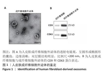
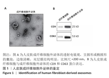
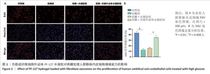
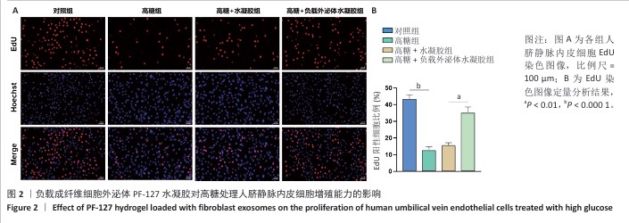
|