Chinese Journal of Tissue Engineering Research ›› 2026, Vol. 30 ›› Issue (21): 5429-5436.doi: 10.12307/2026.125
Previous Articles Next Articles
Finite element analysis of core decompression with ceramic rod implantation in osteonecrosis of the femoral head during the peri-collapse stage
Liang Yingjie, Yuan Lingli, Geng Chunhui, Zhang Zhongchuan, Zheng Wenming, Hu Tengfei, Tang Haoxu, Zhang Kunkun
- Department of Orthopedics of Second Affiliated Hospital of Bengbu Medical University, Key Laboratory of Digital Orthopedics of Bengbu Medical University, Bengbu 233002, Anhui Province, China
-
Accepted:2025-06-17Online:2026-07-28Published:2026-03-04 -
Contact:Yuan Lingli, Chief physician, Associate professor, Department of Orthopedics of Second Affiliated Hospital of Bengbu Medical University, Key Laboratory of Digital Orthopedics of Bengbu Medical University, Bengbu 233002, Anhui Province, China -
About author:Liang Yingjie, Master candidate, Department of Orthopedics of Second Affiliated Hospital of Bengbu Medical University, Key Laboratory of Digital Orthopedics of Bengbu Medical University, Bengbu 233002, Anhui Province, China -
Supported by:Anhui Provincial Key Research Project of Natural Sciences of Higher Education Institutions, No. KJ2021A0756 (to YLL); Anhui Provincial Key Research Project of Natural Sciences of Higher Education Institutions, No. 2024AH051221 (to ZKK)
CLC Number:
Cite this article
Liang Yingjie, Yuan Lingli, Geng Chunhui, Zhang Zhongchuan, Zheng Wenming, Hu Tengfei, Tang Haoxu, Zhang Kunkun. Finite element analysis of core decompression with ceramic rod implantation in osteonecrosis of the femoral head during the peri-collapse stage[J]. Chinese Journal of Tissue Engineering Research, 2026, 30(21): 5429-5436.
share this article
Add to citation manager EndNote|Reference Manager|ProCite|BibTeX|RefWorks
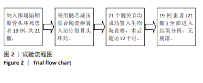
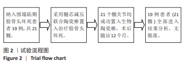
2.1 参与者数量分析 纳入股骨头坏死患者19例,共 21髋,研究过程无脱落。所有患者术后切口经评估均符合Ⅰ期愈合。随访至术后12个月,所有患者均未出现伤口感染、神经血管损伤、股骨颈骨折以及下肢深静脉血栓等并发症。 2.2 试验流程图 见图2。"
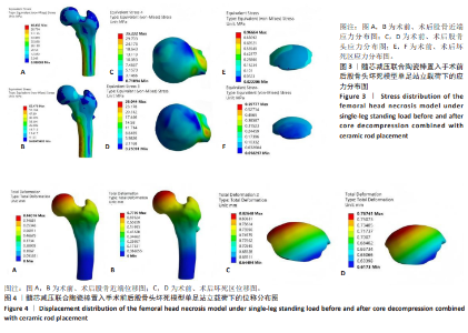
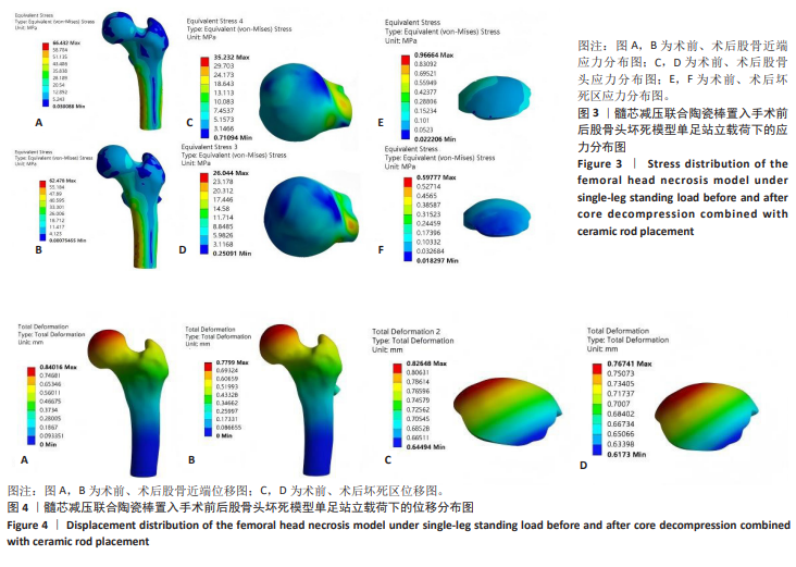
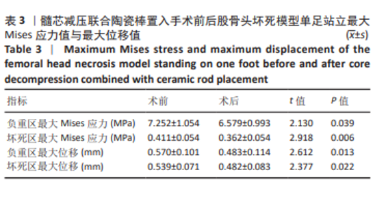
2.3 植入物与宿主的生物相容性 所有患者均未观察到明显的植入物与宿主不良反应发生,如植入物周围感染、切口愈合不良、过敏反应、免疫反应、排斥反应等。 2.4 髓芯减压并陶瓷棒置入前后的有限元分析 2.4.1 加载单足站立位载荷的有限元分析 在单足站位时,股骨头承受最大压力的区域位于坏死部分的前外侧上方,在术前该区域的最大Mises应力值为(7.252±1.054) MPa,术后下降至(6.579±0.993) MPa,差异有显著性意义(P < 0.05);同时,坏死区域的最大Mises应力值也从术前(0.411±0.054) MPa减少到术后(0.362±0.054) MPa,差异有显著性意义(P < 0.05),见图3及表3。"
"
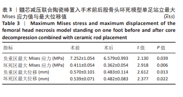
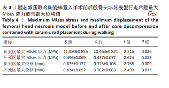
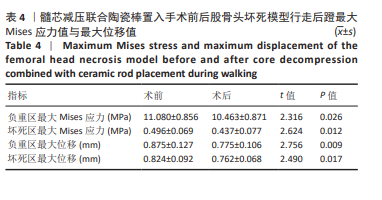
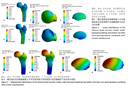
股骨头塌陷值即负重区的位移值[28],术前负重区最大位移值为(0.570±0.101) mm,术后减少至(0.483±0.114) mm,差异有显著性意义(P < 0.05);坏死区的位移值术前为(0.539±0.071) mm,术后减少至(0.482±0.083) mm,差异有显著性意义(P < 0.05),见图4及表3。 2.4.2 加载行走后蹬载荷的有限元分析 在行走后蹬载荷下,术前股骨头负重区的最大Mises应力值为(11.080±0.856) MPa;术后该数值下降至(10.463±0.871) MPa,差异有显著性意义(P < 0.05)。同时,坏死区域的最大Mises应力值也从术前(0.496±0.069) MPa减少到术后(0.437±0.077) MPa,差异有显著性意义(P < 0.05),见图5及表4。 股骨头塌陷值,即负重区的位移值[28],术前负重区最大位移值为(0.875±0.127) mm,术后减少至(0.775±0.106) mm,差异有显著性意义(P < 0.05)。坏死区的最大位移值术前为(0.824±0.092) mm,术后减少至(0.762±0.068) mm,差异有显著性意义(P < 0.05),见图6及表4。"
"

| [1] MA M, TAN Z, LI W, et al. Osteoimmunology and osteonecrosis of the femoral head. Bone Joint Res. 2022;11(1):26-28. [2] WENSI O, YUBO M, GUIMEI G, et al. Efficacy and safety of traditional Chinese medicine in the treatment of osteonecrosis of the femoral head. J Orthop Surg Res. 2023;18(1):600-600. [3] CHEN Z, FENG F, SU X, et al. Experimental study of a 3D-printing technique combined with biphasic calcium phosphates to treat osteonecrosis of the femoral head in a canine model. J Orthop Surg Res. 2023;18(1):693. [4] ZHENG GS, QIU X, WANG BJ, et al. Relationship Between Blood Flow and Collapse of Nontraumatic Osteonecrosis of the Femoral Head. J Bone Joint Surg Am. 2022;104(Suppl 2):13-18. [5] 孙伟, 高福强, 李子荣. 股骨头坏死临床诊疗技术专家共识(2022年)[J].中国修复重建外科杂志,2022,36(11):1319-1326. [6] XU Y, ZENG P. A review and meta-analysis of the surviv-al rate of adult with osteonecrosis of the femoral headtreated with transtrochanteric rotational osteotomy. Medicine(Baltimore). 2022;101(47):e31777. [7] 刘海军, 王前源, 牛存良, 等. C形臂X线定位下多次小直径钻孔联合体外冲击波疗法治疗早期股骨头坏死的疗效观察[J]. 中国骨伤,2023,36(11):1014-1020. [8] YUAN D, WU Z, ZHOU Y, et al. Core decompression assisted by multi-functional minimally invasive instruments for the treatment of early osteonecrosis of the femoral head. Sci Rep. 2025;15(1): 6113. [9] MIGLIORINI F, MAFFULLI N, BARONCINI A, et al. Prognostic factors in the management of osteonecrosis of the femoral head: A systematic review. Surgeon. 2023;21(2):85-98. [10] ZHANG YZ, CAO XY, LI XC, et al. Accuracy of MRI diagnosis of early osteonecrosis of the femoral head: a meta-analysis and systematic review. J Orthop Surg Res. 2018;13(1):167. [11] LYU J, MA T, HUANG X, et al. Core decompression with β-tricalcium phosphate grafts in combination with platelet-rich plasma for the treatment of avascular necrosis of femoral head. BMC Musculoskelet Disord. 2023;24(1):40. [12] 郭玉祺, 李嘉程, 卢博文, 等. 髓芯减压联合不同方法治疗早中期股骨头坏死的网状Meta分析[J]. 中国组织工程研究,2026,30(15): 3993-4009. [13] KURODA Y, NANKAKU M, OKUZU Y, et al. Percutaneous autologous impaction bone graft for advanced femoral head osteonecrosis: a retrospective observational study of unsatisfactory short-term outcomes. J Orthop Surg Res. 2021;16(1):141. [14] BROJENI S, HESARIKIA H, RAHIMNIA A, et al. Treatment of Femoral Head Osteonecrosis (Stages 2B, 3 Ficat) Through Open Direct Core Decompression by Allograft Impaction and Light Bulb Technique. Arch Bone Jt Surg. 2020;8(5):613-619. [15] XIE H, WANG B, TIAN S, et al. Retrospective Long-Term Follow-Up Survival Analysis of the Management of Osteonecrosis of the Femoral Head With Pedicled Vascularized Iliac Bone Graft Transfer. J Arthroplasty. 2019;34(8):1585-1592. [16] WANG Q, LI D, NING W, et al. Short-term clinical outcomes of minimally invasive porous bioceramic rod in treatment of avascular necrosis of the femoral head. Asian J Surg. 2023;46(5):2162-2163. [17] 高永昌, 付彦涛, 赵昕, 等. 步行运动下缺血性坏死股骨头力学性能及塌陷风险预测[J]. 中国组织工程研究,2024,28(33):5265-5269. [18] 倘艳锋, 曹向阳, 岳辰, 等. 围塌陷期股骨头坏死的头颈开窗植骨[J]. 中国矫形外科杂志,2022,30(16):1512-1515. [19] 中国医师协会骨科医师分会显微修复工作委员会, 中国修复重建外科专业委员会骨缺损及骨坏死学组, 中华医学会骨科分会显微修复学组. 成人股骨头坏死临床诊疗指南(2016)[J]. 中华骨科杂志, 2016,36(15):945-954. [20] ZHANG Z, LIN T, ZHONG Y, et al. Effect of femoral head necrosis cystic area on femoral head collapse and stress distribution in femoral head: A clinical and finite element study. Open Med (Wars). 2022;17(1): 1282-1291. [21] 李博. β-磷酸三钙生物陶瓷棒系统治疗早期股骨头坏死的临床观察与生物力学研究[D]. 南宁:广西医科大学,2017. [22] 陆舜, 林天烨, 何敏聪, 等. 基于前外侧保留角预测股骨头坏死塌陷的有限元分析[J]. 中国修复重建外科杂志,2023,37(11): 1394-1402. [23] 杨宾宾, 刘耀升, 刘蜀彬, 等. 多种髓芯减压术治疗股骨头坏死的有限元研究[J]. 中华损伤与修复杂志(电子版),2017,12(1):39-45. [24] YANG P, LIN TY, XU JL, et al. Finite element modeling of proximal femur with quantifiable weight-bearing area in standing position. J Orthop Surg Res. 2020;15(1):384. [25] HECKELMAN LN, KRATZER AL, SPRITZER CE, et al. Influence of running on femoroacetabular joint bone‐to‐bone distances. J Orthop Res. 2024;42(4):837-842. [26] MCCARTHY JC, NOBLE PC, SCHUCK MR, et al. The role of labral lesions to development of early degenerative hip disease. Clin Orthop Relat Res. 2001;(393):25-37. [27] TAUVIQIRRAHMAN M, AMMARULLAH MI, JAMARI J, et al. Analysis of contact pressure in a 3D model of dual-mobility hip joint prosthesis under a gait cycle. Sci Rep. 2023;13(1):3564. [28] BYRD JW, JONES KS. Prospective analysis of hip arthroscopy with 10-year followup. Clin Orthop Relat Res. 2010;468(3):741-746. [29] LU J, WANG QY, SHENG JG, et al. A 3D-printed, personalized,biomechanics-specific beta-tricalcium phosphate bioceramic rod system: personalized treatment strategy for patients with femoral shaft non-union based on finite element analysis. BMC Musculoskelet Disord. 2020;21(1):421. [30] WEN MT, LIANG XZ, LUO D, et al. The effect of the hip flexion angle in osteonecrosis of the femoral head based on China-Japan Friendship Hospital Classification - A finite element study. Orthop Surg. 2023;15(10):2689-2700. [31] WANG P, WANG C, MENG H, et al. The role of structural deteriora- tion and biomechanical changes of the necrotic lesion in collapse mechanism of osteonecrosis of the femoral head. Orthop Surg. 2022; 14(5):831-839. [32] 魏腾飞, 何晓铭, 韦雨柔, 等. Piezo1在激素性和酒精性股骨头坏死骨组织中的差异表达[J]. 中国组织工程研究,2023,27(2):270- 275. [33] LU S, LIN T, HAN L, et al. Location or size? A finite element analysis study of necrotic lesion impact on femoral head collapse. J Orthop Surg Res. 2025;20(1):48. [34] BAKIRCIOGLU S, ATILLA B. Hip preserving procedures for osteonecrosis of the femoral head after collapse. J Clin Orthop Trauma. 2021;23: 101636. [35] 刘钊, 徐西林, 申意伟, 等. 塌陷预测方法联合分期分型对股骨头坏死治疗的指导作用与前景[J]. 中国组织工程研究,2021,25(6): 929-934. [36] ZHANG Y, WANG X, JIANG C, et al. Biomechanical research of medial femoral circumflex vascularized bone-graſting in the treatment of early-to-mid osteonecrosis of the femoral head: a finite element analysis. J Orthop Surg Res. 2022;17(1):441. [37] CHEN Y, MIAO Y, LIU K, et al. Evolutionary course of the femoral head osteonecrosis: Histopathological - radiologic characteristics and clinical staging systems. J Orthop Translat. 2021;32:28-40. [38] BEHESHTIZADEH N, AZAMI M, ABBASI H, et al. Applying extrusion-based 3D printing technique accelerates fabricating complex biphasic calcium phosphate-based scaffolds for bone tissue regeneration. J Adv Res. 2022;40:69-94. [39] XU L, KAZEZIAN Z, PITSILLIDES AA, et al. A synoptic literature review of animal models for investigating the biomechanics of knee osteoarthritis. Front Bioeng Biotechnol. 2024;12:1408015. [40] LU Y, CHEN X, LU X, et al. Reconstructing avascular necrotic femoral head through a bioactive β-TCP system: From design to application. Bioact Mater. 2023;28:495-510. [41] WEN P, ZHANG Y, HAO L, et al. The effect of the necrotic area on the biomechanics of the femoral head - a finite element study. BMC Musculoskelet Disord. 2020;21(1):211. [42] WANG SL, HU YB, CHEN H, et al. Efficacy of bone marrow stem cells combined with core decompression in the treatment of osteonecrosis of the femoral head: APRISMA-compliant meta-analysis. Medicine(Baltimore). 2020;99(25):e20509. [43] 鲁亚杰, 王臻, 卢霄, 等. 生物陶瓷系统微创治疗ARCOⅡ、Ⅲ期股骨头坏死[J]. 中国修复重建外科杂志,2019,33(10):1291-1298. [44] 范亚楠, 陈俊名, 何沛霖, 等. 多孔陶瓷生物材料治疗早中期非创伤性股骨头坏死的临床研究[J]. 中华骨与关节外科杂志,2022, 15(2):81-86. [45] BOONTANAPIBUL K, STEERE JT, AMANATULLAH DF, et al. Diagnosis of Osteonecrosis of the Femoral Head: Too Little, Too Late, and Independent of Etiology. J Arthroplasty. 2020;35(9):2342-2349. [46] WANG Q, LI D, NING W, et al. Short-term clinical outcomes of minimally invasive porous bioceramic rod in treatment of avascular necrosis of the femoral head. Asian J Surg. 2023;46(5):2162-2163. |
| [1] | Zhou Daobin, Wang Kehao, Xie Yang, Ning Rende. Biomechanical characteristics of volar locking plate only versus combined dorsal mini-plate fixation of distal radius fractures with dorsal ulnar fragment [J]. Chinese Journal of Tissue Engineering Research, 2026, 30(9): 2255-2261. |
| [2] | Liu Wenlong, Dong Lei, Xiao Zhengzheng, Nie Yu. Finite element analysis of tibial prosthesis loosening after fixed-bearing unicompartmental knee arthroplasty for osteoporosis [J]. Chinese Journal of Tissue Engineering Research, 2026, 30(9): 2191-2198. |
| [3] | Zheng Wangyang, Fei Ji, Yang Di, Zhao Lang, Wang Lingli, Liu Peng, Li Haiyang. Finite element analysis of the force changes of the supraspinatus tendon and glenohumeral joint during the abduction and flexion of the humerus [J]. Chinese Journal of Tissue Engineering Research, 2026, 30(9): 2199-2207. |
| [4] | Cai Qirui, Dai Xiaowei, Zheng Xiaobin, Jian Sili, Lu Shaoping, Liu Texi, Liu Guoke, Lin Yuanfang. Mechanical effects of Long’s traction orthopedic method on cervical functional units: quantitative analysis of biomechanical model of head and neck [J]. Chinese Journal of Tissue Engineering Research, 2026, 30(9): 2208-2216. |
| [5] | Rao Jingcheng, Li Yuwan, Zheng Hongbing, Xu Zhi, Zhu Aixiang, Shi Ce, Wang Bing, Yang Chun, Kong Xiangru, Zhu Dawei. Biomechanical differences between the new proximal femoral stable intramedullary nail and traditional intramedullary nail#br# [J]. Chinese Journal of Tissue Engineering Research, 2026, 30(9): 2217-2225. |
| [6] | Chen Long, Wang Xiaozhen, Xi Jintao, Lu Qilin. Biomechanical performance of short-segment screw fixation combined with expandable polyetheretherketone vertebral body replacement in osteoporotic vertebrae [J]. Chinese Journal of Tissue Engineering Research, 2026, 30(9): 2226-2235. |
| [7] | Yan Xiangning, Chen Lei, Chen Yonghuan, Wang Chao, Li Xiaosheng. Influence of different depths and loads on knee joint mechanics and peripheral muscle force characteristics during squatting [J]. Chinese Journal of Tissue Engineering Research, 2026, 30(9): 2236-2247. |
| [8] | Zhang Nan, Meng Qinghua, Bao Chunyu. Characteristics and clinical application of ankle joint finite element models [J]. Chinese Journal of Tissue Engineering Research, 2026, 30(9): 2343-2349. |
| [9] | Chen Huiting, Zeng Weiquan, Zhou Jianhong, Wang Jie, Zhuang Congying, Chen Peiyou, Liang Zeqian, Deng Weiming. Tail anchoring technique of vertebroplasty in treatment of osteoporotic vertebral compression fractures with intravertebral cleft: a finite element analysis [J]. Chinese Journal of Tissue Engineering Research, 2026, 30(9): 2145-2152. |
| [10] | Zeng Xuan, Weng Rui, Ye Shicheng, Tang Jiadong, Mo Ling, Li Wenchao. Two lumbar rotary manipulation techniques in treating lumbar disc herniation: a finite element analysis of biomechanical differences [J]. Chinese Journal of Tissue Engineering Research, 2026, 30(9): 2153-2161. |
| [11] | Cheng Qisheng, Julaiti·Maitirouzi, Xiao Yang, Zhang Chenwei, Paerhati·Rexiti. Finite element analysis of novel variable-diameter screws in modified cortical bone trajectory of lumbar vertebrae [J]. Chinese Journal of Tissue Engineering Research, 2026, 30(9): 2162-2171. |
| [12] | Wu Hongxu, Liu Xuanyu, Wang Taoyu, Wang Shiyao, Cheng Jingyi, Zhang Mingwen, Zhang Yinxia, Liu Zhihua, Wang Xiaojie. Finite element simulation of scoliosis with muscle unit introduction: verification of correction effect under bidirectional load [J]. Chinese Journal of Tissue Engineering Research, 2026, 30(9): 2172-2181. |
| [13] | Liu Jiafu, Ren Ruxia, Liao Zhouwei, Zhou Xiali, Wu Yihong, Zhang Shaoqun. Three-dimensional finite element analysis of cervical spine biomechanical characteristics in a rat model of cervical vertigo [J]. Chinese Journal of Tissue Engineering Research, 2026, 30(9): 2182-2190. |
| [14] | Zhang Zizheng, Luo Wang, Liu Changlu. Application value of finite element analysis on unicompartmental knee arthroplasty for medial knee compartmental osteoarthritis [J]. Chinese Journal of Tissue Engineering Research, 2026, 30(9): 2313-2322. |
| [15] | Zhao Feifan, Cao Yujing. Risk factors and coping strategies of internal fixation failure in treatment of intertrochanteric fracture with proximal femoral nail antirotation [J]. Chinese Journal of Tissue Engineering Research, 2026, 30(9): 2323-2333. |
| Viewed | ||||||
|
Full text |
|
|||||
|
Abstract |
|
|||||