Chinese Journal of Tissue Engineering Research ›› 2026, Vol. 30 ›› Issue (1): 101-110.doi: 10.12307/2025.567
Previous Articles Next Articles
Contribution and interaction of various cells in bone marrow microenvironment to exosomal circular RNA associated with multiple myeloma bone disease
Yu Manya1, Cui Xing2
- 1First School of Clinical Medicine, Shandong University of Traditional Chinese Medicine, Jinan 250014, Shandong Province, China; 2Cancer Center, Second Affiliated Hospital of Shandong University of Traditional Chinese Medicine, Jinan 250001, Shandong Province, China
-
Received:2024-09-20Accepted:2024-11-14Online:2026-01-08Published:2025-07-02 -
Contact:Cui Xing, MD, Chief physician, Professor, Doctoral supervisor, Cancer Center, Second Affiliated Hospital of Shandong University of Traditional Chinese Medicine, Jinan 250001, Shandong Province, China -
About author:Yu Manya, Master candidate, First School of Clinical Medicine, Shandong University of Traditional Chinese Medicine, Jinan 250014, Shandong Province, China -
Supported by:National Natural Science Foundation of China, No. 82074348, 82274491 (to CX); Shandong Natural Science Foundation Innovation and Development Joint Fund, No. ZR2023LZL009 (to CX)
CLC Number:
Cite this article
Yu Manya, Cui Xing. Contribution and interaction of various cells in bone marrow microenvironment to exosomal circular RNA associated with multiple myeloma bone disease[J]. Chinese Journal of Tissue Engineering Research, 2026, 30(1): 101-110.
share this article
Add to citation manager EndNote|Reference Manager|ProCite|BibTeX|RefWorks
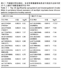
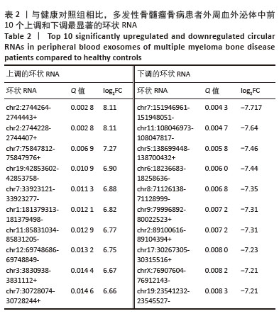
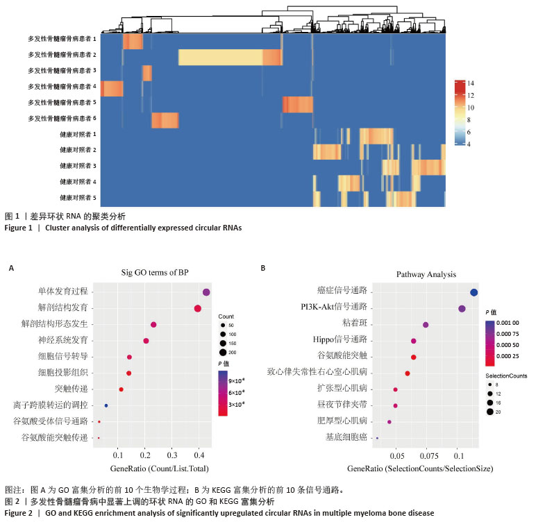
2.1 差异环状RNA的筛选 以差异倍数对数的绝对值(log2|fold change (FC)|)> 1且矫正后的P值< 0.05(Q值)为阈值,筛选并鉴定出2 052个差异表达的环状RNA,聚类分析图展示了每个样本中差异基因的表达概况,见图1。与健康志愿者相比,多发性骨髓瘤骨病患者表达上调的环状RNA有1 265个,下调的环状RNA有787个。从中获得差异表达最显著的上调及下调的环状RNA各10个,相关信息见表2。"
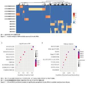
2.2 GO分析和KEGG分析 对多发性骨髓瘤骨病患者中表达水平显著上调的环状RNA进行GO和KEGG富集分析(图2A,B),结果显示差异环状RNA的亲本基因主要富集在神经系统发育、谷氨酸受体信号通路、细胞信号转导、癌症通路、PI3K-Akt信号通路、Hippo信号通路、致心律失常性右心室心肌病、扩张型心肌病、昼夜节律同步、肥厚型心肌病通路。相关研究表明,PI3K-Akt信号通路和Hippo信号通路与骨髓瘤细胞的存活、增殖、迁移和代谢密切相关[11-12]。此外,PI3K-Akt信号通路在骨重塑中发挥重要作用,抑制PI3K可以促进矿化,并抑制破骨细胞生成[13];Hippo信号通路可以与Wnt通路相互作用调节骨生成,其通路成分如YAP/TAZ/TEAD 复合物、RASSF2、MST2等也与破骨细胞成熟和功能有关,是多发性骨髓瘤骨病的重要调节因子[14-15]。 "
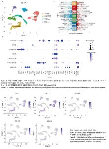
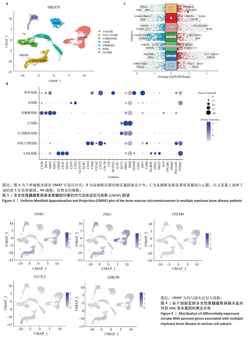
2.3 单细胞降维、聚类分析 为了推测与多发性骨髓瘤骨病相关的差异环状RNA的来源细胞,选取GSE271107数据集中4例多发性骨髓瘤患者的骨髓单细胞RNA测序数据。通过对单细胞RNA测序数据的质控、UMAP降维,获得了21 868个细胞的转录组数据。根据基因的表达对细胞进行自动注释和手动矫正,分别是T细胞/自然杀伤细胞(7 351个)、单核细胞/巨噬细胞(4 077个)、红细胞祖细胞(2 849个)、红细胞(2 585个)、骨髓瘤细胞(2 508个)、B细胞(2 380个)、树突状细胞(118个),见图3A,B。通过对7个注释出的细胞亚群进行差异化分析,并绘制火山图,可以看出在不同细胞亚群中显著上调和下调的前3位差异基因,见图3C。 2.4 外泌体环状RNA溯源 将多发性骨髓瘤骨病相关差异环状RNA的亲本基因(参与PI3K-Akt通路和Hippo通路的差异基因)通过单细胞RNA测序的聚类特征图(图4)可视化,发现GNB1、JAK1、THEM4、TCF7L2、GSK3B分布在特定的细胞亚群中,GNB1和JAK1主要分布在T细胞/自然杀伤细胞、B细胞和单核/巨噬细胞中,GSK3B主要分布在T细胞/自然杀伤细胞和单核/巨噬细胞中,TCF7L2主要分布在单核/巨噬细胞中,THEM4主要分布在T细胞/自然杀伤细胞中,由此推测多发性骨髓瘤骨病相关差异环状RNA主要来源于骨髓微环境中的T细胞/自然杀伤细胞、B细胞和单核细胞/巨噬细胞。 "
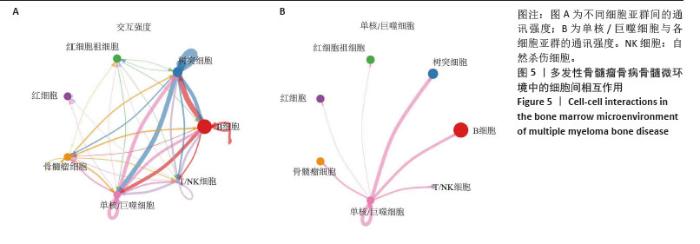
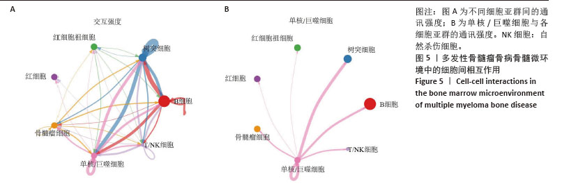
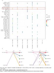
2.5 单核/巨噬细胞与其他细胞的交互作用 单核/巨噬细胞作为破骨细胞的前体细胞,其向破骨细胞的分化在多发性骨髓瘤骨病的病理过程中发挥重要作用。为了在单细胞水平上探究单核/巨噬细胞与骨髓微环境中其他细胞类型的交互作用,利用CellChat检测不同细胞间的配体-受体对及分子相互作用,细胞间互作强度网络见图5A,B,单核/巨噬细胞与红细胞祖细胞、红细胞、骨髓瘤细胞、B细胞、树突状细胞、T细胞/自然杀伤细胞均有交互作用,其与树突状细胞和B细胞交互作用较强,这表明骨髓微环境中的多种细胞均与破骨细胞的前体细胞间存在细胞通讯。 "
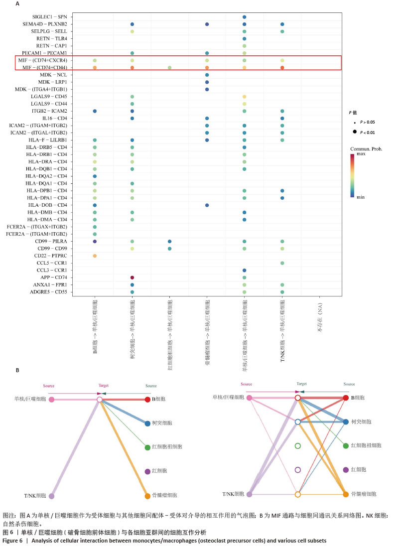
2.6 配体受体互作分析 为进一步明确细胞间相互作用的分子途径,利用CellChat探究单核/巨噬细胞作为受体参与的信号通路(图6A),其主要参与的MIF通路在单核/巨噬细胞向破骨细胞分化过程中发挥重要作用。作用于单核/巨噬细胞的MIF主要来源于T细胞/自然杀伤细胞、树突状细胞、骨髓瘤细胞、B细胞以及单核/巨噬细胞(图6B)。"
| [1] MALARD F, NERI P, BAHLIS NJ, et al. Multiple myeloma. Nat Rev Dis Primers. 2024;10(1):45. [2] MUKKAMALLA SKR, MALIPEDDI D. Myeloma Bone Disease: A Comprehensive Review. Int J Mol Sci. 2021;22(12):6208. [3] YU D, LI Y, WANG M, et al. Exosomes as a new frontier of cancer liquid biopsy. Mol Cancer. 2022;21(1):56. [4] BERNSTEIN ZS, KIM EB, RAJE N. Bone Disease in Multiple Myeloma: Biologic and Clinical Implications. Cells. 2022;11(15): 2308. [5] MENU E, VANDERKERKEN K. Exosomes in multiple myeloma: from bench to bedside. Blood. 2022;140(23):2429-2442. [6] LIU R, ZHONG Y, CHEN R, et al. m6A reader hnRNPA2B1 drives multiple myeloma osteolytic bone disease. Theranostics. 2022; 12(18):7760-7774. [7] LI B, XU H, HAN H, et al. Exosome-mediated transfer of lncRUNX2-AS1 from multiple myeloma cells to MSCs contributes to osteogenesis. Oncogene. 2018;37(41): 5508-5519. [8] UNTI MJ, JAFFREY SR. Highly efficient cellular expression of circular mRNA enables prolonged protein expression. Cell Chem Biol. 2024; 31(1):163-176.e5. [9] ZHANG F, JIANG J, QIAN H, et al. Exosomal circRNA: emerging insights into cancer progression and clinical application potential. J Hematol Oncol. 2023;16(1):67. [10] ARAN D, LOONEY AP, LIU L, et al. Reference-based analysis of lung single-cell sequencing reveals a transitional profibrotic macrophage. Nat Immunol. 2019;20(2):163-172. [11] KIKUCHI H, AMOFA E, MCENERY M, et al. Inhibition of PI3K Class IA Kinases Using GDC-0941 Overcomes Cytoprotection of Multiple Myeloma Cells in the Osteoclastic Bone Marrow Microenvironment Enhancing the Efficacy of Current Clinical Therapeutics. Cancers (Basel). 2023;15(2):462. [12] LI H, ZHANG Y, MOU X, et al. Interference with PLA2G16 promotes cell cycle arrest and apoptosis and inhibits the reprogramming of glucose metabolism in multiple myeloma cells by modulating the Hippo/YAP signaling pathway. Anticancer Drugs. 2024;35(10):902-911. [13] TSUBAKI M, KATO C, MANNO M, et al. Macrophage inflammatory protein-1alpha (MIP-1alpha) enhances a receptor activator of nuclear factor kappaB ligand (RANKL) expression in mouse bone marrow stromal cells and osteoblasts through MAPK and PI3K/Akt pathways. Mol Cell Biochem. 2007;304(1-2):53-60. [14] PAN JX, XIONG L, ZHAO K, et al. YAP promotes osteogenesis and suppresses adipogenic differentiation by regulating β-catenin signaling. Bone Res. 2018;6:18. [15] YANG W, HAN W, QIN A, et al. The emerging role of Hippo signaling pathway in regulating osteoclast formation. J Cell Physiol. 2018;233(6): 4606-4617. [16] MIAOMIAO S, XIAOQIAN W, YUWEI S, et al. Cancer-associated fibroblast-derived exosome microRNA-21 promotes angiogenesis in multiple myeloma. Sci Rep. 2023;13(1):9671. [17] MIZUHARA K, SHIMURA Y, TSUKAMOTO T, et al. Tumour-derived exosomes promote the induction of monocytic myeloid-derived suppressor cells from peripheral blood mononuclear cells by delivering miR-106a-5p and miR-146a-5p in multiple myeloma. Br J Haematol. 2023;203(3):426-438. [18] WANG Z, HE J, BACH DH, et al. Induction of m6A methylation in adipocyte exosomal LncRNAs mediates myeloma drug resistance. J Exp Clin Cancer Res. 2022;41(1):4. [19] YU M, JI L, LI S, et al. Exosomal circ-CACNG2 promotes cardiomyocyte apoptosis in multiple myeloma via modulating miR-197-3p/caspase3 axis. Exp Cell Res. 2022;417(2):113229. [20] ALIMOHAMMADI M, RAHIMZADEH P, KHORRAMI R, et al. A comprehensive review of the PTEN/PI3K/Akt axis in multiple myeloma: From molecular interactions to potential therapeutic targets. Pathol Res Pract. 2024;260:155401. [21] HEINEMANN L, MÖLLERS KM, AHMED HMM, et al. Inhibiting PI3K-AKT-mTOR Signaling in Multiple Myeloma-Associated Mesenchymal Stem Cells Impedes the Proliferation of Multiple Myeloma Cells. Front Oncol. 2022;12:874325. [22] KYRIAZOGLOU A, NTANASIS-STATHOPOULOS I, TERPOS E, et al. Emerging Insights Into the Role of the Hippo Pathway in Multiple Myeloma and Associated Bone Disease. Clin Lymphoma Myeloma Leuk. 2020;20(2):57-62. [23] YANG J, ZHANG X, WANG J, et al. Anti beta2-microglobulin monoclonal antibodies induce apoptosis in myeloma cells by recruiting MHC class I to and excluding growth and survival cytokine receptors from lipid rafts. Blood. 2007;110(8):3028-3035. [24] MENG B, WU D, CHENG Y, et al. Interleukin-20 differentially regulates bone mesenchymal stem cell activities in RANKL-induced osteoclastogenesis through the OPG/RANKL/RANK axis and the NF-κB, MAPK and AKT signalling pathways. Scand J Immunol. 2020;91(5): e12874. [25] ZHANG Z, ZHANG X, ZHAO D, et al. TGF‑β1 promotes the osteoinduction of human osteoblasts via the PI3K/AKT/mTOR/S6K1 signalling pathway. Mol Med Rep. 2019;19(5):3505-3518. [26] MARTIN SK, GAN ZY, FITTER S, et al. The effect of the PI3K inhibitor BKM120 on tumour growth and osteolytic bone disease in multiple myeloma. Leuk Res. 2015;39(3):380-387. [27] SEO E, BASU-ROY U, GUNARATNE PH, et al. SOX2 regulates YAP1 to maintain stemness and determine cell fate in the osteo-adipo lineage. Cell Rep. 2013;3(6):2075-2087. [28] MATSUMOTO Y, LA ROSE J, KENT OA, et al. Reciprocal stabilization of ABL and TAZ regulates osteoblastogenesis through transcription factor RUNX2. J Clin Invest. 2016;126(12):4482-4496. [29] PARK H, NOH AL, KANG JH, et al. Peroxiredoxin II negatively regulates lipopolysaccharide-induced osteoclast formation and bone loss via JNK and STAT3. Antioxid Redox Signal. 2015;22(1):63-77. [30] LU J, YE C, HUANG Y, et al. Corilagin suppresses RANKL-induced osteoclastogenesis and inhibits oestrogen deficiency-induced bone loss via the NF-κB and PI3K/AKT signalling pathways. J Cell Mol Med. 2020;24(18):10444-10457. [31] HUANG F, WONG P, LI J, et al. Osteoimmunology: The correlation between osteoclasts and the Th17/Treg balance in osteoporosis. J Cell Mol Med. 2022;26(13):3591-3597. [32] WU LZ, DUAN DM, LIU YF, et al. Nicotine favors osteoclastogenesis in human periodontal ligament cells co-cultured with CD4(+) T cells by upregulating IL-1β. Int J Mol Med. 2013;31(4):938-942. [33] NAGAI S, KUREBAYASHI Y, KOYASU S. Role of PI3K/Akt and mTOR complexes in Th17 cell differentiation. Ann N Y Acad Sci. 2013;1280: 30-34. [34] FU M, HU Y, LAN T, et al. The Hippo signalling pathway and its implications in human health and diseases. Signal Transduct Target Ther. 2022;7(1):376. [35] SÖDERSTRÖM K, STEIN E, COLMENERO P, et al. Natural killer cells trigger osteoclastogenesis and bone destruction in arthritis. Proc Natl Acad Sci U S A. 2010;107(29):13028-13033. [36] FENG P, LUO L, YANG Q, et al. Hippo kinases Mst1 and Mst2 maintain NK cell homeostasis by orchestrating metabolic state and transcriptional activity. Cell Death Dis. 2024;15(6):430. [37] ALI AK, NANDAGOPAL N, LEE SH. IL-15-PI3K-AKT-mTOR: A Critical Pathway in the Life Journey of Natural Killer Cells. Front Immunol. 2015;6:355. [38] CHEN Y, WANG H, NI Q, et al. B-Cell-Derived TGF-β1 Inhibits Osteogenesis and Contributes to Bone Loss in Periodontitis. J Dent Res. 2023;102(7):767-776. [39] FISCHER V, HAFFNER-LUNTZER M. Interaction between bone and immune cells: Implications for postmenopausal osteoporosis. Semin Cell Dev Biol. 2022;123:14-21. [40] GRČEVIĆ D, SANJAY A, LORENZO J. Interactions of B-lymphocytes and bone cells in health and disease. Bone. 2023;168:116296. [41] HODSON DJ, TURNER M. The role of PI3K signalling in the B cell response to antigen. Adv Exp Med Biol. 2009;633:43-53. [42] ZHENG L, GAO J, JIN K, et al. Macrophage migration inhibitory factor (MIF) inhibitor 4-IPP suppresses osteoclast formation and promotes osteoblast differentiation through the inhibition of the NF-κB signaling pathway. FASEB J. 2019;33(6):7667-7683. [43] HE J, ZHENG L, LI X, et al. Obacunone targets macrophage migration inhibitory factor (MIF) to impede osteoclastogenesis and alleviate ovariectomy-induced bone loss. J Adv Res. 2023;53:235-248. |
| [1] | Wen Guangwei, Zhen Yinghao, Zheng Taikeng, Zhou Shuyi, Mo Guoye, Zhou Tengpeng, Li Haishan, Lai Yiyi. Effects and mechanisms of isoginkgetin on osteoclastogenesis [J]. Chinese Journal of Tissue Engineering Research, 2026, 30(6): 1348-1358. |
| [2] | Xu Jiamu, Yang Cheng, Li Weimin, Wang Chunqing. Role and pathogenesis of pyroptosis and inflammatory factors in osteoporosis [J]. Chinese Journal of Tissue Engineering Research, 2026, 30(3): 691-700. |
| [3] | Zuo Na, Tang Qi, Yu Meng, Tao Kai. Effect of miR-196b-5p in adipose-derived stem cell exosomes on burn wound healing in rats [J]. Chinese Journal of Tissue Engineering Research, 2026, 30(1): 43-49. |
| [4] | Yuan Weiyuan, Lei Qinhui, Li Xiuqi, Lu Tiezhu, Fu Ziwen, Liang Zhili, Ji Shaoyang, Li Yijia, Ren Yu . Therapeutic effects of adipose-derived mesenchymal stem cells and their exosomes on dexamethasone-induced sarcopenia in mice [J]. Chinese Journal of Tissue Engineering Research, 2026, 30(1): 58-67. |
| [5] | Zhang Tingting, Li Yalong, Yue Haodi, Li Yanjun, Geng Xiwen, Zhang Yuwei, Liu Xiaozhuan. Protection of exosomes derived from bone marrow mesenchymal stem cells of different mouse ages on radiation-induced lung injury [J]. Chinese Journal of Tissue Engineering Research, 2026, 30(1): 1-9. |
| [6] | Zhang Zhaowei, Chen Ouzile, Bai Mingru, Wang Chenglin. Therapeutic potential of bioactive substances secreted by dental mesenchymal stem cells for bone repair [J]. Chinese Journal of Tissue Engineering Research, 2026, 30(1): 163-174. |
| [7] | Liu Nian, Dong Xinyue, Wang Songpeng, Xu Yingjiang, Zhang Xiaoming. Stem cell exosomes and biomaterial-assisted exosomes in bone defect repair [J]. Chinese Journal of Tissue Engineering Research, 2026, 30(1): 175-183. |
| [8] | Lyu Ruyue, Gu Lulu, Liu Qian, Zhou Siyi, Li Beibei, Xue Letian, Sun Peng. Regulatory mechanisms of exosome secretion and its application prospects in biomedicine [J]. Chinese Journal of Tissue Engineering Research, 2026, 30(1): 184-193. |
| [9] | Liu Yu, Gong Senyi, Yang Lihua, Li Weifeng, Hu Yuwen, Yan Qinbiao, Guo Meijin. Isolation, identification, and application of exosomes derived from mesenchymal stem cells [J]. Chinese Journal of Tissue Engineering Research, 2026, 30(1): 194-203. |
| [10] | Luo Wenbin, Li Ruoyun, Pan Chaofan, Luo Changjiang. Engineered exosomes for repairing tissue damage: application potential, excellent biological stability, and targeting specificity [J]. Chinese Journal of Tissue Engineering Research, 2026, 30(1): 204-217. |
| [11] | Lai Pengyu, Liang Ran, Shen Shan. Tissue engineering technology for repairing temporomandibular joint: problems and challenges [J]. Chinese Journal of Tissue Engineering Research, 2025, 29(在线): 1-9. |
| [12] | Han Haihui, Ran Lei, Meng Xiaohui, Xin Pengfei, Xiang Zheng, Bian Yanqin, Shi Qi, Xiao Lianbo. Targeting fibroblast growth factor receptor 1 signaling to improve bone destruction in rheumatoid arthritis [J]. Chinese Journal of Tissue Engineering Research, 2025, 29(9): 1905-1912. |
| [13] | Zhao Jiyu, Wang Shaowei. Forkhead box transcription factor O1 signaling pathway in bone metabolism [J]. Chinese Journal of Tissue Engineering Research, 2025, 29(9): 1923-1930. |
| [14] | Wang Wentao, Hou Zhenyang, Wang Yijun, Xu Yaozeng. Apelin-13 alleviates systemic inflammatory bone loss by inhibiting macrophage M1 polarization [J]. Chinese Journal of Tissue Engineering Research, 2025, 29(8): 1548-1555. |
| [15] | Yu Jingbang, Wu Yayun. Regulatory effect of non-coding RNA in pulmonary fibrosis [J]. Chinese Journal of Tissue Engineering Research, 2025, 29(8): 1659-1666. |
| Viewed | ||||||
|
Full text |
|
|||||
|
Abstract |
|
|||||