Chinese Journal of Tissue Engineering Research ›› 2025, Vol. 29 ›› Issue (30): 6391-9397.doi: 10.12307/2025.915
Previous Articles Next Articles
Vanillic acid inhibits inflammatory response and extracellular matrix degradation of endplate chondrocytes
Yu Qinghe1, Cai Ziming2, Wu Jintao2, Ma Pengfei1, Zhang Xin1, Zhou Longqian2, Wang Yakun2, Lin Xiaoqin2, Lin Wenping1
- 1Department of Spine Surgery, Shenzhen Pingle Orthopedic Hospital, Affiliated Hospital of Guangzhou University of Chinese Medicine, Shenzhen 518118, Guangdong Province, China; 2Shenzhen Pingle Orthopedic Hospital, Affiliated Hospital of Guangzhou University of Chinese Medicine, Shenzhen 518118, Guangdong Province, China
-
Received:2024-08-26Accepted:2024-10-23Online:2025-10-28Published:2025-03-27 -
Contact:Lin Wenping, MD, Chief physician, Doctoral supervisor, Department of Spine Surgery, Shenzhen Pingle Orthopedic Hospital, Affiliated Hospital of Guangzhou University of Chinese Medicine, Shenzhen 518118, Guangdong Province, China -
About author:Yu Qinghe, MS, Physician, Department of Spine Surgery, Shenzhen Pingle Orthopedic Hospital, Affiliated Hospital of Guangzhou University of Chinese Medicine, Shenzhen 518118, Guangdong Province, China Cai Ziming, Master candidate, Shenzhen Pingle Orthopedic Hospital, Affiliated Hospital of Guangzhou University of Chinese Medicine, Shenzhen 518118, Guangdong Province, China Yu Qinghe and Cai Ziming contributed equally to this article. -
Supported by:National Natural Science Foundation of China, No. 81771323 (to LWP); Natural Science Foundation of Guangdong Province of China, No. 2021A1515010722, 2024A1515010445 (to LWP); Natural Science Foundation of Shenzhen Municipality, No. JCYJ20190813112401660(to LWP)
CLC Number:
Cite this article
Yu Qinghe, Cai Ziming, Wu Jintao, Ma Pengfei, Zhang Xin, Zhou Longqian, Wang Yakun, Lin Xiaoqin, Lin Wenping. Vanillic acid inhibits inflammatory response and extracellular matrix degradation of endplate chondrocytes[J]. Chinese Journal of Tissue Engineering Research, 2025, 29(30): 6391-9397.
share this article
Add to citation manager EndNote|Reference Manager|ProCite|BibTeX|RefWorks

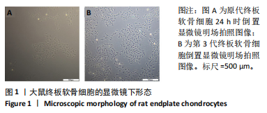
2.1 终板软骨细胞形态观察 原代终板软骨细胞在培养皿中24 h后显微镜下观察到有较多细胞贴壁,形态呈梭形或三角形(图1A);第3代终板软骨细胞增殖能力旺盛,状态较好(图1B),用于后续实验。"

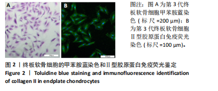
2.2 终板软骨细胞鉴定 第3代终板软骨细胞经甲苯胺蓝染色后,细胞核呈蓝紫色,细胞质呈浅紫色,细胞周围出现少许蓝紫色异染颗粒(图2A)。第3代终板软骨细胞Ⅱ型胶原蛋白免疫荧光示所培养细胞Ⅱ型胶原蛋白高表达(图2B)。根据形态学和甲苯胺蓝染色、Ⅱ型胶原蛋白免疫荧光阳性,结合取材部位,表明实验提取的细胞为终板软骨细胞。"
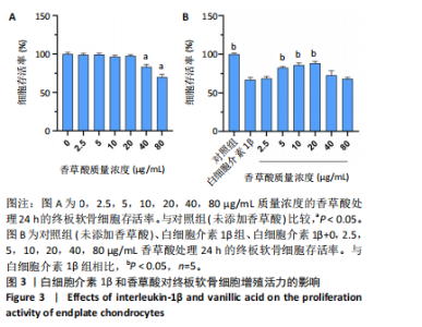
2.3 白细胞介素1β和香草酸对终板软骨细胞增殖活力的影响 CCK-8结果显示,与对照组(未添加香草酸)比较,2.5,5,10,20 μg/mL香草酸处理24 h的终板软骨细胞活力无显著差异(P > 0.05),40,80 μg/mL香草酸处理24 h的终板软骨细胞活力显著降低(P < 0.05),见图3A。与白细胞介素1β组(培养基中添加10 ng/mL白细胞介素1β)比较,用白细胞介素1β和5,10,20 μg/mL香草酸处理24 h的终板软骨细胞活力显著提高(P < 0.05),见图3B,因此后续选用5,10,20 μg/mL香草酸为低、中、高剂量组处理终板软骨细胞。"

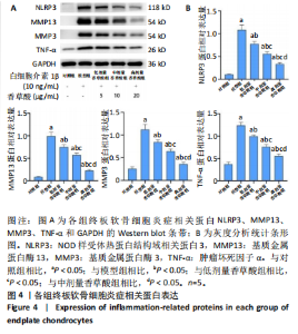
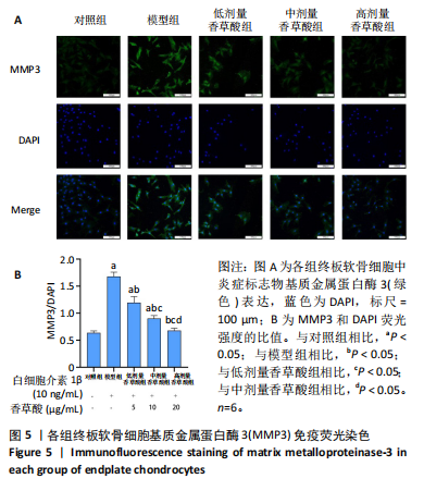
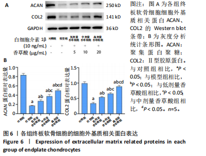
2.4 香草酸对终板软骨细胞炎症相关蛋白的影响 Western blot结果显示,终板软骨细胞经白细胞介素1β和香草酸处理24 h后,与模型组相比,香草酸低、中、高剂量组NLRP3、基质金属蛋白酶13、基质金属蛋白酶3、肿瘤坏死因子α蛋白表达均明显降低(P < 0.05),见图4。免疫荧光结果显示,终板软骨细胞应用白细胞介素1β和香草酸处理24 h后,与模型组相比,香草酸低、中、高剂量组基质金属蛋白酶3蛋白表达显著降低(P < 0.05),见图5。以上结果表明,香草酸减轻了白细胞介素1β引起的终板软骨细胞的炎症反应。 2.5 香草酸对终板软骨细胞细胞外基质相关蛋白的影响 Western blot结果显示,终板软骨细胞经白细胞介素1β"


"

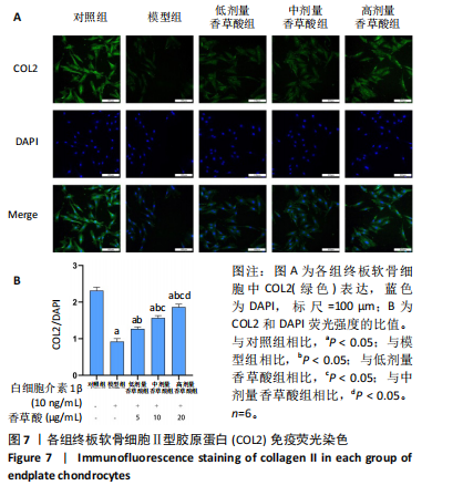
和香草酸处理24 h后,与模型组相比,香草酸低、中、高剂量组聚集蛋白聚糖、Ⅱ型胶原蛋白表达均明显上升(P < 0.05),见图6。免疫荧光结果显示,终板软骨细胞应用白细胞介素1β和香草酸处理24 h后,与模型组相比,香草酸低、中、高剂量组Ⅱ型胶原蛋白表达显著上升(P < 0.05),见图7。以上结果表明,香草酸减轻了白细胞介素1β引起的终板软骨细胞的细胞外基质降解。"

"
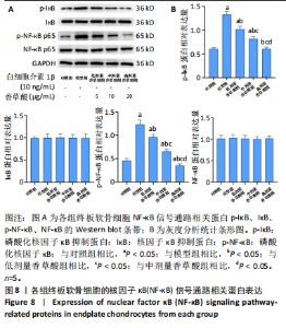
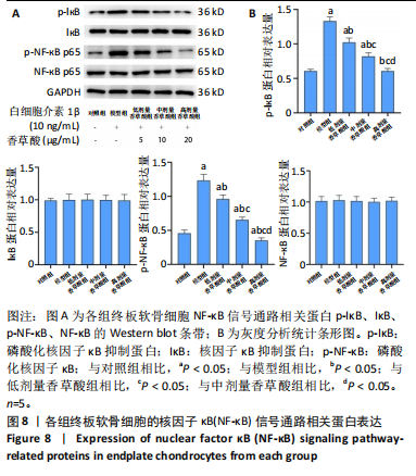
2.6 香草酸对终板软骨细胞中NF-κB信号通路相关蛋白表达的影响 Western blot结果显示,终板软骨细胞经白细胞介素1β和香草酸处理24 h后,与模型组相比,香草酸低、中、高剂量组中p-NF-κB、p-IκB的蛋白表达水平逐渐降低(P < 0.05),而NF-κB、IκB的表达无明显差异(P > 0.05),这说明香草酸抑制了终板软骨细胞中因白细胞介素1β处理而激活的NF-κB信号通路,见图8。"
| [1] SALMAN A, KHAWAJA AS, BAIG K, et al. Comparing contribution of bony and cartilaginous endplate changes to intervertebral disc degeneration. Pak J Med Sci. 2024;40(7):1516-1522. [2] LI K, YANG W, CHEN X, et al. A structured biomimetic nanoparticle as inflammatory factor sponge and autophagy-regulatory agent against intervertebral disc degeneration and discogenic pain. J Nanobiotechnology. 2024;22(1):486. [3] SUN R, WANG F, ZHONG C, et al. The regulatory mechanism of cyclic GMP-AMP synthase on inflammatory senescence of nucleus pulposus cell. J Orthop Surg Res. 2024;19(1):421. [4] ZHANG K, DU L, LI Z, et al. M2 macrophage-derived small extracellular vesicles ameliorate pyroptosis and intervertebral disc degeneration. Biomater Res. 2024;28:47. [5] YU L, HAO YJ, REN ZN, et al. Ginsenoside Rg1 relieves rat intervertebral disc degeneration and inhibits IL-1β-induced nucleus pulposus cell apoptosis and inflammation via NF-κB signaling pathway. In Vitro Cell Dev Biol Anim. 2024;60(3):287-299. [6] XIA Q, ZHAO Y, DONG H, et al. Progress in the study of molecular mechanisms of intervertebral disc degeneration. Biomed Pharmacother. 2024;174:116593. [7] YANG G, LIU X, JING X, et al. Astaxanthin suppresses oxidative stress and calcification in vertebral cartilage endplate via activating Nrf-2/HO-1 signaling pathway. Int Immunopharmacol. 2023;119:110159. [8] 张伟业, 展嘉文, 朱立国, 等. 循环牵张力作用下髓核外泌体对终板软骨细胞的影响[J]. 中国组织工程研究,2023,27(2):223-229. [9] QUAN H, ZUO X, HUAN Y, et al. A systematic morphology study on the effect of high glucose on intervertebral disc endplate degeneration in mice. Heliyon. 2023;9(2):e13295. [10] TIAN X, ZHAO H, YANG S, et al. The effect of diabetes mellitus on lumbar disc degeneration: an MRI-based study. Eur Spine J. 2024;33(5): 1999-2006. [11] ZHANG Z, LIN J, TIAN N, et al. Melatonin protects vertebral endplate chondrocytes against apoptosis and calcification via the Sirt1-autophagy pathway. J Cell Mol Med. 2019;23(1):177-193. [12] QUE Y, WONG C, QIU J, et al. Maslinic acid alleviates intervertebral disc degeneration by inhibiting the PI3K/AKT and NF-κB signaling pathways. Acta Biochim Biophys Sin (Shanghai). 2024;56(5):776-788. [13] CHEN E, LI M, LIAO Z, et al. Phillyrin reduces ROS production to alleviate the progression of intervertebral disc degeneration by inhibiting NF-κB pathway. J Orthop Surg Res. 2024;19(1):308. [14] CHEN L, ZHONG Y, SUN S, et al. HTRA1 from OVX rat osteoclasts causes detrimental effects on endplate chondrocytes through NF-κB. Heliyon. 2023;9(6):e17595. [15] WANG X, ZENG Q, GE Q, et al. Protective effects of Shensuitongzhi formula on intervertebral disc degeneration via downregulation of NF-κB signaling pathway and inflammatory response. J Orthop Surg Res. 2024;19(1):80. [16] 谢越, 翟伟峰, 郭际, 等. 椎间盘退变相关焦亡基因的综合分析[J]. 中国组织工程研究 ,2024,28(21):3438-3444. [17] ZOU K, YING J, XU H, et al. Aucubin alleviates intervertebral disc degeneration by repressing NF-κB-NLRP3 inflammasome activation in endplate chondrocytes. J Inflamm Res. 2023;16:5899-5913. [18] CHOI GY, LEE IS, MOON E, et al. Ameliorative effect of vanillic acid against scopolamine-induced learning and memory impairment in rat via attenuation of oxidative stress and dysfunctional synaptic plasticity. Biomed Pharmacother. 2024;177:117000. [19] GHADERI S, GHOLIPOUR P, KOMAKI A, et al. Underlying mechanisms behind the neuroprotective effect of vanillic acid against diabetes-associated cognitive decline: An in vivo study in a rat model. Phytother Res. 2024;38(3):1262-1277. [20] SWEED NM, ZAAFAN MA, EL-BISHBISHY MH, et al. The pulmonary protective potential of vanillic acid-loaded TPGS-liposomes: modulation of miR-217/MAPK/NF-κb signalling pathway. J Microencapsul. 2024; 41(4):255-268. [21] ZIADLOU R, BARBERO A, MARTIN I, et al. Anti-inflammatory and chondroprotective effects of vanillic acid and Epimedin C in human osteoarthritic chondrocytes. Biomolecules. 2020;10(6):932. [22] HUANG X, XI Y, MAO Z, et al. Vanillic acid attenuates cartilage degeneration by regulating the MAPK and PI3K/AKT/NF-κB pathways. Eur J Pharmacol. 2019;859:172481. [23] NI J, ZHANG L, FENG G, et al. Vanillic acid restores homeostasis of intestinal epithelium in colitis through inhibiting CA9/STIM1-mediated ferroptosis. Pharmacol Res. 2024;202:107128. [24] BAI H, ZHANG Z, LI Y, et al. L-theanine reduced the development of knee osteoarthritis in rats via its anti-inflammation and anti-matrix degradation actions: in vivo and in vitro study. Nutrients. 2020; 12(7):1988. [25] CHEN X, ZHANG A, ZHAO K, et al. The role of oxidative stress in intervertebral disc degeneration: Mechanisms and therapeutic implications. Ageing Res Rev. 2024;98:102323. [26] BAHAR ME, HWANG JS, LAI TH, et al. The survival of human intervertebral disc nucleus pulposus cells under oxidative stress relies on the autophagy triggered by delphinidin. Antioxidants (Basel). 2024; 13(7):759. [27] LI L, ZHANG G, YANG Z, et al. Stress-activated protein kinases in intervertebral disc degeneration: unraveling the impact of JNK and p38 MAPK. Biomolecules. 2024;14(4):393. [28] SONG C, HU P, PENG R, et al. Bioenergetic dysfunction in the pathogenesis of intervertebral disc degeneration. Pharmacol Res. 2024;202:107119. [29] WANG J, JING X, LIU X, et al. Naringin safeguards vertebral endplate chondrocytes from apoptosis and NLRP3 inflammasome activation through SIRT3-mediated mitophagy. Int Immunopharmacol. 2024; 140:112801. [30] JING X, WANG W, HE X, et al. HIF-2α/TFR1 mediated iron homeostasis disruption aggravates cartilage endplate degeneration through ferroptotic damage and mtDNA release: A new mechanism of intervertebral disc degeneration. J Orthop Translat. 2024;46:65-78. [31] WU W, TANG J, BAO W, et al. Thiols-rich peptide from water buffalo horn keratin alleviates oxidative stress and inflammation through co-regulating Nrf2/Hmox-1 and NF-κB signaling pathway. Free Radic Biol Med. 2024;223:131-143. [32] HUANG C, ZOU K, WANG Y, et al. Esculetin alleviates IL-1β-evoked nucleus pulposus cell death, extracellular matrix remodeling, and inflammation by activating Nrf2/HO-1/NF-κb. ACS Omega. 2024;9(1): 817-827. [33] BAINS M, KAUR J, AKHTAR A, et al. Anti-inflammatory effects of ellagic acid and vanillic acid against quinolinic acid-induced rat model of Huntington’s disease by targeting IKK-NF-κB pathway. Eur J Pharmacol. 2022;934:175316. [34] KHALIL HE, ABDELWAHAB MF, EMEKA PM, et al. Brassica oleracea L. var. botrytis leaf extract alleviates gentamicin-induced hepatorenal injury in rats-possible modulation of IL-1β and NF-κB activity assisted with computational approach. Life (Basel). 2022;12(9):1370. [35] SINGH B, KUMAR A, SINGH H, et al. Protective effect of vanillic acid against diabetes and diabetic nephropathy by attenuating oxidative stress and upregulation of NF-κB, TNF-α and COX-2 proteins in rats. Phytother Res. 2022;36(3):1338-1352. |
| [1] | Yu Shuai, Liu Jiawei, Zhu Bin, Pan Tan, Li Xinglong, Sun Guangfeng, Yu Haiyang, Ding Ya, Wang Hongliang. Hot issues and application prospects of small molecule drugs in treatment of osteoarthritis [J]. Chinese Journal of Tissue Engineering Research, 2025, 29(9): 1913-1922. |
| [2] | Li Kaiying, Wei Xiaoge, Song Fei, Yang Nan, Zhao Zhenning, Wang Yan, Mu Jing, Ma Huisheng. Mechanism of Lijin manipulation regulating scar formation in skeletal muscle injury repair in rabbits [J]. Chinese Journal of Tissue Engineering Research, 2025, 29(8): 1600-1608. |
| [3] | Qian Kun, Li Ziqing, Sun Shui . Endoplasmic reticulum stress in the occurrence and development of common degenerative bone diseases [J]. Chinese Journal of Tissue Engineering Research, 2025, 29(6): 1285-1295. |
| [4] | Bai Jing, Zhang Xue, Ren Yan, Li Yuehui, Tian Xiaoyu. Effect of lncRNA-TNFRSF13C on hypoxia-inducible factor 1alpha in periodontal cells by modulation of #br# miR-1246 #br# [J]. Chinese Journal of Tissue Engineering Research, 2025, 29(5): 928-935. |
| [5] | Zhi Fang, Zhu Manhua, Xiong Wei, Lin Xingzhen. Analgesic effect of acupuncture in a rat model of lumbar disc herniation [J]. Chinese Journal of Tissue Engineering Research, 2025, 29(5): 936-941. |
| [6] | Xiang Pan, Che Yanjun, Luo Zongping. Compressive stress induces degeneration of cartilaginous endplate cells through the SOST/Wnt/beta-catenin pathway [J]. Chinese Journal of Tissue Engineering Research, 2025, 29(5): 951-957. |
| [7] | Ding Zhili, Huang Jie, Jiang Qiang, Li Tusheng, Liu Jiang, Ding Yu. Constructing rabbit intervertebral disc degeneration models by different methods under X-ray guidance: a comparative study [J]. Chinese Journal of Tissue Engineering Research, 2025, 29(5): 995-1002. |
| [8] | Wang Yuru, Li Siyuan, Xu Ye, Zhang Yumeng, Liu Yang, Hao Huiqin. Effects of wogonin on joint inflammation in collagen-induced arthritis rats via the endoplasmic reticulum stress pathway [J]. Chinese Journal of Tissue Engineering Research, 2025, 29(5): 1026-1035. |
| [9] | Zheng Yitong, Wang Yongxin, Liu Wen, Amujite, Qin Hu. Action mechanism of intrathecal transplantation of human umbilical cord mesenchymal stem cell-derived exosomes for repair of spinal cord injury under neuroendoscopy [J]. Chinese Journal of Tissue Engineering Research, 2025, 29(36): 7743-7751. |
| [10] | Yu Hui, Yang Yang, Wei Ting, Li Wenli, Luo Wenqian, Liu Bin. Gadd45b alleviates white matter damage in chronic ischemic rats by modulating astrocyte phenotype [J]. Chinese Journal of Tissue Engineering Research, 2025, 29(36): 7797-7803. |
| [11] | Pan Chun, Fan Zhencheng, Hong Runyang, Shi Yujie, Chen Hao. Effect and mechanism of polystyrene microplastics on prostate in male mice [J]. Chinese Journal of Tissue Engineering Research, 2025, 29(34): 7353-7361. |
| [12] | Su Yongkun, Sun Hong, Liu Miao, Yang Hua, Li Qingsong. Development of novel antioxidants and antioxidant combination carried by nano-hydrogel systems in treatment of intervertebral disc degeneration [J]. Chinese Journal of Tissue Engineering Research, 2025, 29(34): 7376-7384. |
| [13] | Tian Yushi, Fu Qiang, Li Ji . Bioinformatics identification and validation of mitochondrial genes related to acute myocardial infarction [J]. Chinese Journal of Tissue Engineering Research, 2025, 29(31): 6697-6707. |
| [14] | Ren Shutong, Hao Miao, Liu Yue, Hou Ping, Quan Juanhua. Effect of human umbilical cord mesenchymal stem cells co-culture combined with ginsenoside Rg1 on heart failure cell model [J]. Chinese Journal of Tissue Engineering Research, 2025, 29(31): 6625-6633. |
| [15] | Fan Jiaxin, Jia Xiang, Xu Tianjie, Liu Kainan, Guo Xiaoling, Zhang Hui, Wang Qian . Metformin inhibits ferroptosis and improves cartilage damage in osteoarthritis model rats [J]. Chinese Journal of Tissue Engineering Research, 2025, 29(30): 6398-6408. |
| Viewed | ||||||
|
Full text |
|
|||||
|
Abstract |
|
|||||