Chinese Journal of Tissue Engineering Research ›› 2024, Vol. 28 ›› Issue (20): 3150-3156.doi: 10.12307/2024.342
Previous Articles Next Articles
Mechanism by which strength training improves bone injury in ovariectomized rats
Yang Mengxiao1, Fu Changxi2
- 1School of Physical Education, China University of Mining and Technology, Xuzhou 221116, Jiangsu Province, China; 2Department of Physical Education, Xuzhou University of Technology, Xuzhou 221008, Jiangsu Province, China
-
Received:2023-05-10Accepted:2023-06-05Online:2024-07-18Published:2023-09-09 -
Contact:Fu Changxi, PhD candidate, Associate professor, Department of Physical Education, Xuzhou University of Technology, Xuzhou 221008, Jiangsu Province, China -
About author:Yang Mengxiao, Master, Assistant, School of Physical Education, China University of Mining and Technology, Xuzhou 221116, Jiangsu Province, China -
Supported by:the Social Science Foundation of Jiangsu Province, No. 22TYD001 (to FCX)
CLC Number:
Cite this article
Yang Mengxiao, Fu Changxi. Mechanism by which strength training improves bone injury in ovariectomized rats[J]. Chinese Journal of Tissue Engineering Research, 2024, 28(20): 3150-3156.
share this article
Add to citation manager EndNote|Reference Manager|ProCite|BibTeX|RefWorks
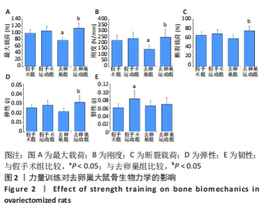
2.1 力量训练对去卵巢大鼠骨生物力学的影响 与假手术组比较,假手术运动组骨韧性增加(P < 0.05),去卵巢组最大载荷和刚度下降(P < 0.05)。与去卵巢组比较,去卵巢运动组最大载荷、刚度、断裂载荷和弹性均升高(P < 0.05),见图2。"


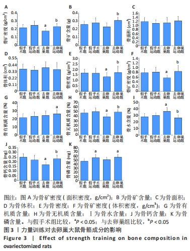
2.2 力量训练对去卵巢大鼠骨骼成分(生物物理学和生物化学)的影响 与假手术组比较,假手术运动组骨磷含量增加(P < 0.05);去卵巢组骨矿密度(面积密度)、骨密度、骨矿密度(体积密度)、骨无机质含量、骨钙含量下降(P < 0.05),骨水含量升高(P < 0.05);去卵巢运动组骨有机质含量和骨磷含量增加(P < 0.05)。与去卵巢组比较,去卵巢运动组骨矿密度(面积密度)、骨矿含量、骨密度、骨矿密度(体积密度)、骨无机质含量、骨钙含量升高(P < 0.05),骨水含量下降(P < 0.05)。各组骨面积和骨体积比较无统计学意义(P > 0.05),见图3。"

2.3 力量训练对去卵巢大鼠骨微结构的影响 与假手术组比较,去卵巢组骨小梁体积分数、骨小梁连接密度、骨小梁数量下降(P < 0.05),骨小梁分离度、结构模型指数升高(P < 0.05);去卵巢运动组骨小梁体积分数、骨小梁连接密度、骨小梁数量、骨髓面积下降(P < 0.05),骨小梁分离度、骨小梁厚度、结构模型指数、皮质骨体积分数、皮质骨面积分数、皮质骨厚度升高(P < 0.05)。与去卵巢组比较,去卵巢运动组骨小梁分离度、骨髓面积下降(P < 0.05),骨小梁厚度、皮质骨体积分数、皮质骨面积分数、皮质骨厚度、皮质骨孔隙度增加(P < 0.05),见图4。"


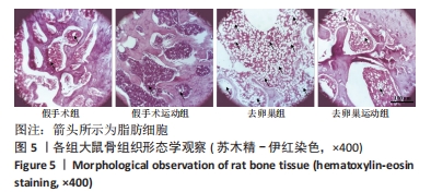
2.4 力量训练对去卵巢大鼠骨组织形态学的影响 骨组织苏木精-伊红染色显示:假手术组和假手术运动组骨小梁排列紧密、厚度均匀;去卵巢组骨细胞缺失,脂肪细胞(箭头所示)明显增加,骨小梁间隙较大,排列紊乱;去卵巢运动组骨细胞数增多,脂肪细胞显著减少,排列较紧密,骨小梁厚度增加,见图5。"

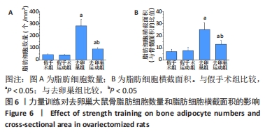
与假手术组比较,去卵巢组和去卵巢运动组骨脂肪细胞数量和横截面积均增加(P < 0.05)。与去卵巢组比较,去卵巢运动组骨脂肪细胞数量和横截面积减少(P < 0.05),见图6。"
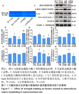
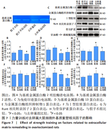
2.5 力量训练对去卵巢大鼠细胞外基质重塑相关因子的影响 与假手术组比较,假手术运动组基质金属蛋白酶2活性和Runt相关转录因子2蛋白表达增加(P < 0.05);去卵巢组基质金属蛋白酶2活性和基质金属蛋白酶13蛋白表达下降(P < 0.05),金属蛋白酶组织抑制物1、Runt相关转录因子2和骨保护素蛋白表达升高(P < 0.05);去卵巢运动组基质金属蛋白酶2活性升高(P < 0.05),基质金属蛋白酶13蛋白表达量下降(P < 0.05)。与去卵巢组比较,去卵巢运动组基质金属蛋白酶2活性升高(P < 0.05),金属蛋白酶组织抑制物1、Runt相关转录因子2、骨保护素蛋白表达下降(P < 0.05),见图7。"
| [1] MINAKOVIĆ I, ZVEKIć-SVORCAN J, JANKOVIĆ T, et al. Early menopause and risk of fractures-a preventable gap. Iran J Public Health. 2023;52(3):534-541. [2] HäNDEL MN, CARDOSO I, VON BÜLOW C, et al. Fracture risk reduction and safety by osteoporosis treatment compared with placebo or active comparator in postmenopausal women: systematic review, network meta-analysis, and meta-regression analysis of randomised clinical trials. BMJ. 2023;381:e068033. [3] XU X, LI X, LIANG Y, et al. Estrogen Modulates Cartilage and subchondral bone remodeling in an ovariectomized rat model of postmenopausal osteoarthritis. Med Sci Monit. 2019;25:3146-3153. [4] JOUSHEGHAN SS, SAJJADI MM, JOUSHEGHAN SS, et al. Extracellular vesicles as novel approaches for the treatment of osteoarthritis: a narrative review on potential mechanisms. J Mol Histol. 2021;52(5):879-891. [5] KOU L, JIANG X, LIN X, et al. Matrix Metalloproteinase inspired therapeutic strategies for bone diseases. Curr Pharm Biotechnol. 2021;22(4):451-467. [6] DE FIGUEIREDO F, AZEVEDO JP, LARA MG, et al. Recombinant human MMP-2 associated with monoolein improves bone repair. Bratisl Lek Listy. 2020;121(8):571-579. [7] GUIMARAES-STABILI MR, DE MEDEIROS MC, ROSSI D, et al. Silencing matrix metalloproteinase-13 (Mmp-13) reduces inflammatory bone resorption associated with LPS-induced periodontal disease in vivo. Clin Oral Investig. 2021;25(5):3161-3172. [8] GRAHAM S, ARCHER DF, SIMON JA, et al. Review of menopausal hormone therapy with estradiol and progesterone versus other estrogens and progestins. Gynecol Endocrinol. 2022;38(11):891-910. [9] YONG EL, LOGAN S. Menopausal osteoporosis: screening, prevention and treatment. Singapore Med J. 2021;62(4):159-166. [10] DING R, ZHANG N, WANG Q, et al. Alterations of the subchondral bone in osteoarthritis: complying with Wolff’s Law. Curr Rheumatol Rev. 2022;18(3):178-185. [11] SANCHEZ-TRIGO H, RITTWEGER J, SAÑUDO B. Effects of non-supervised exercise interventions on bone mineral density in adult women: a systematic review and meta‑analysis. Osteoporos Int. 2022;33(7):1415-1427. [12] SÁ K, DA SILVA GR, MARTINS UK, et al. Resistance training for postmenopausal women: systematic review and meta-analysis. Menopause. 2023;30(1):108-116. [13] YUAN Y, CHEN X, ZHANG L, et al. The roles of exercise in bone remodeling and in prevention and treatment of osteoporosis. Prog Biophys Mol Biol. 2016;122(2):122-130. [14] WANG Z, DUAN R, JIN Y, et al. Effects of ferric ammonium citrate on iron accumulation, bone turnover and bone density in ovariectomized rat models with osteoporosis. Cell Mol Biol (Noisy-le-grand). 2022;68(8):163-166. [15] LEE S, KIM K, LAMBRECHT NJ, et al. Interaction of resistance training, electroacupuncture and Huang Qi supplementation on skeletal muscle function and GLUT4 protein concentration in rats. Acupunct Med. 2016;34(5):380-385. [16] 付鹏宇,于晶晶,龚丽景.叉头框蛋白O1介导的自噬途径在抗阻训练缓解低氧诱导大鼠肌萎缩中的作用[J].中国病理生理杂志,2022,38(9):1667-1676. [17] 刘芃,马刚,何瑞波,等.有氧运动干预慢性肾功能衰竭模型大鼠的肾脏纤维化[J].中国组织工程研究,2023,27(20):3209-3215. [18] GENG Q, GAO H, YANG R, et al. Pyrroloquinoline quinone prevents estrogen deficiency-induced osteoporosis by inhibiting oxidative stress and osteocyte senescence. Int J Biol Sci. 2019;15(1):58-68. [19] 郑鹏骥,陆杨,张象,等.吡咯喹啉醌通过降低氧化应激和RANKL/OPG比率减少去卵巢大鼠骨量流失研究[J].中国骨质疏松杂志,2020,26(10):1431-1435. [20] 杨芳芳,葸慧荣,高玉海,等.丹参酮ⅡA对去卵巢大鼠骨密度及骨形态计量学的影响[J].中华骨质疏松和骨矿盐疾病杂志,2018,11(2):166-171. [21] UNAL M, CREECY A, NYMAN JS. The role of matrix composition in the mechanical behavior of bone. Curr Osteoporos Rep. 2018;16(3):205-215. [22] ZHANG N, MAGLAND JF, RAJAPAKSE CS, et al. Assessment of trabecular bone yield and post-yield behavior from high-resolution MRI-based nonlinear finite element analysis at the distal radius of premenopausal and postmenopausal women susceptible to osteoporosis. Acad Radiol. 2013;20(12):1584-1591. [23] WESCOTT DJ. Postmortem change in bone biomechanical properties: Loss of plasticity. Forensic Sci Int. 2019;300:164-169. [24] SALAS-RAMIREZ M, LASSMANN M, TRAN-GIA J. Quantification of the volume fraction of fat, water and bone mineral in spongiosa for red marrow dosimetry in molecular radiotherapy by using a dual-energy (SPECT/)CT. Z Med Phys. 2022;32(4):428-437. [25] JU YI, SONE T, OHNARU K, et al. Effect of swimming exercise on three-dimensional trabecular bone microarchitecture in ovariectomized rats. J Appl Physiol (1985). 2015; 119(9):990-997. [26] DIAZ BRINTON R. Minireview: translational animal models of human menopause: challenges and emerging opportunities. Endocrinology. 2012;153(8):3571-3578. [27] MEAKIN LB, UDEH C, GALEA GL, et al. Exercise does not enhance aged bone’s impaired response to artificial loading in C57Bl/6 mice. Bone. 2015;81:47-52. [28] TANG L, GAO X, YANG X, et al. Ladder-climbing training prevents bone loss and microarchitecture deterioration in diet-induced obese rats. Calcif Tissue Int. 2016;98(1): 85-93. [29] LITTLE-LETSINGER SE, PAGNOTTI GM, MCGRATH C, et al. Exercise and diet: uncovering prospective mediators of skeletal fragility in bone and marrow adipose tissue. Curr Osteoporos Rep. 2020;18(6):774-789. [30] HUANG X, CHEN W, GU C, et al. Melatonin suppresses bone marrow adiposity in ovariectomized rats by rescuing the imbalance between osteogenesis and adipogenesis through SIRT1 activation. J Orthop Translat. 2023;38:84-97. [31] SHIGUEMOTO GE, PRESTES J, LEITE RD, et al. Effects of resistance training on matrix metalloproteinase-2 activity and biomechanical and physical properties of bone in ovariectomized and intact rats. Scand J Med Sci Sports. 2012;22(5):607-617. [32] TANG SY, HERBER RP, HO SP, et al. Matrix metalloproteinase-13 is required for osteocytic perilacunar remodeling and maintains bone fracture resistance. J Bone Miner Res. 2012;27(9):1936-1950. [33] JEMTLAND R, HOLDEN M, REPPE S, et al. Molecular disease map of bone characterizing the postmenopausal osteoporosis phenotype. J Bone Miner Res. 2011;26(8):1793-1801. [34] SCHILTZ C, MARTY C, DE VERNEJOUL MC, et al. Inhibition of osteoblastic metalloproteinases in mice prevents bone loss induced by oestrogen deficiency. J Cell Biochem. 2008;104(5):1803-1817. [35] SOBUE T, HAKEDA Y, KOBAYASHI Y, et al. Tissue inhibitor of metalloproteinases 1 and 2 directly stimulate the bone-resorbing activity of isolated mature osteoclasts. J Bone Miner Res. 2001;16(12):2205-2214. [36] DE FARIAS JUNIOR GC, DE SOUSA NETO IV, GUZZONI V, et al. Remodeling process in bone of aged rats in response to resistance training. Life Sci. 2020;256:118-129. [37] GAMSJAEGER S, BROZEK W, RECKER R, et al. Transmenopausal changes in trabecular bone quality. J Bone Miner Res. 2014;29(3):608-617. [38] RUIZ P, HANDAN BA, DE MOURA C, et al. Protective effect of grape or apple juices in bone tissue of rats exposed to cadmium: role of RUNX-2 and RANK/L expression. Environ Sci Pollut Res Int. 2018;25(16):15785-15792. [39] ORTH M, SHENAR AK, SCHEUER C, et al. VEGF-loaded mineral-coated microparticles improve bone repair and are associated with increased expression of epo and RUNX-2 in murine non-unions. J Orthop Res. 2019;37(4):821-831. [40] HAYAMI T, KAPILA YL, KAPILA S. Divergent upstream osteogenic events contribute to the differential modulation of MG63 cell osteoblast differentiation by MMP-1 (collagenase-1) and MMP-13 (collagenase-3). Matrix Biol. 2011;30(4):281-289. [41] 刘晨涛,张娟娟,苗华,等.负重爬梯运动调节去卵巢大鼠OPG/Ikkα/Runx2基因表达以及对股骨结构和生物力学的影响[J].中国运动医学杂志,2019,38(4):296-304. [42] GHARIBI B, CAMA G, CAPURRO M, et al. Gene expression responses to mechanical stimulation of mesenchymal stem cells seeded on calcium phosphate cement. Tissue Eng Part A. 2013;19(21-22):2426-2438. [43] KLEIN-NULEND J, BACABAC RG, BAKKER AD. Mechanical loading and how it affects bone cells: the role of the osteocyte cytoskeleton in maintaining our skeleton. Eur Cell Mater. 2012;24:278-291. [44] ROCHETTE L, MELOUX A, RIGAL E, et al. The Role of osteoprotegerin in vascular calcification and bone metabolism: the basis for developing new therapeutics. Calcif Tissue Int. 2019;105(3):239-251. [45] 王强,杨民,王剑.跑台运动对去卵巢大鼠骨组织中OPG、RANKL和RUNX2 mRNA表达的影响[J].中国骨伤,2013,26(11):940-943. |
| [1] | Li Zhifei, Yang Yin, Chen Hualong, Liang Qinqiu, Zhong Yuanming, Zhang Yisheng. Finite element analysis of the correlation between tilt angle of titanium cage and postoperative subsidence of titanium cage after anterior subtotal cervical corpectomy, decompression and fusion [J]. Chinese Journal of Tissue Engineering Research, 2024, 28(9): 1313-1319. |
| [2] | Chen Mengmeng, Bao Li, Chen Hao, Jia Pu, Feng Fei, Shi Guan, Tang Hai. Biomechanical characteristics of a novel interspinous distraction fusion device BacFuse for the repair of lumbar degenerative disease [J]. Chinese Journal of Tissue Engineering Research, 2024, 28(9): 1325-1329. |
| [3] | Liang Cheng, Zhang Linqi, Wang Guan, Li Wen, Duan Ke, Li Zhong, Lu Xiaobo, Zhuo Naiqiang. Finite element and biomechanical analysis of different implants in repair for unilateral unstable pelvic posterior ring injury [J]. Chinese Journal of Tissue Engineering Research, 2024, 28(9): 1336-1341. |
| [4] | Guo Sutong, Feng Dehong, Guo Yu, Wang Ling, Ding Yujian, Liu Yi, Qian Zhengying, Li Mingyang. Construction and finite element analysis of normal and osteoporotic hip models [J]. Chinese Journal of Tissue Engineering Research, 2024, 28(9): 1342-1346. |
| [5] | Yang Junliang, Lu Tan, Xu Biao, Jiang Yaqiong, Wang Fucheng. Three-dimensional finite element analysis of effects of partial anterior cruciate ligament rupture on knee joint stress [J]. Chinese Journal of Tissue Engineering Research, 2024, 28(9): 1347-1353. |
| [6] | Weng Rui, Lin Dongxin, Guo Haiwei, Zhang Wensheng, Song Yuke, Lin Hongheng, Li Wenchao, Ye Linqiang. Abnormal types of intervertebral disc structure and related mechanical loading with biomechanical factors [J]. Chinese Journal of Tissue Engineering Research, 2024, 28(9): 1436-1442. |
| [7] | Zhang Xiaoyun, Liu Hua, Chai Yuan, Chen Feng, Zeng Hao, Gao Zhengang, Huang Yourong. Effect of Yishen Gushu Formula on bone metabolic markers and clinical efficacyn in patients with osteoporosis of kidney deficiency and blood stasis type [J]. Chinese Journal of Tissue Engineering Research, 2024, 28(8): 1155-1160. |
| [8] | Dai Yuexing, Zheng Liqin, Wu Minhui, Li Zhihong, Li Shaobin, Zheng Desheng, Lin Ziling. Effect of vessel number on computational fluid dynamics in vascular networks [J]. Chinese Journal of Tissue Engineering Research, 2024, 28(8): 1206-1210. |
| [9] | Wang Qiang, Li Shiyun, Xiong Ying, Li Tiantian. Biomechanical changes of the cervical spine in internal fixation with different anterior cervical interbody fusion systems [J]. Chinese Journal of Tissue Engineering Research, 2024, 28(6): 821-826. |
| [10] | Wei Yuanbiao, Lin Zhan, Chen Yanmei, Yang Tenghui, Zhao Xiao, Chen Yangsheng, Zhou Yanhui, Yang Minchao, Huang Feiqi. Finite element analysis of effects of sagittal cervical manipulation on intervertebral disc and facet joints [J]. Chinese Journal of Tissue Engineering Research, 2024, 28(6): 827-832. |
| [11] | Zhang Rui, Wang Kun, Shen Zicong, Mao Lu, Wu Xiaotao. Effects of endoscopic foraminoplasty and laminoplasty on biomechanical properties of intervertebral disc and isthmus [J]. Chinese Journal of Tissue Engineering Research, 2024, 28(6): 833-839. |
| [12] | Zhang Min, Peng Jing, Zhang Qiang, Chen Dewang. Mechanical properties of L3/4 laminar decompression and intervertebral fusion in elderly osteoporosis patients analyzed by finite element method [J]. Chinese Journal of Tissue Engineering Research, 2024, 28(6): 847-851. |
| [13] | Xue Xiaofeng, Wei Yongkang, Qiao Xiaohong, Du Yuyong, Niu Jianjun, Ren Lixin, Yang Huifeng, Zhang Zhimin, Guo Yuan, Chen Weiyi. Finite element analysis of osteoporosis in proximal femur after cannulated screw fixation for femoral neck fracture [J]. Chinese Journal of Tissue Engineering Research, 2024, 28(6): 862-867. |
| [14] | Huang Peizhen, Dong Hang, Cai Qunbin, Lin Ziling, Huang Feng. Finite element analysis of anterograde and retrograde intramedullary nail for different areas of femoral shaft fractures [J]. Chinese Journal of Tissue Engineering Research, 2024, 28(6): 868-872. |
| [15] | Wang Mingming, Zhang Zhong, Sun Jianhua, Zhao Gang, Song Hua, Yan Huadong, Lyu Bin. Finite element analysis of three different minimally invasive fixation methods for distal tibial fractures with soft tissue injury [J]. Chinese Journal of Tissue Engineering Research, 2024, 28(6): 879-885. |
| Viewed | ||||||
|
Full text |
|
|||||
|
Abstract |
|
|||||