Chinese Journal of Tissue Engineering Research ›› 2015, Vol. 19 ›› Issue (51): 8241-8246.doi: 10.3969/j.issn.2095-4344.2015.51.008
Previous Articles Next Articles
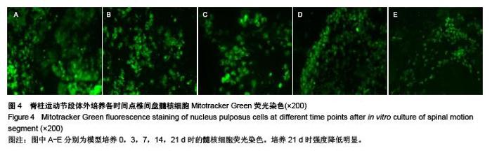
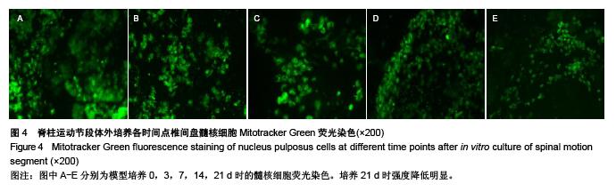
Changes of the nucleus pulposus after in vitro culture of rabbit spinal motion segments
Feng Min-shan1, Zhan Jia-wen1, Zhu Li-guo1, Zhang Ping2, Wang Yuan2, Bai Jian-qi2
- 1Second Department of Spine, Beijing Key Laboratory of Palasy Technology, Wangjing Hospital of China Academy of Chinese Medical Sciences, Beijing 100102, China; 2Department of Pathology, Wangjing Hospital of China Academy of Chinese Medical Sciences, Beijing 100102, China
-
Received:2015-10-07Online:2015-12-10Published:2015-12-10 -
Contact:Zhan Jia-wen, M.D., Physician, Second Department of Spine, Beijing Key Laboratory of Palasy Technology, Wangjing Hospital of China Academy of Chinese Medical Sciences, Beijing 100102, China -
About author:Feng Min-shan, M.D., Attending physician, Second Department of Spine, Beijing Key Laboratory of Palasy Technology, Wangjing Hospital of China Academy of Chinese Medical Sciences, Beijing 100102, China -
Supported by:TCM Innovation Team of Prevention and Control Osteoarticular Degenerative Diseases, No. YS1304; Bone and Joint Degenerative Diseases Subject Research, No. WJYY2014-ZX-06
CLC Number:
Cite this article
Feng Min-shan, Zhan Jia-wen, Zhu Li-guo, Zhang Ping, Wang Yuan, Bai Jian-qi. Changes of the nucleus pulposus after in vitro culture of rabbit spinal motion segments [J]. Chinese Journal of Tissue Engineering Research, 2015, 19(51): 8241-8246.
share this article
"

"

"
"
| [1] 杨松波,高春华,庞晓东,等.椎间盘退变模型的研究进展[J].脊柱外科杂志,2012,10 (5): 315-317.
[2] Risbud MV, Izzo MW, Adams CS, et al.An organ culture system for the study of the nucleus pulposus: description of the system and evaluation of the cells. Spine (Phila Pa 1976), 2003;28(24): 2652-2658; discussion 2658-2659.
[3] Richardson SM, Walker RV, Parker S, et al.Intervertebral disc cell-mediated mesenchymal stem cell differentiation.Stem Cells.2006;24(3): 707-716.
[4] 牛朋彦,熊伟,李锋,等.渗透压负荷对兔椎间盘器官培养模型的影响[J].中国脊柱脊髓杂志,2009,19 (10): 729-734.
[5] Korecki CL, Costi JJ,Iatridis JC.Needle puncture injury affects intervertebral disc mechanics and biology in an organ culture model. Spine (Phila Pa 1976).2008;33(3): 235-241.
[6] Boden SD, McCowin PR, Davis DO, et al.Abnormal magnetic-resonance scans of the cervical spine in asymptomatic subjects. A prospective investigation. J Bone Joint Surg Am.1990; 72(8): 1178-1184.
[7] Chan SC, Gantenbein-Ritter B, Leung VY, et al. Cryopreserved intervertebral disc with injected bone marrow-derived stromal cells: a feasibility study using organ culture. Spine J.2010;10(6): 486-496.
[8] Rannou F, Lee TS, Zhou RH, et al.Intervertebral disc degeneration: the role of the mitochondrial pathway in annulus fibrosus cell apoptosis induced by overload. Am J Pathol.2004; 164(3): 915-924.
[9] Jim B, Steffen T, Moir J, et al.Development of an intact intervertebral disc organ culture system in which degeneration can be induced as a prelude to studying repair potential. Eur Spine J.2011;20(8): 1244-1254.
[10] 张聪明,刘强,李钢,等.大鼠椎间盘器官整体培养条件下髓核组织的变化[J].中国医疗前沿,2010,5(6): 19-20.
[11] Gantenbein B, Grunhagen T, Lee CR, et al.An in vitro organ culturing system for intervertebral disc explants with vertebral endplates: a feasibility study with ovine caudal discs. Spine (Phila Pa 1976).2006;31(23): 2665-2673.
[12] Gruber HE,Hanley EN, Jr.Human disc cells in monolayer vs 3D culture: cell shape, division and matrix formation.BMC Musculoskelet Disord.2000;1: 1.
[13] Zhang Y, Phillips FM, Thonar EJ, et al.Cell therapy using articular chondrocytes overexpressing BMP-7 or BMP-10 in a rabbit disc organ culture model. Spine (Phila Pa 1976).2008; 33(8): 831-838.
[14] Ariga K,Yonenobu K,Nakase T, et al.Mechanical stress-induced apoptosis of endplate chondrocytes in organ-cultured mouse intervertebral discs: an ex vivo study. Spine (Phila Pa 1976),2003. 28(14): 1528-1533.
[15] Kim KW, Lim TH, Kim JG, et al.The origin of chondrocytes in the nucleus pulposus and histologic findings associated with the transition of a notochordal nucleus pulposus to a fibrocartilaginous nucleus pulposus in intact rabbit intervertebral discs. Spine (Phila Pa 1976).2003;28(10): 982-990.
[16] Gawri R, Mwale F, Ouellet J, et al.Development of an organ culture system for long-term survival of the intact human intervertebral disc[J]. Spine (Phila Pa 1976),2011. 36(22): 1835-1842.
[17] 李钢,范顺武,赵凤东,等.大鼠椎间盘器官整体培养条件下髓核组织的变化[J].中国修复重建外科杂志,2010,5 (4): 385-390.
[18] Haschtmann D, Stoyanov J V, Ettinger L, et al.Establishment of a novel intervertebral disc/endplate culture model: analysis of an ex vivo in vitro whole-organ rabbit culture system. Spine (Phila Pa 1976).2006;31(25): 2918-2925.
[19] Ohshima H, Urban J P,Bergel DH.Effect of static load on matrix synthesis rates in the intervertebral disc measured in vitro by a new perfusion technique. J Orthop Res.1995;13(1): 22-29.
[20] Hutton WC, Ganey TM, Elmer WA, et al.Does long-term compressive loading on the intervertebral disc cause degeneration?. Spine (Phila Pa 1976).2000;25(23): 2993-3004.
[21] Paul CP, Zuiderbaan HA, Zandieh Doulabi B,et al.Simulated-physiological loading conditions preserve biological and mechanical properties of caprine lumbar intervertebral discs in ex vivo culture. PLoS One.2012;7(3): e33147.
[22] Oshima H, Ishihara H, Urban J P, et al.The use of coccygeal discs to study intervertebral disc metabolism. J Orthop Res. 1993; 11(3): 332-338.
[23] Lee CR, Iatridis JC, Poveda L, et al.In vitro organ culture of the bovine intervertebral disc: effects of vertebral endplate and potential for mechanobiology studies[J]. Spine (Phila Pa 1976).2006;31(5): 515-522.
[24] Korecki CL, MacLean JJ,Iatridis JC.Characterization of an in vitro intervertebral disc organ culture system. Eur Spine J. 2007; 16(7): 1029-1037.
[25] Hutton WC, Elmer WA, Boden SD, et al.The effect of hydrostatic pressure on intervertebral disc metabolism. Spine (Phila Pa 1976).1999;24(15): 1507-1015.
[26] Chiba K, Andersson GB, Masuda K, et al.A new culture system to study the metabolism of the intervertebral disc in vitro. Spine (Phila Pa 1976).1998;23(17): 1821-1827; discussion 1828.
[27] Lim TH, Ramakrishnan PS, Kurriger GL, et al.Rat spinal motion segment in organ culture: a cell viability study. Spine (Phila Pa 1976).2006;31(12): 1291-1297; discussion 1298.
[28] Seol D, Choe H, Ramakrishnan P S, et al.Organ culture stability of the intervertebral disc: rat versus rabbit. J Orthop Res.2013; 31(6): 838-46.
[29] Urban J P,Maroudas A.Swelling of the intervertebral disc in vitro. Connect Tissue Res.1981;9(1): 1-10.
[30] 徐宏光,章平治,宋俊兴,等.退变大鼠椎间盘器官培养模型的建立及其意义[J].中国骨与关节外科,2012,5(3): 233-237,197.
[31] 牛朋彦,熊伟,李锋,等.软骨终板通透性对体外培养大鼠髓核细胞生物学特性的影响[J].中国脊柱脊髓杂志,2011,21(7): 597-602.
[32] 董妙珠,肖萍,叶于薇,等.PAS、AB染色法在软骨蛋白黏多糖检测中的运用[J].上海预防医学杂志,2004,16(9): 419-420.
[33] 徐无忌,李悦,原超.六味地黄丸含药血清对椎间盘Ⅰ型和Ⅱ型胶原表达的影响[J]. 中国组织工程研究,2013,17 (26): 4857-4864.
[34] 徐宏光,章平治,宋俊兴,等.循环机械压力诱导下兔椎间盘退变器官模型的建立及意义[J]. 中国骨与关节外科,2014,7(1): 45-51.
[35] Poot M, Zhang YZ, Kramer JA, et al.Analysis of mitochondrial morphology and function with novel fixable fluorescent stains. J Histochem Cytochem.1996;44(12): 1363-1372.
[36] Isenberg JS,Klaunig JE.Role of the mitochondrial membrane permeability transition (MPT) in rotenone-induced apoptosis in liver cells. Toxicol Sci,2000. 53(2): 340-351.
[37] Haschtmann D, Stoyanov J V,Ferguson S J.Influence of diurnal hyperosmotic loading on the metabolism and matrix gene expression of a whole-organ intervertebral disc model. J Orthop Res.2006;24(10): 1957-1966.
[38] Hutton WC, Murakami H, Li J, et al.The effect of blocking a nutritional pathway to the intervertebral disc in the dog mode. J Spinal Disord Tech.2004;17(1): 53-63.
|
| [1] | Chen Ziyang, Pu Rui, Deng Shuang, Yuan Lingyan. Regulatory effect of exosomes on exercise-mediated insulin resistance diseases [J]. Chinese Journal of Tissue Engineering Research, 2021, 25(25): 4089-4094. |
| [2] | Chen Yang, Huang Denggao, Gao Yuanhui, Wang Shunlan, Cao Hui, Zheng Linlin, He Haowei, Luo Siqin, Xiao Jingchuan, Zhang Yingai, Zhang Shufang. Low-intensity pulsed ultrasound promotes the proliferation and adhesion of human adipose-derived mesenchymal stem cells [J]. Chinese Journal of Tissue Engineering Research, 2021, 25(25): 3949-3955. |
| [3] | Yang Junhui, Luo Jinli, Yuan Xiaoping. Effects of human growth hormone on proliferation and osteogenic differentiation of human periodontal ligament stem cells [J]. Chinese Journal of Tissue Engineering Research, 2021, 25(25): 3956-3961. |
| [4] | Sun Jianwei, Yang Xinming, Zhang Ying. Effect of montelukast combined with bone marrow mesenchymal stem cell transplantation on spinal cord injury in rat models [J]. Chinese Journal of Tissue Engineering Research, 2021, 25(25): 3962-3969. |
| [5] | Gao Shan, Huang Dongjing, Hong Haiman, Jia Jingqiao, Meng Fei. Comparison on the curative effect of human placenta-derived mesenchymal stem cells and induced islet-like cells in gestational diabetes mellitus rats [J]. Chinese Journal of Tissue Engineering Research, 2021, 25(25): 3981-3987. |
| [6] | Hao Xiaona, Zhang Yingjie, Li Yuyun, Xu Tao. Bone marrow mesenchymal stem cells overexpressing prolyl oligopeptidase on the repair of liver fibrosis in rat models [J]. Chinese Journal of Tissue Engineering Research, 2021, 25(25): 3988-3993. |
| [7] | Liu Jianyou, Jia Zhongwei, Niu Jiawei, Cao Xinjie, Zhang Dong, Wei Jie. A new method for measuring the anteversion angle of the femoral neck by constructing the three-dimensional digital model of the femur [J]. Chinese Journal of Tissue Engineering Research, 2021, 25(24): 3779-3783. |
| [8] | Meng Lingjie, Qian Hui, Sheng Xiaolei, Lu Jianfeng, Huang Jianping, Qi Liangang, Liu Zongbao. Application of three-dimensional printing technology combined with bone cement in minimally invasive treatment of the collapsed Sanders III type of calcaneal fractures [J]. Chinese Journal of Tissue Engineering Research, 2021, 25(24): 3784-3789. |
| [9] | Qian Xuankun, Huang Hefei, Wu Chengcong, Liu Keting, Ou Hua, Zhang Jinpeng, Ren Jing, Wan Jianshan. Computer-assisted navigation combined with minimally invasive transforaminal lumbar interbody fusion for lumbar spondylolisthesis [J]. Chinese Journal of Tissue Engineering Research, 2021, 25(24): 3790-3795. |
| [10] | Hu Jing, Xiang Yang, Ye Chuan, Han Ziji. Three-dimensional printing assisted screw placement and freehand pedicle screw fixation in the treatment of thoracolumbar fractures: 1-year follow-up [J]. Chinese Journal of Tissue Engineering Research, 2021, 25(24): 3804-3809. |
| [11] | Shu Qihang, Liao Yijia, Xue Jingbo, Yan Yiguo, Wang Cheng. Three-dimensional finite element analysis of a new three-dimensional printed porous fusion cage for cervical vertebra [J]. Chinese Journal of Tissue Engineering Research, 2021, 25(24): 3810-3815. |
| [12] | Wang Yihan, Li Yang, Zhang Ling, Zhang Rui, Xu Ruida, Han Xiaofeng, Cheng Guangqi, Wang Weil. Application of three-dimensional visualization technology for digital orthopedics in the reduction and fixation of intertrochanteric fracture [J]. Chinese Journal of Tissue Engineering Research, 2021, 25(24): 3816-3820. |
| [13] | Sun Maji, Wang Qiuan, Zhang Xingchen, Guo Chong, Yuan Feng, Guo Kaijin. Development and biomechanical analysis of a new anterior cervical pedicle screw fixation system [J]. Chinese Journal of Tissue Engineering Research, 2021, 25(24): 3821-3825. |
| [14] | Lin Wang, Wang Yingying, Guo Weizhong, Yuan Cuihua, Xu Shenggui, Zhang Shenshen, Lin Chengshou. Adopting expanded lateral approach to enhance the mechanical stability and knee function for treating posterolateral column fracture of tibial plateau [J]. Chinese Journal of Tissue Engineering Research, 2021, 25(24): 3826-3827. |
| [15] | Zhu Yun, Chen Yu, Qiu Hao, Liu Dun, Jin Guorong, Chen Shimou, Weng Zheng. Finite element analysis for treatment of osteoporotic femoral fracture with far cortical locking screw [J]. Chinese Journal of Tissue Engineering Research, 2021, 25(24): 3832-3837. |
| Viewed | ||||||
|
Full text |
|
|||||
|
Abstract |
|
|||||