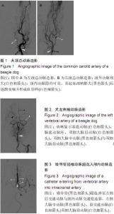
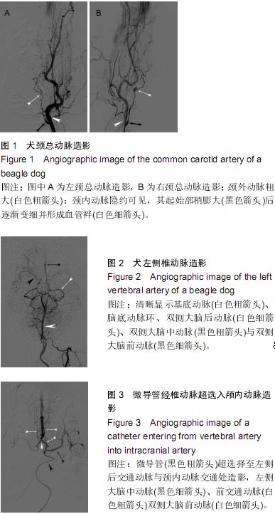
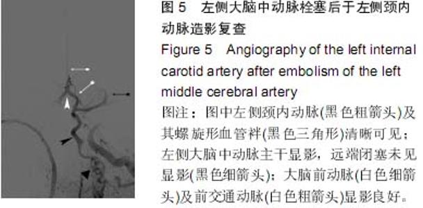
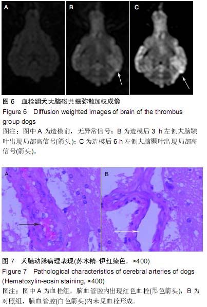
| [1] Sudlow CL, Warlow CP. Comparable studies of the incidence of stroke and its pathological types: results from an international collaboration. International Stroke Incidence Collaboration. Stroke. 1997;28(3):491-499.
[2] Grunwald IQ, Wakhloo AK, Walter S, et al. Endovascular stroke treatment today. AJNR Am J Neuroradiol. 2011; 32(2):238-243.
[3] Widimský P, Ko?nar B, Štětká?ová I. Modern treatment of acute ischemic stroke. Vnitr Lek. 2014;60(12):1086-1089.
[4] Hayakawa M. Intravenous thrombolysis for acute ischemic stroke: past, present and future. Rinsho Shinkeigaku. 2014; 54(12):1197-1199.
[5] Pierot L, Soize S, Benaissa A, et al. Techniques for endovascular treatment of acute ischemic stroke: from intra-arterial fibrinolytics to stent-retrievers. Stroke. 2015; 46(3):909-914.
[6] Comai A, Haglmüller T, Ferro F, et al. Sequential endovascular thrombectomy approach (SETA) to acute ischemic stroke: preliminary single-centre results and cost analysis. Radiol Med. 2015. in press.
[7] Hlavica M, Diepers M, Garcia-Esperon C, et al. Pharmacological recanalization therapy in acute ischemic stroke - Evolution, current state and perspectives of intravenous and intra-arterial thrombolysis. J Neuroradiol. 2015;42(1):30-46.
[8] Alkhalili K, Chalouhi N, Tjoumakaris S, et al. Endovascular intervention for acute ischemic stroke in light of recent trials. ScientificWorldJournal. 2014;2014:429549.
[9] Iwata T. Current and future prospects of endovascular treatment for acute ischemic stroke. Rinsho Shinkeigaku. 2014;54(12):1200-1202.
[10] Turc G, Isabel C, Calvet D. Intravenous thrombolysis for acute ischemic stroke. Diagn Interv Imaging. 2014;95(12): 1129-1133.
[11] Mokin M, Khalessi AA, Mocco J, et al. Endovascular treatment of acute ischemic stroke: the end or just the beginning? Neurosurg Focus. 2014;36(1):E5.
[12] Yamagami H, Sakai N. Current status of endovascular therapy for acute ischemic stroke. Rinsho Shinkeigaku. 2013;53(11):1166-1168.
[13] Kang BT, Lee JH, Jung DI, et al. Canine model of ischemic stroke with permanent middle cerebral artery occlusion: clinical and histopathological findings. J Vet Sci. 2007;8(4):369-376.
[14] Lu SS, Liu S, Zu QQ, et al. Multimodal magnetic resonance imaging for assessing lacunar infarction after proximal middle cerebral artery occlusion in a canine model. Chin Med J (Engl). 2013;126(2):311-317.
[15] Liu S, Hu WX, Zu QQ, et al. A novel embolic stroke model resembling lacunar infarction following proximal middle cerebral artery occlusion in beagle dogs. J Neurosci Methods. 2012;209(1):90-96.
[16] Borsody MK, Yamada C, Bielawski D, et al. Effects of noninvasive facial nerve stimulation in the dog middle cerebral artery occlusion model of ischemic stroke. Stroke. 2014;45(4):1102-1107.
[17] Zu QQ, Liu S, Xu XQ, et al. An endovascular canine stroke model: middle cerebral artery occlusion with autologous clots followed by ipsilateral internal carotid artery blockade. Lab Invest. 2013;93(7):760-767.
[18] Boulos AS, Deshaies EM, Dalfino JC, et al. Tamoxifen as an effective neuroprotectant in an endovascular canine model of stroke. J Neurosurg. 2011;114(4):1117-1126.
[19] 刘圣,施海彬,胡卫星,等.毕格犬和杂种犬脑血管及脑组织的对照研究[J].介入放射学杂志,2011,20(9):717-722.
[20] Purdy PD, Horowitz MB, Mathews D, et al. Calcium 45 autoradiography and dual-isotope single-photon emission CT in a canine model of cerebral ischemia and middle cerebral artery occlusion. AJNR Am J Neuroradiol. 1996;17(6):1161- 1170.
[21] Rink C, Christoforidis G, Abduljalil A, et al. Minimally invasive neuroradiologic model of preclinical transient middle cerebral artery occlusion in canines. Proc Natl Acad Sci U S A. 2008; 105(37):14100-14105.
[22] Dwyer MG, Bergsland N, Saluste E, et al. Application of hidden Markov random field approach for quantification of perfusion/diffusion mismatch in acute ischemic stroke. Neurol Res. 2008;30(8):827-834.
[23] González RG, Schaefer PW, Buonanno FS, et al. Diffusion-weighted MR imaging: diagnostic accuracy in patients imaged within 6 hours of stroke symptom onset. Radiology. 1999;210(1):155-162.
[24] Romero JM, Schaefer PW, Grant PE, et al. Diffusion MR imaging of acute ischemic stroke. Neuroimaging Clin N Am. 2002;12(1):35-53.
[25] 孙静华,刘海霞,付英杰,等.头颈部CTA、DWI及ABCD2评分在短暂性脑缺血发作中的应用[J].实用放射学杂志,2012,28(4): 504-508.
[26] Audebert HJ, Fiebach JB. Brain imaging in acute ischemic stroke—MRI or CT? Curr Neurol Neurosci Rep. 2015;15(3):6.
[27] Yoo RE, Yun TJ, Rhim JH, et al. Bright vessel appearance on arterial spin labeling MRI for localizing arterial occlusion in acute ischemic stroke. Stroke. 2015;46(2):564-567.
[28] Madai VI, Galinovic I, Grittner U, et al. DWI intensity values predict FLAIR lesions in acute ischemic stroke. PLoS One. 2014;9(3):e92295.
[29] Lettau M, Laible M. 3-T high-b-value diffusion-weighted MR imaging in hyperacute ischemic stroke. J Neuroradiol. 2013; 40(3):149-157.
[30] Nagashima H, Doi K, Ogura T, et al. Automated adjustment of display conditions in brain MR images: diffusion-weighted MRIs and apparent diffusion coefficient maps for hyperacute ischemic stroke. Radiol Phys Technol. 2013;6(1):202-209.
[31] Vymazal J, Rulseh AM, Keller J, et al. Comparison of CT and MR imaging in ischemic stroke. Insights Imaging. 2012;3(6): 619-627.
[32] González RG. Clinical MRI of acute ischemic stroke. J Magn Reson Imaging. 2012;36(2):259-271.
[33] Mitomi M, Kimura K, Aoki J, et al. Comparison of CT and DWI findings in ischemic stroke patients within 3 hours of onset. J Stroke Cerebrovasc Dis. 2014;23(1):37-42.
[34] 罗中华,宦怡,贺洪德,等.急性缺血性脑卒中介入取栓器血栓模型的机械特性比较[J].介入放射学杂志,2013,22(4):317-321.
[35] Rath DP, Little CM, Zhang H, et al. Sodium pentobarbital versus alpha-chloralose anesthesia. Experimental production of substantially different slopes in the transmural CP/ATP ratios within the left ventricle of the canine myocardium. Circulation. 1995;91(2):471-475.
[36] Muñoz-Islas E, Gupta S, Jiménez-Mena LR, et al. Donitriptan, but not sumatriptan, inhibits capsaicin-induced canine external carotid vasodilatation via 5-HT1B rather than 5-HT1D receptors. Br J Pharmacol. 2006;149(1):82-91.
[37] Ogün CO, Ta?tekin G, Kire?i D, et al. A new reversible ischemic neurologic deficit model in dogs. Med Sci Monit. 2008;14(10):BR214-218. |