Chinese Journal of Tissue Engineering Research ›› 2026, Vol. 30 ›› Issue (31): 8108-8115.doi: 10.12307/2026.838
Previous Articles Next Articles
Human umbilical cord-derived mesenchymal stem cells to treat osteoarthritis in rats: dual effect of relieving pain and protecting cartilage
Liu Fang, Hu Haimin, Song Dan, Nan Jie, Cai Xiaoxiao, He Qingrui, Lian Ruijiao, Luo Lin, Yuan Chunhui
- Beijing Regenerative Biotechnology Research Institute Co., Ltd., Beijing 102609, China
-
Received:2025-09-17Accepted:2026-01-12Online:2026-11-08Published:2026-05-23 -
Contact:Yuan Chunhui, MS, Beijing Regenerative Biotechnology Research Institute Co., Ltd., Beijing 102609, China -
About author:Liu Fang, MS, Beijing Regenerative Biotechnology Research Institute Co., Ltd., Beijing 102609, China

CLC Number:
Cite this article
Liu Fang, Hu Haimin, Song Dan, Nan Jie, Cai Xiaoxiao, He Qingrui, Lian Ruijiao, Luo Lin, Yuan Chunhui. Human umbilical cord-derived mesenchymal stem cells to treat osteoarthritis in rats: dual effect of relieving pain and protecting cartilage[J]. Chinese Journal of Tissue Engineering Research, 2026, 30(31): 8108-8115.
share this article
Add to citation manager EndNote|Reference Manager|ProCite|BibTeX|RefWorks
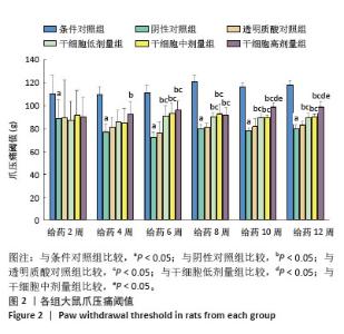
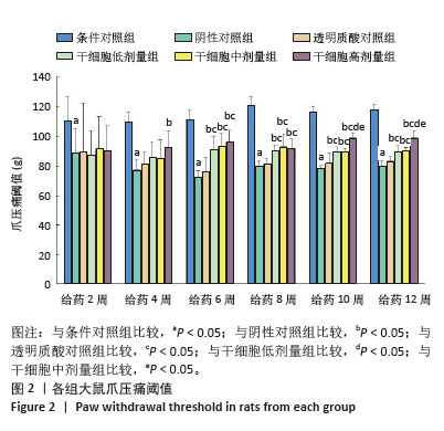
2.1 实验动物数量分析 该研究共使用96只SD大鼠,所有大鼠均进入结果分析。 2.2 行为学检查结果 2.2.1 爪压痛阈值 图2显示,给药2-12周时,阴性对照组的爪压痛阈值均低于条件对照组,差异有显著性意义(P < 0.05)。给药4周时,干细胞高剂量组的爪压痛阈值高于阴性对照组,差异有显著性意义(P < 0.05)。给药6,8,10,12周时,干细胞低、中、高剂量组的爪压痛阈值均高于阴性对照组和透明质酸对照组,差异有显著性意义(P < 0.05)。给药10,12周时,干细胞高剂量组的爪压痛阈值高于干细胞低、中剂量组,差异有显著性意义(P < 0.05);其余时间点干细胞各剂量组的爪压痛阈值无明显差异(P > 0.05)。"
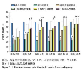
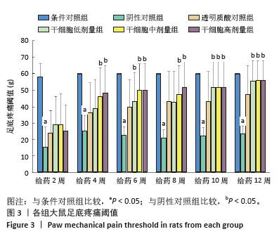
2.2.2 足底疼痛阈值 图3显示,给药2-12周时,阴性对照组的足底疼痛阈值均低于条件对照组,差异有显著性意义(P < 0.05)。给药4周时,干细胞中、高剂量组的足底疼痛阈值高于阴性对照组,差异有显著性意义(P < 0.05)。给药6周时,干细胞低、中、高剂量组的足底疼痛阈值高于阴性对照组,差异有显著性意义(P < 0.05)。给药8周时,干细胞中、高剂量组的足底疼痛阈值高于阴性对照组,差异有显著性意义(P < 0.05)。给药10,12周时,干细胞低、中、高剂量组的足底疼痛阈值高于阴性对照组,差异有显著性意义(P < 0.05)。在所有检测时间点,干细胞各剂量组的足底疼痛阈值无明显差异(P > 0.05)。 "
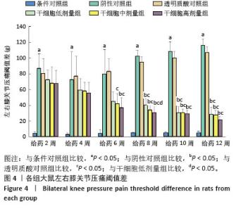
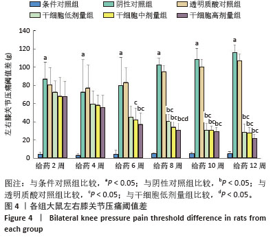
2.2.3 左右膝关节压痛阈值差 图4显示,给药2-12周时,阴性对照组的左右膝关节压痛阈值差高于条件对照组,差异有显著性意义(P < 0.05)。给药6周时,干细胞高剂量组的左右膝关节压痛阈值差低于阴性对照组,差异有显著性意义(P < 0.05);干细胞中、高剂量组的左右膝关节压痛阈值差低于透明质酸对照组,差异有显著性意义(P < 0.05)。给药8,10,12周时,干细胞低、中、高剂量组的左右膝关节压痛阈值差均低于阴性对照组、透明质酸对照组,差异有显著性意义(P < 0.05)。给药8周时,干细胞高剂量组的左右膝关节压痛阈值差低于干细胞低剂量组,差异有显著性意义(P < 0.05);其余时间点干细胞各剂量组的左右膝关节压痛阈值差无明显差异(P > 0.05)。 "
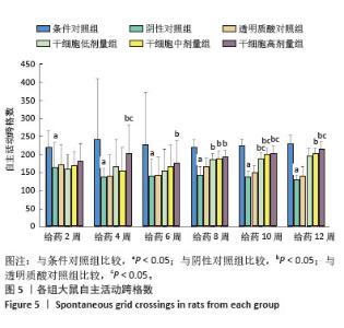
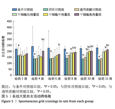
2.2.4 自主活动能力 自主活动跨格数检测结果(图5)显示,给药2-12周时,阴性对照组的跨格数低于条件对照组,差异有显著性意义(P < 0.05)。给药4周时,干细胞高剂量组的跨格数高于阴性对照组、透明质酸对照组,差异有显著性意义(P < 0.05)。给药6周时,干细胞高剂量组的跨格数高于阴性对照组,差异有显著性意义(P < 0.05)。给药8周时,干细胞低、中、高剂量组的跨格数高于阴性对照组,差异有显著性意义(P < 0.05)。给药10周时,干细胞低、中、高剂量组的跨格数高于阴性对照组、透明质酸对照组,差异有显著性意义(P < 0.05)。给药12周时,干细胞中剂量组的跨格数高于阴性对照组(P < 0.05);干细胞高剂量组的跨格数高于阴性对照组和透明质酸对照组,差异有显著性意义(P < 0.05)。在所有检测时间点,干细胞各剂量组的自发活动跨格数无明显差异(P > 0.05)。 "
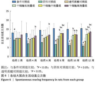
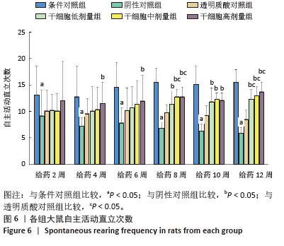
自主活动直立次数检测结果(图6)显示,给药2-12周时,阴性对照组的直立次数低于条件对照组,差异有显著性意义(P < 0.05)。给药4周和6周时,干细胞高剂量组的直立次数高于阴性对照组,差异有显著性意义(P < 0.05)。给药8周时,干细胞低、中、高剂量组的直立次数高于阴性对照组,差异有显著性意义(P < 0.05);干细胞中、高剂量组的直立次数高于透明质酸对照组,差异有显著性意义(P < 0.05)。给药10周时,干细胞低、中、高剂量组的直立次数高于阴性对照组,差异有显著性意义(P < 0.05)。给药12周时,干细胞低、中、高剂量组的直立次数高于阴性对照组、透明质酸对照组,差异有显著性意义(P < 0.05)。在所有检测时间点,干细胞各剂量组的自发活动直立次数无明显差异(P > 0.05)。 "
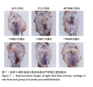
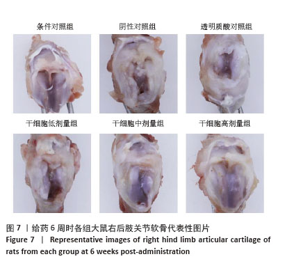
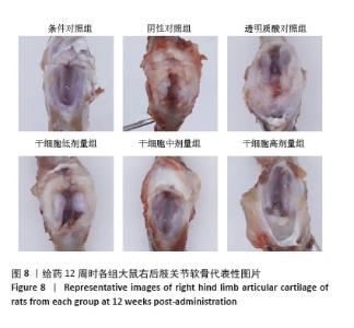
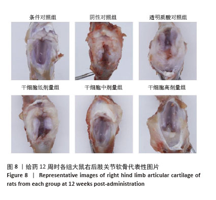
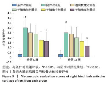
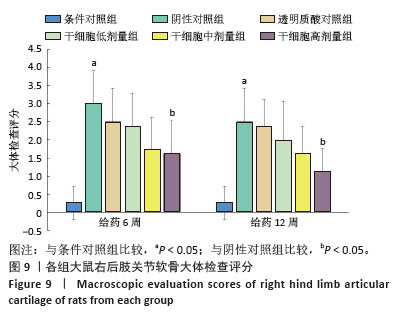
2.3 关节软骨大体检查结果 图7,8显示,给药6,12周时,条件对照组大鼠的关节软骨表面光滑,玻璃样外观,无明显滑膜增生;阴性对照组、透明质酸对照组及干细胞低、中剂量组大鼠的关节软骨表面粗糙且不透明,可见明显磨损、纤维化组织和滑膜增生;而干细胞高剂量组大鼠的上述情况有所改善。分级评分结果(图9)显示,给药6,12周时,阴性对照组的关节软骨损伤评分高于条件对照组,差异有显著性意义(P < 0.05);干细胞高剂量组的关节软骨损伤评分低于阴性对照组,差异有显著性意义(P < 0.05)。在所有检测时间点,干细胞各剂量组的关节软骨损伤评分无明显差异(P > 0.05)。 "
"
"
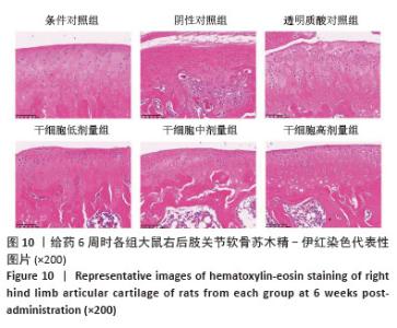
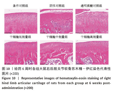
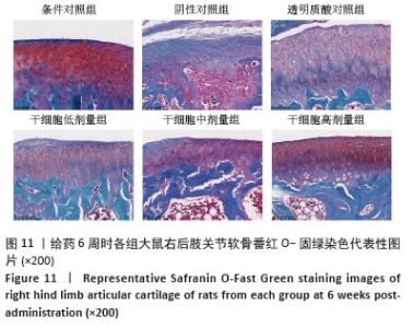
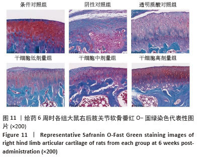
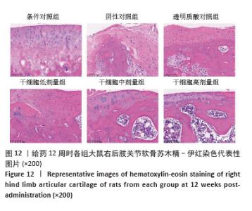
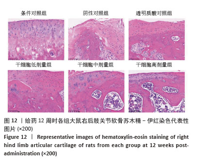
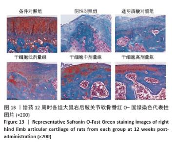
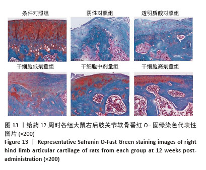
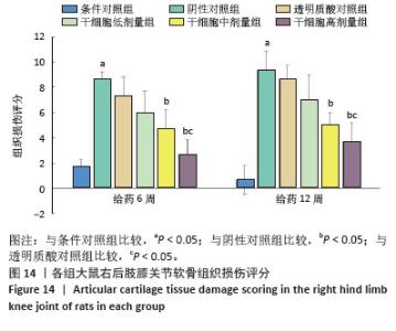
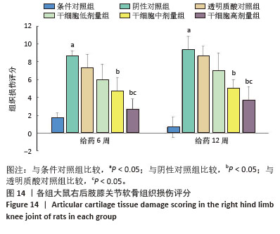
2.4 苏木精-伊红染色和番红O-固绿染色结果 苏木精-伊红染色和番红-固绿染色(图10-13)显示,给药6,12周时,条件对照组大鼠关节软骨表面规则,基质较厚,潮线完整;阴性对照组、透明质酸对照组、干细胞低剂量组大鼠表面不平整,具有不规则裂隙,基质较薄且番红O染色较浅,潮线不完整;干细胞中、高剂量组上述病理改变有所改善。分级评分结果(图14)显示,给药6,12周时,阴性对照组的组织损伤评分高于条件对照组,差异有显著性意义(P < 0.05);干细胞中、高剂量组的组织损伤评分低于阴性对照组,差异有显著性意义(P < 0.05);干细胞高剂量组的组织损伤评分低于透明质酸对照组(P < 0.05)。在所有检测时间点,干细胞各剂量组的组织损伤评分无明显差异(P > 0.05)。 "
"
"
"
"
| [1] KWON DG, KIM MK, JEON YS, et al. State of the Art: The Immunomodulatory Role of MSCs for Osteoarthritis. Int J Mol Sci. 2022;23(3):1618. [2] ZHANG P, DONG B, YUAN P, et al. Human umbilical cord mesenchymal stem cells promoting knee joint chondrogenesis for the treatment of knee osteoarthritis: a systematic review. J Orthop Surg Res. 2023;18(1):639. [3] XIANG XN, ZHU SY, HE HC, et al. Mesenchymal stromal cell-based therapy for cartilage regeneration in knee osteoarthritis. Stem Cell Res Ther. 2022;13(1):14. [4] ZHU Y, YE L, CAI X, et al. Icariin-Loaded Hydrogel Regulates Bone Marrow Mesenchymal Stem Cell Chondrogenic Differentiation and Promotes Cartilage Repair in Osteoarthritis. Front Bioeng Biotechnol. 2022;10:755260. [5] HE L, HE T, XING J, et al. Bone marrow mesenchymal stem cell-derived exosomes protect cartilage damage and relieve knee osteoarthritis pain in a rat model of osteoarthritis. Stem Cell Res Ther. 2020;11(1):276. [6] CARNEIRO DC, ARAÚJO LT, SANTOS GC, et al. Clinical Trials with Mesenchymal Stem Cell Therapies for Osteoarthritis: Challenges in the Regeneration of Articular Cartilage. Int J Mol Sci. 2023;24(12):9939. [7] HAMDALLA HM, AHMED RR, GALALY SR, et al. Assessment of the Efficacy of Bone Marrow-Derived Mesenchymal Stem Cells against a Monoiodoacetate-Induced Osteoarthritis Model in Wistar Rats. Stem Cells Int. 2022;2022:1900403. [8] COPP G, ROBB KP, VISWANATHAN S. Culture-expanded mesenchymal stromal cell therapy: does it work in knee osteoarthritis? A pathway to clinical success. Cell Mol Immunol. 2023;20(6):626-650. [9] 陈驹,郑锦畅,梁振,等.β-石竹烯对小鼠膝骨关节炎的作用及机制[J].中国组织工程研究,2026,30(6):1341-1347. [10] DE SOUSA VALENTE J. The Pharmacology of Pain Associated With the Monoiodoacetate Model of Osteoarthritis. Front Pharmacol. 2019;10:974. [11] PAN X, LI X, ZHANG L, et al. Umbilical cord mesenchymal stem cells relieve osteoarthritis in rats through immunoregulation and inhibition of chondrocyte apoptosis. Sci Rep. 2023;13(1):14975. [12] LIU X, LIU Y, HE H, et al. Human adipose and synovial mesenchymal stem cells improve osteoarthritis in rats by reducing chondrocyte reactive oxygen species and inhibiting inflammatory response. J Clin Lab Anal. 2022;36(5):e24353. [13] WANG G, XING D, LIU W, et al. Preclinical studies and clinical trials on mesenchymal stem cell therapy for knee osteoarthritis: A systematic review on models and cell doses. Int J Rheum Dis. 2022;25(5):532-562. [14] MOHAND-KACI F, ASSOUL N, MARTELLY I, et al. Optimized hyaluronic acid-hydrogel design and culture conditions for preservation of mesenchymal stem cell properties. Tissue Eng Part C Methods. 2013;19(4):288-298. [15] GERWIN N, BENDELE AM, GLASSON S, et al. The OARSI histopathology initiative - recommendations for histological assessments of osteoarthritis in the rat. Osteoarthritis Cartilage. 2010;18 Suppl 3:S24-34. [16] COURTIES A, KOUKI I, SOLIMAN N, et al. Osteoarthritis year in review 2024: Epidemiology and therapy. Osteoarthritis Cartilage. 2024;32(11):1397-1404. [17] GIORGINO R, ALBANO D, FUSCO S, et al. Knee Osteoarthritis: Epidemiology, Pathogenesis, and Mesenchymal Stem Cells: What Else Is New? An Update. Int J Mol Sci. 2023;24(7):6405. [18] WATSON N, DIVERS R, KEDAR R, et al. Discarded Wharton jelly of the human umbilical cord: a viable source for mesenchymal stromal cells. Cytotherapy. 2015;17(1):18-24. [19] WU C, XU H, WU Z, et al. Subchondral injection of human umbilical cord mesenchymal stem cells ameliorates knee osteoarthritis by inhibiting osteoblast apoptosis and TGF-beta activity. Stem Cell Res Ther. 2025;16(1):235. [20] PERRY J, MENNAN C, COOL P, et al. Intra-Articular Injection of Human Umbilical Cord-Derived Mesenchymal Stromal Cells Reduces Radiographic Osteoarthritis in an Ovine Model. Cartilage. 2024:19476035241287832. [21] AO Y, DUAN J, XIONG N, et al. Repeated intra-articular injections of umbilical cord-derived mesenchymal stem cells for knee osteoarthritis: a phase I, single-arm study. BMC Musculoskelet Disord. 2023;24(1):488. [22] PICO OA, ESPINOZA F, CÁDIZ MI, et al. Efficacy of a single dose of cryopreserved human umbilical cord mesenchymal stromal cells for the treatment of knee osteoarthritis:a randomized, controlled, double-blind pilot study. Cytotherapy. 2025;27(2):188-200. [23] SONG Y, ZHANG J, XU H, et al. Mesenchymal stem cells in knee osteoarthritis treatment: A systematic review and meta-analysis. J Orthop Translat. 2020;24:121-130. [24] YU H, HUANG T, LU WW, et al. Osteoarthritis Pain. Int J Mol Sci. 2022;23(9):4642. [25] KUAH D, SIVELL S, LONGWORTH T, et al. Safety, tolerability and efficacy of intra-articular Progenza in knee osteoarthritis: a randomized double-blind placebo-controlled single ascending dose study. J Transl Med. 2018;16(1):49. [26] GUPTA PK, CHULLIKANA A, RENGASAMY M, et al. Efficacy and safety of adult human bone marrow-derived, cultured, pooled, allogeneic mesenchymal stromal cells (Stempeucel®): preclinical and clinical trial in osteoarthritis of the knee joint. Arthritis Res Ther. 2016;18(1):301. [27] HO KK, LEE WY, GRIFFITH JF, et al. Randomized control trial of mesenchymal stem cells versus hyaluronic acid in patients with knee osteoarthritis - A Hong Kong pilot study. J Orthop Translat. 2022;37:69-77. [28] JIN WS, YIN LX, SUN HQ, et al. Mesenchymal Stem Cells Injection Is More Effective Than Hyaluronic Acid Injection in the Treatment of Knee Osteoarthritis With Similar Safety: A Systematic Review and Meta-analysis. Arthroscopy. 2025;41(2):318-332. [29] COACCIOLI S, SARZI-PUTTINI P, ZIS P, et al. Osteoarthritis: New Insight on Its Pathophysiology. J Clin Med. 2022;11(20):6013. [30] ABDEL JALEEL GA, SALEH DO, AL-AWDAN SW, et al. Impact of type III collagen on monosodium iodoacetate-induced osteoarthritis in rats. Heliyon. 2020;6(6):e04083. [31] SAMARA O, JAFAR H, HAMDAN M, et al. Ultrasound-guided intra-articular injection of expanded umbilical cord mesenchymal stem cells in knee osteoarthritis: a safety/efficacy study with MRI data. Regen Med. 2022;17(5):299-312. [32] GÜNAY AE, KARAMAN I, GUNEY A, et al. Assessment of clinical, biochemical, and radiological outcomes following intra-articular injection of Wharton jelly-derived mesenchymal stromal cells in patients with knee osteoarthritis: A prospective clinical study. Medicine (Baltimore). 2022;101(37):e30628. [33] LIM HC, PARK YB, HA CW, et al. Allogeneic Umbilical Cord Blood-Derived Mesenchymal Stem Cell Implantation Versus Microfracture for Large, Full-Thickness Cartilage Defects in Older Patients: A Multicenter Randomized Clinical Trial and Extended 5-Year Clinical Follow-up. Orthop J Sports Med. 2021;9(1):2325967120973052. [34] MATAS J, GARCÍA C, POBLETE D, et al. A Phase I Dose-Escalation Clinical Trial to Assess the Safety and Efficacy of Umbilical Cord-Derived Mesenchymal Stromal Cells in Knee Osteoarthritis. Stem Cells Transl Med. 2024;13(3):193-203. [35] 闫文鑫,张烨,郭景玥,等.间充质干细胞注射液对大鼠膝骨关节炎的影响[J].药物评价研究,2023,46(10):2179-2185. [36] CHEN CF, HU CC, WU CT, et al. Treatment of knee osteoarthritis with intra-articular injection of allogeneic adipose-derived stem cells (ADSCs) ELIXCYTE®: a phase I/II, randomized, active-control, single-blind, multiple-center clinical trial. Stem Cell Res Ther. 2021;12(1):562. [37] NAIR AB, JACOB S. A simple practice guide for dose conversion between animals and human. J Basic Clin Pharm. 2016;7(2):27-31. [38] FANG Z, LI X, LEI S, et al. Protective effects of Pudilan Tablets against osteoarthritis in mice induced by monosodium iodoacetate. Sci Rep. 2023;13(1):2760. [39] SAMVELYAN HJ, HUGHES D, STEVENS C, et al. Models of Osteoarthritis: Relevance and New Insights. Calcif Tissue Int. 2021;109(3):243-256. [40] KWON M, NAM D, KIM J. Pathological Characteristics of Monosodium Iodoacetate-Induced Osteoarthritis in Rats. Tissue Eng Regen Med. 2023;20(3):435-446. [41] ENOMOTO T, AKAGI R, OGAWA Y, et al. Timing of Intra-Articular Injection of Synovial Mesenchymal Stem Cells Affects Cartilage Restoration in a Partial Thickness Cartilage Defect Model in Rats. Cartilage. 2020;11(1):122-129. [42] SATUÉ M, SCHÜLER C, GINNER N, et al. Intra-articularly injected mesenchymal stem cells promote cartilage regeneration, but do not permanently engraft in distant organs. Sci Rep. 2019;9(1):10153. [43] TOUPET K, MAUMUS M, LUZ-CRAWFORD P, et al. Survival and biodistribution of xenogenic adipose mesenchymal stem cells is not affected by the degree of inflammation in arthritis. PLoS One. 2015;10(1):e0114962. [44] TOUPET K, MAUMUS M, PEYRAFITTE JA, et al. Long-term detection of human adipose-derived mesenchymal stem cells after intraarticular injection in SCID mice. Arthritis Rheum. 2013;65(7):1786-1794. [45] SHIM G, LEE S, HAN J, et al. Pharmacokinetics and in vivo fate of intra-articularly transplanted human bone marrow-derived clonal mesenchymal stem cells. Stem Cells Dev. 2015;24(9):1124-1132. |
| [1] | Fang Fengchao, Yang Wenwu, Wu Hongtao, Peng Peng, Liang Zhiye, Jiang Tao, Chen Weijian, Liu Wengang, Zhao Chuanxi. Association between mitochondrial dynamics imbalance and cartilage degeneration in osteoarthritis [J]. Chinese Journal of Tissue Engineering Research, 2026, 30(在线): 1-8. |
| [2] | Yu Cenqi, Liu Yang, Yu Jiangfeng, Kang Kang, Deng Yaoge, Xia Xiaowei, Zhang Yijian, Zhu Xuesong. Biomimetic Black Phosphorus Nanosystem Regulates Synovial Macrophage Polarization for Osteoarthritis Treatment [J]. Chinese Journal of Tissue Engineering Research, 2026, 30(在线): 1-13. |
| [3] | Wu Hongxu, Liu Xuanyu, Wang Taoyu, Wang Shiyao, Cheng Jingyi, Zhang Mingwen, Zhang Yinxia, Liu Zhihua, Wang Xiaojie. Finite element simulation of scoliosis with muscle unit introduction: verification of correction effect under bidirectional load [J]. Chinese Journal of Tissue Engineering Research, 2026, 30(9): 2172-2181. |
| [4] | Liu Jinlong, Abuduwupuer·Haibier, Bai Zhen, Su Danyang, Miao Xin, Li Fei, Yang Xiaopeng. Efficacy of different nonsurgical treatments for adolescent idiopathic scoliosis: a systematic review and network meta-analysis [J]. Chinese Journal of Tissue Engineering Research, 2026, 30(9): 2370-2379. |
| [5] | Zhu Xiaolong, Zhang Wei, Yang Yang. Visualization analysis of research hotspots and cutting-edge information in the field of intervertebral disc regeneration and repair [J]. Chinese Journal of Tissue Engineering Research, 2026, 30(9): 2391-2402. |
| [6] | Wen Fayan, Li Yan, Qiang Tianming, Yang Chen, Shen Linming, Li Yadong, Liu Yongming. Unilateral biportal endoscopic technology for treatment of lumbar degenerative diseases: global research status and changing trends [J]. Chinese Journal of Tissue Engineering Research, 2026, 30(9): 2380-2390. |
| [7] | Zhang Nan, Meng Qinghua, Bao Chunyu. Characteristics and clinical application of ankle joint finite element models [J]. Chinese Journal of Tissue Engineering Research, 2026, 30(9): 2343-2349. |
| [8] | Zhao Jiaqing, Liu Dong, Lu Suni, Wang Dawei, Geng Xiaopeng, Ning Huaxiu. One-hole split endoscope surgery with the aid of digital 3D technology for highly migrated lumbar disc herniation [J]. Chinese Journal of Tissue Engineering Research, 2026, 30(9): 2278-2285. |
| [9] | Chen Qiuhan, Yang Long, Yuan Daizhu, Wu Zhanyu, Zou Zihao, Ye Chuan. Peri-knee osteotomy for treatment of knee osteoarthritis: optimization of treatment strategies [J]. Chinese Journal of Tissue Engineering Research, 2026, 30(9): 2303-2312. |
| [10] | Zhang Zizheng, Luo Wang, Liu Changlu. Application value of finite element analysis on unicompartmental knee arthroplasty for medial knee compartmental osteoarthritis [J]. Chinese Journal of Tissue Engineering Research, 2026, 30(9): 2313-2322. |
| [11] | Zhang Xianxu, Ma Zhong, Liu Xin, Huang Lei, Shen Wenxiang, Luo Zhiqiang . Lumbar fusion combined with unilateral fixation for lumbar degenerative diseases: biomechanics, technical evolution, and clinical applications [J]. Chinese Journal of Tissue Engineering Research, 2026, 30(9): 2334-2342. |
| [12] | Wang Qisa, Lu Yuzheng, Han Xiufeng, Zhao Wenling, Shi Haitao, Xu Zhe. Cytocompatibility of 3D printed methyl acrylated hyaluronic acid/decellularized skin hydrogel scaffolds [J]. Chinese Journal of Tissue Engineering Research, 2026, 30(8): 1912-1920. |
| [13] | Gao Yanguo, Guo Xu, Li Xiaohan, Chen Shiqi, Zhu Haitao, Huang Liangyong, Ye Fang, Lu Wei Wang Qibin, Zheng Tao, Chen Li. Optimization of prescription ratio of “Honghuangbai” gel by orthogonal test in diabetic skin wound mouse models [J]. Chinese Journal of Tissue Engineering Research, 2026, 30(8): 1921-1928. |
| [14] | Tan Jing, Li Li, Wang Liangliang, Qin Xiangyu. Bionic functional coating improves the integration of titanium implants and skin tissue interface [J]. Chinese Journal of Tissue Engineering Research, 2026, 30(8): 2014-2022. |
| [15] | Sun Lei, Zhang Qi, Zhang Yu. Pro-osteoblastic effect of chlorogenic acid protein microsphere/polycaprolactone electrospinning membrane [J]. Chinese Journal of Tissue Engineering Research, 2026, 30(8): 1877-1884. |
| Viewed | ||||||
|
Full text |
|
|||||
|
Abstract |
|
|||||