Chinese Journal of Tissue Engineering Research ›› 2026, Vol. 30 ›› Issue (13): 3288-3297.doi: 10.12307/2026.322
Previous Articles Next Articles
Regulation of antler stem cell exosomes miRNA-145 on inflammatory chondrocytes
Jiang Yidi, Zhao Jianwei, Zhou Jue, Lyu Jinpeng, Wang Datao, Li Xunsheng, Yue Zhigang, Cui Bo, Sun Hongmei
- Institute of Special Animal and Plant Sciences, Chinese Academy of Agricultural Sciences, Changchun 130000, Jilin Province, China
-
Received:2025-04-03Revised:2025-07-11Accepted:2025-07-30Online:2026-05-08Published:2025-12-25 -
Contact:Sun Hongmei, PhD, Associate researcher, Master’s supervisor, Institute of Special Animal and Plant Sciences, Chinese Academy of Agricultural Sciences, Changchun 130000, Jilin Province, China -
About author:Jiang Yidi, Master candidate, Institute of Special Animal and Plant Sciences, Chinese Academy of Agricultural Sciences, Changchun 130000, Jilin Province, China -
Supported by:Natural Science Foundation of Jilin Province, No. YDZJ202401459ZYTS (to SHM); Science and Technology Innovation Project of Chinese Academy of Agricultural Sciences, No. CAAS-ASTIP-2021-ISAPS (to SHM)
CLC Number:
Cite this article
Jiang Yidi, Zhao Jianwei, Zhou Jue, Lyu Jinpeng, Wang Datao, Li Xunsheng, Yue Zhigang, Cui Bo, Sun Hongmei. Regulation of antler stem cell exosomes miRNA-145 on inflammatory chondrocytes[J]. Chinese Journal of Tissue Engineering Research, 2026, 30(13): 3288-3297.
share this article
Add to citation manager EndNote|Reference Manager|ProCite|BibTeX|RefWorks
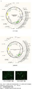
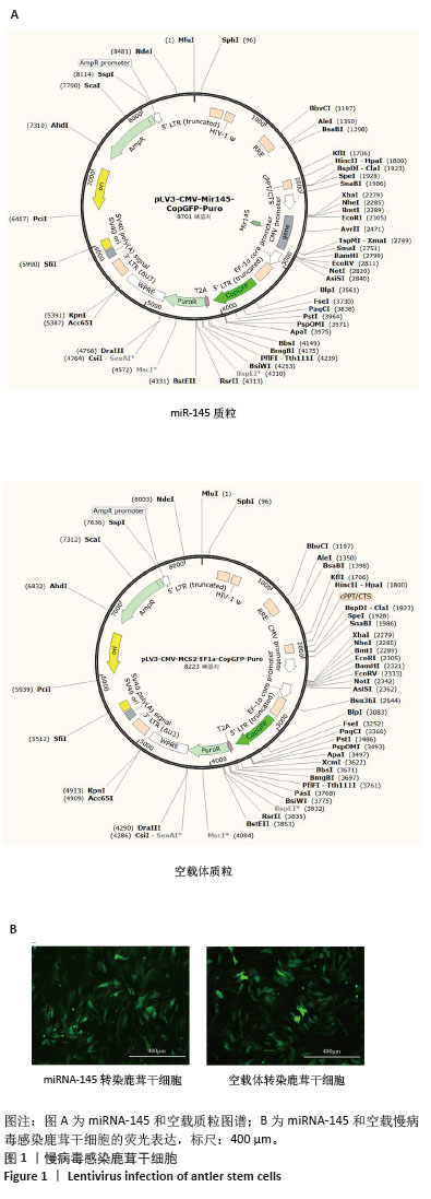
2.1 过表达miRNA-145鹿茸干细胞株建立 通过慢病毒感染鹿茸干细胞,获得稳定表达miRNA-145的鹿茸干细胞以及空载鹿茸干细胞,如图1所示,在慢病毒感染48 h后约90%的鹿茸干细胞表达绿色荧光,细胞大多呈现均一的长梭形,多次传代后细胞状态良好且绿色荧光不减少。qPCR检测3种细胞miRNA-145表达量,如图2所示,相较于未转染鹿茸干细胞和空载体转染鹿茸干细胞,miRNA-145转染鹿茸干细胞的miRNA-145表达量显著升高,提高约30倍,表明鹿茸干细胞稳转细胞系构建成功。"
"

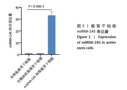
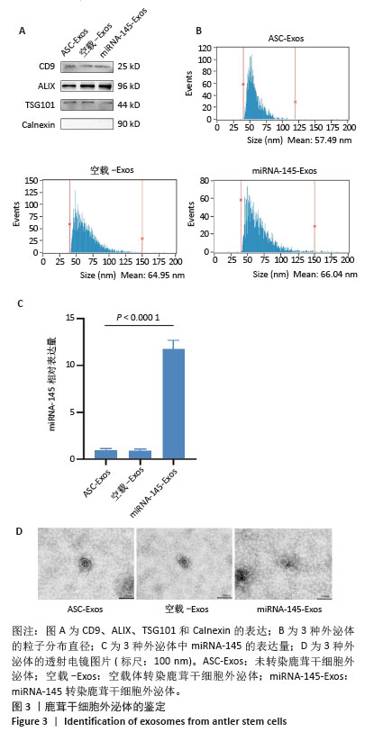
2.2 外泌体鉴定结果 如图3所示,透射电镜观察显示提取的外泌体呈现典型一侧凹陷的圆形或椭圆形双层囊泡样结构。粒径分析结果显示3种外泌体的直径在50-150 nm之间,符合外泌体的直径大小。Western blot结果显示3种外泌体均表达CD9、ALIX和TSG101,且不表达Calnexin。qPCR结果显示miRNA-145转染外泌体的miRNA-145表达量显著高于单纯外泌体和空载外泌体,这表明miRNA-145成功在鹿茸干细胞中过表达,且能够在鹿茸干细胞外泌体中富集。"
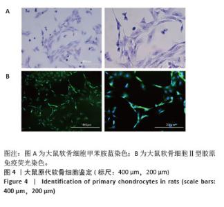
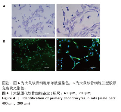
2.3 大鼠软骨细胞分离与鉴定 通过甲苯胺蓝染色、Ⅱ型胶原免疫荧光染色鉴定提取的大鼠原代软骨细胞,如图4A所示,甲苯胺蓝染色后大鼠软骨细胞基质呈蓝色,细胞核呈深蓝色,呈现三角形或短梭形,细胞核为圆形,随着培养时间延长逐渐变为圆形或近圆的多边形。如图4B所示,Ⅱ型胶原免疫荧光染色显示,90%以上的第2代大鼠软骨细胞发出明显的绿色荧光,呈阳性。"
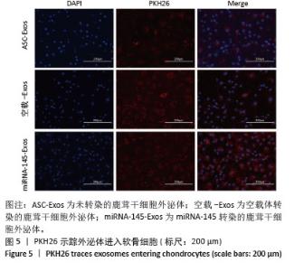
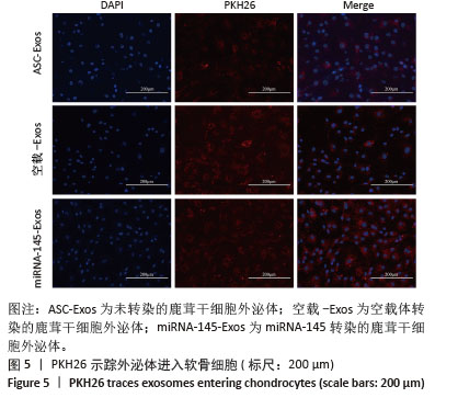
2.4 软骨细胞摄取外泌体情况 用PKH26(红色荧光)标记外泌体后,与大鼠软骨细胞共培养24 h,如图5显示,软骨细胞内聚集了大量红色颗粒样物质,这表明软骨细胞成功摄取3种外泌体。"
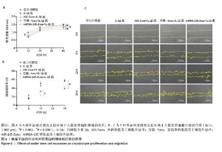
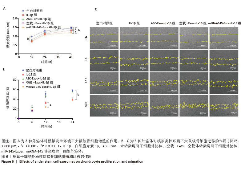
2.5 鹿茸干细胞外泌体促进软骨细胞增殖和迁移 CCK-8实验结果显示加入白细胞介素1β后软骨细胞活力显著下降(P < 0.05);与外泌体+白细胞介素1β组和空载外泌体+白细胞介素1β组相比,miRNA-145转染外泌体+白细胞介素1β组培养48 h后软骨细胞活力显著下降(P < 0.05);与白细胞介素1β组相比,外泌体+白细胞介素1β组和空载外泌体+白细胞介素1β组培养48 h后软骨细胞活力显著升高(P < 0.05),见图6A。结果表明鹿茸干细胞外泌体对炎性环境下软骨细胞增殖有促进作用,而过表达miRNA-145的鹿茸干细胞外泌体对软骨细胞增殖有抑制作用。 划痕实验结果显示加入白细胞介素1β 12 h后软骨细胞迁移率显著下降(P < 0.05);与白细胞介素1β组相比,miRNA-145转染外泌体+白细胞介素1β组细胞迁移率显著下降(P < 0.05);培养24 h后,与白细胞介素1β组相比,外泌体+白细胞介素1β组和空载外泌体+白细胞介素1β组软骨细胞迁移率显著升高(P < 0.05),见图6B,C。结果表明鹿茸干细胞外泌体对炎性环境下软骨细胞的迁移有促进作用,但过表达miRNA-145后鹿茸干细胞外泌体会抑制软骨细胞的迁移。"
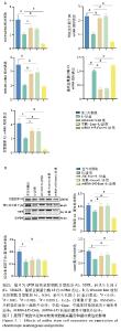
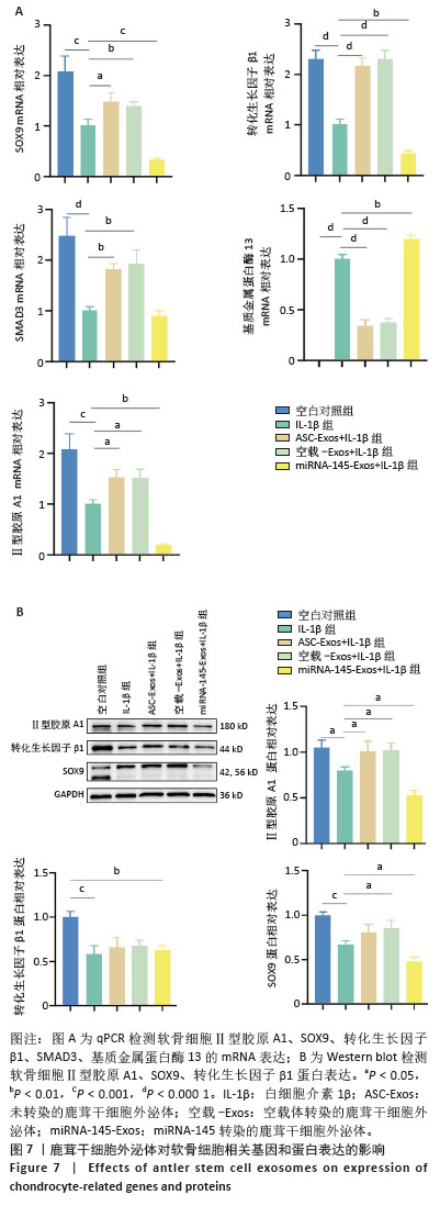
2.6 鹿茸干细胞外泌体抑制白细胞介素1β诱导的软骨细胞损伤 qPCR结果显示,加入白细胞介素1β后,软骨细胞Ⅱ型胶原A1、SOX9、转化生长因子β1、SMAD3 mRNA表达显著降低(P < 0.05),基质金属蛋白酶13 mRNA表达显著升高(P < 0.05);与白细胞介素1β组相比,外泌体+白细胞介素1β组和空载外泌体+白细胞介素1β组软骨细胞Ⅱ型胶原A1、SOX9、转化生长因子1β、SMAD3 mRNA表达显著升高(P < 0.05),基质金属蛋白酶13 mRNA表达显著降低(P < 0.05);与白细胞介素1β组相比,miRNA-145转染外泌体+白细胞介素1β组软骨细胞Ⅱ型胶原A1、SOX9、转化生长因子β1 mRNA表达显著降低(P < 0.05),基质金属蛋白酶13 mRNA表达显著升高(P < 0.05),见图7A。 Western blot结果同样表明,白细胞介素1β处理后,软骨细胞Ⅱ型胶原A1、SOX9、转化生长因子β1蛋白表达显著降低(P < 0.05);与白细胞介素1β组相比,外泌体+白细胞介素1β组和空载外泌体+白细胞介素1β组Ⅱ型胶原A1蛋白表达显著升高,空载外泌体+白细胞介素1β组SOX9蛋白显著升高(P < 0.05);与白细胞介素1β组相比,miRNA-145转染外泌体+白细胞介素1β组Ⅱ型胶原A1、SOX9蛋白表达显著下降(P < 0.05),见图7B。 上述结果表明鹿茸干细胞外泌体可以有效逆转炎性环境下大鼠软骨细胞Ⅱ型胶原A1的降解,降低基质金属蛋白酶13的表达水平,从而保护软骨细胞细胞外基质平衡。鹿茸干细胞外泌体可以上调转化生长因子β1/SMAD3通路,有效促进软骨细胞Ⅱ型胶原A1、SOX9表达。而miRNA-145则起到抑制作用,miRNA-145可靶向降低SOX9的表达,提高基质金属蛋白酶13的表达,使炎症环境下软骨细胞的损伤进一步加剧。"
| [1] 中华医学会物理医学与康复学分会,四川大学华西医院.中国膝骨关节炎康复治疗指南(2023版)[J].中国循证医学杂志,2024,24(1): 1-14. [2] ZHANG L, YANG H, LIU J, et al. Metabolomics-based Approach to Analyze the Therapeutic Targets and Metabolites of a Synovitis Ointment for Knee Osteoarthritis. Current Pharmaceutical Analysis. 2023;19(3):222-234. [3] XIA Z, MA P, WU N, et al. Altered function in cartilage derived mesenchymal stem cell leads to OA-related cartilage erosion. Am J Transl Res. 2016;8(2):433-446. [4] IM GI, HENROTIN Y. Regenerative medicine for early osteoarthritis. Ther Adv Musculoskelet Dis. 2023;15:1759720X231194813. [5] RAPOSO G, STOORVOGEL W. Extracellular vesicles: exosomes, microvesicles, and friends. J Cell Biol. 2013;200(4):373-383. [6] LOBB RJ, BECKER M, WEN SW, et al. Optimized exosome isolation protocol for cell culture supernatant and human plasma. J Extracell Vesicles. 2015;4:27031. [7] TIAN X, HE X, QIAN S, et al. Immunoregulatory effects of human amniotic mesenchymal stem cells and their exosomes on human peripheral blood mononuclear cells. BIOCELL.2023;47(5):1085-1093. [8] PEKÁČOVÁ A, BALOUN J, ŠVEC X, et al. Non-coding RNAs in diseases with a focus on osteoarthritis. Wiley Interdiscip Rev RNA. 2023;14(3): e1756. [9] YAMAZAKI A, TOMO Y, ETO H, et al. A pilot study of microRNA assessment as a means to identify novel biomarkers of spontaneous osteoarthritis in dogs. Sci Rep. 2022;12(1):18152. [10] 周珏.鹿茸干细胞分泌蛋白促进大鼠软骨缺损修复[D].北京:中国农业科学院,2023. [11] 秦涛.鹿茸四区段mRNA-microRNA表达谱测定、分析及蜡片区调控网络构建[D].北京:中国农业科学院,2020. [12] LI C, LITTLEJOHN RP, SUTTIE JM. Effects of insulin-like growth factor 1 and testosterone on the proliferation of antlerogenic cells in vitro. J Exp Zool. 1999;284(1):82-90. [13] FAN B, LI C, SZALAD A, et al. Mesenchymal stromal cell-derived exosomes ameliorate peripheral neuropathy in a mouse model of diabetes. Diabetologia. 2020;63(2):431-443. [14] KIM J, TRAN AN, LEE JY, et al. Human Fetal Cartilage-Derived Progenitor Cells Exhibit Anti-Inflammatory Effect on IL-1β-Mediated Osteoarthritis Phenotypes In Vitro. Tissue Eng Regen Med. 2022;19(6):1237-1250. [15] 刘冬阳,黄昕彤,赖晋锋,等.中国中老年人慢性病共病流行趋势研究[J].中国慢性病预防与控制,2024,32(4):244-249. [16] 王伟康,刘晓冬,周长林,等.MiRNAs在骨关节炎发生发展中的调控作用[J].中国组织工程研究,2021,25(35):5709-5715. [17] JIANG S, LIU Y, XU B, et al. Noncoding RNAs: New regulatory code in chondrocyte apoptosis and autophagy. Wiley Interdiscip Rev RNA. 2020;11(4):e1584. [18] PALAMÀ MEF, COCO S, SHAW GM, et al. Xeno-free cultured mesenchymal stromal cells release extracellular vesicles with a “therapeutic” miRNA cargo ameliorating cartilage inflammation in vitro. Theranostics. 2023;13(5):1470-1489. [19] YIN B, NI J, WITHEREL CE, et al. Harnessing Tissue-derived Extracellular Vesicles for Osteoarthritis Theranostics. Theranostics. 2022;12(1): 207-231. [20] ZHOU J, ZHAO J, WANG Y, et al. Repair of Mechanical Cartilage Damage Using Exosomes Derived from Deer Antler Stem Cells. Front Biosci (Landmark Ed). 2024;29(8):309. [21] KIM M, SHIN DI, CHOI BH, et al. Exosomes from IL-1β-Primed Mesenchymal Stem Cells Inhibited IL-1β- and TNF-α-Mediated Inflammatory Responses in Osteoarthritic SW982 Cells. Tissue Eng Regen Med. 2021;18(4):525-536. [22] LI S, STÖCKL S, LUKAS C, et al. Curcumin-primed human BMSC-derived extracellular vesicles reverse IL-1β-induced catabolic responses of OA chondrocytes by upregulating miR-126-3p. Stem Cell Res Ther. 2021;12(1):252. [23] GAO JY, WANG XJ, YU JP, et al. Loss of autophagy in condylar chondrocytes causes increased apoptosis rate in temporomandibular joint osteoarthritis of rats. Zhonghua Kou Qiang Yi Xue Za Zhi. 2020; 55(5):343-347. [24] ABO-ZALAM HB, ABDELSALAM RM, ABDEL-RAHMAN RF, et al. In Vivo Investigation of the Ameliorating Effect of Tempol against MIA-Induced Knee Osteoarthritis in Rats: Involvement of TGF-β1/SMAD3/NOX4 Cue. Molecules. 2021;26(22):6993. [25] PU P, QINGYUAN M, WEISHAN W, et al. Protein-Degrading Enzymes in Osteoarthritis. Z Orthop Unfall. 2021;159(1):54-66. [26] 杨均,李澎.转化生长因子β诱导骨髓间充质干细胞分化为半月板纤维软骨细胞[J].中国组织工程研究,2023,27(15):2412-2419. [27] FONGSODSRI K, TIYASATKULKOVIT W, CHAISRI U, et al. Sericin promotes chondrogenic proliferation and differentiation via glycolysis and Smad2/3 TGF-β signaling inductions and alleviates inflammation in three-dimensional models. Sci Rep. 2024;14(1):11553. [28] KISHTA MS, KHAMIS A, AM H, et al. Exploring the tumor-suppressive role of miRNA-200c in head and neck squamous cell carcinoma: Potential and mechanisms of exosome-mediated delivery for therapeutic applications. Transl Oncol. 2025;51:102216. [29] WANG Z, WANG W, ZUO B, et al. Identification of potential pathogenic genes related to osteoporosis and osteoarthritis. Technol Health Care. 2024;32(6):4431-4444. [30] MUÑOZ EL, FUENTES FB, FELMER RN, et al. Extracellular vesicles in mammalian reproduction: a review. Zygote. 2022;30(4):440-463. [31] YANG F, XIONG WQ, LI CZ, et al. Extracellular vesicles derived from mesenchymal stem cells mediate extracellular matrix remodeling in osteoarthritis through the transport of microRNA-29a. World J Stem Cells. 2024;16(2):191-206. [32] LIN T, WU N, WANG L, et al. Inhibition of chondrocyte apoptosis in a rat model of osteoarthritis by exosomes derived from miR‑140‑5p‑overexpressing human dental pulp stem cells. Int J Mol Med. 2021;47(3):7. [33] JOUNG S, YOON DS, CHO S, et al. Downregulation of MicroRNA-495 Alleviates IL-1β Responses among Chondrocytes by Preventing SOX9 Reduction. Yonsei Med J. 2021;62(7):650-659. [34] LIU H, LIU P. Kartogenin Promotes the BMSCs Chondrogenic Differentiation in Osteoarthritis by Down-Regulation of miR-145-5p Targeting Smad4 Pathway. Tissue Eng Regen Med. 2021;18(6): 989-1000. [35] MARTINEZ-SANCHEZ A, LAZZARANO S, SHARMA E, et al. High-Throughput Identification of MiR-145 Targets in Human Articular Chondrocytes. Life (Basel). 2020;10(5):58. [36] WU M, LIU F, YAN L, et al. MiR-145-5p restrains chondrogenic differentiation of synovium-derived mesenchymal stem cells by suppressing TLR4. Nucleosides Nucleotides Nucleic Acids. 2022;41(7): 625-642. |
| [1] | Zhang Nan, Meng Qinghua, Bao Chunyu. Characteristics and clinical application of ankle joint finite element models [J]. Chinese Journal of Tissue Engineering Research, 2026, 30(9): 2343-2349. |
| [2] | Zhang Zizheng, Luo Wang, Liu Changlu. Application value of finite element analysis on unicompartmental knee arthroplasty for medial knee compartmental osteoarthritis [J]. Chinese Journal of Tissue Engineering Research, 2026, 30(9): 2313-2322. |
| [3] | Chen Qiuhan, Yang Long, Yuan Daizhu, Wu Zhanyu, Zou Zihao, Ye Chuan. Peri-knee osteotomy for treatment of knee osteoarthritis: optimization of treatment strategies [J]. Chinese Journal of Tissue Engineering Research, 2026, 30(9): 2303-2312. |
| [4] | Li Qingbin, Lin Jianhui, Huang Wenjie, Wang Mingshuang, Du Jiankai, Lao Yongqiang. Bone cement filling after enlarged curettage of giant cell tumor around the knee joint: a comparison of subchondral bone grafting and non-grafting [J]. Chinese Journal of Tissue Engineering Research, 2026, 30(8): 1896-1902. |
| [5] | Song Puzhen, Ma Hebin, Chen Hongguang, Zhang Yadong. Effect of bone marrow mesenchymal stem cell-derived exosomes combined with transforming growth factor beta 1 on macrophages [J]. Chinese Journal of Tissue Engineering Research, 2026, 30(7): 1616-1623. |
| [6] | He Jiale, Huang Xi, Dong Hongfei, Chen Lang, Zhong Fangyu, Li Xianhui. Acellular dermal matrix combined with adipose-derived stem cell exosomes promotes burn wound healing [J]. Chinese Journal of Tissue Engineering Research, 2026, 30(7): 1699-1710. |
| [7] | Xia Linfeng, Wang Lu, Long Qianfa, Tang Rongwu, Luo Haodong, Tang Yi, Zhong Jun, Liu Yang. Human umbilical cord mesenchymal stem cell-derived exosomes alleviate blood-brain barrier damage in mice with septic encephalopathy [J]. Chinese Journal of Tissue Engineering Research, 2026, 30(7): 1711-1719. |
| [8] | Chen Yulin, He Yingying, Hu Kai, Chen Zhifan, Nie Sha Meng Yanhui, Li Runzhen, Zhang Xiaoduo , Li Yuxi, Tang Yaoping. Effect and mechanism of exosome-like vesicles derived from Trichosanthes kirilowii Maxim. in preventing and treating atherosclerosis [J]. Chinese Journal of Tissue Engineering Research, 2026, 30(7): 1768-1781. |
| [9] | Han Teng, Ma Hong, Yang Ruoyi, Luo Yi, Li Chao. Oral squamous cell carcinoma-derived exosomal delivery of angiopoietin-2 is involved in tumor angiogenesis [J]. Chinese Journal of Tissue Engineering Research, 2026, 30(7): 1755-1767. |
| [10] | Tao Daiju, Su Haiyu, Wang Yuqi, Shen Zhiqiang, He Bo . Construction and identification of stable PC12 cell lines with high/low expression of miR-122-5p [J]. Chinese Journal of Tissue Engineering Research, 2026, 30(7): 1790-1799. |
| [11] | Huang Jiawen, Pan Zhiyi, Xue Wenjun, Lian Yuanpei, Xu Jianda. Plant-derived vesicles and malignant tumor therapy: cross-species communication and modulation of host cell responses [J]. Chinese Journal of Tissue Engineering Research, 2026, 30(7): 1828-1838. |
| [12] | Wang Baiyan, Yang Shu, Wang Yiming, Wu Mengqing, Xiao Yu, Guo Zixuan, Zhang Boyi, Feng Shuying. Exosome-delivered CRISPR/Cas system enables gene editing in target cells [J]. Chinese Journal of Tissue Engineering Research, 2026, 30(7): 1839-1849. |
| [13] | Wang Zhenze, Liu Fende, Zhang Rui, Li Wujun. Mesenchymal stem cells in treatment of arteriosclerosis obliterans of lower extremities: systematic review and meta-analysis [J]. Chinese Journal of Tissue Engineering Research, 2026, 30(7): 1869-1876. |
| [14] | Li Hao, Tao Hongcheng, Zeng Ping, Liu Jinfu, Ding Qiang, Niu Chicheng, Huang Kai, Kang Hongyu. Mitogen-activated protein kinase signaling pathway regulates the development of osteoarthritis: guiding targeted therapy with traditional Chinese medicine [J]. Chinese Journal of Tissue Engineering Research, 2026, 30(6): 1476-1485. |
| [15] | Li Linzhen, Jiao Hongzhuo, Chen Weinan, Zhang Mingzhe, Wang Jianlong, Zhang Juntao. Effect of icariin-containing serum on lipopolysaccharide-induced inflammatory damage in human chondrocytes [J]. Chinese Journal of Tissue Engineering Research, 2026, 30(6): 1368-1374. |
| Viewed | ||||||
|
Full text |
|
|||||
|
Abstract |
|
|||||