Chinese Journal of Tissue Engineering Research ›› 2025, Vol. 29 ›› Issue (36): 7839-7847.doi: 10.12307/2025.749
Previous Articles Next Articles
Regenerative effects of engineered extracellular vesicles on repairing bone defects
Zhou Yang1, 2, Liu Kexin3, Wang Deli1, Sun Zhang2
- 1School of Stomatology, Mudanjiang Medical University, Mudanjiang 157011, Heilongjiang Province, China; 2Department of Stomatology, 3Department of Orthopedics, Affiliated Hongqi Hospital of Mudanjiang Medical University, Mudanjiang 157011, Heilongjiang Province, China
-
Received:2024-08-05Accepted:2024-10-31Online:2025-12-28Published:2025-03-17 -
Contact:Sun Zhang, MM, Attending physician, Department of Stomatology, Affiliated Hongqi Hospital of Mudanjiang Medical University, Mudanjiang 157011, Heilongjiang Province, China -
About author:Zhou Yang, MS, School of Stomatology, Mudanjiang Medical University, Mudanjiang 157011, Heilongjiang Province, China; Department of Stomatology, Affiliated Hongqi Hospital of Mudanjiang Medical University, Mudanjiang 157011, Heilongjiang Province, China -
Supported by:Heilongjiang Provincial Health Commission Research Project, No. 20230808030122 (to ZY); 2020 Basic Research Business Expenses Research Project of Heilongjiang Provincial Higher Education Institutions, No. 2020-KYYWFMY-0766 (to ZY)
CLC Number:
Cite this article
Zhou Yang, Liu Kexin, Wang Deli, Sun Zhang. Regenerative effects of engineered extracellular vesicles on repairing bone defects[J]. Chinese Journal of Tissue Engineering Research, 2025, 29(36): 7839-7847.
share this article
Add to citation manager EndNote|Reference Manager|ProCite|BibTeX|RefWorks

2.1 细胞外囊泡的分离与表征 几乎所有细胞类型都可以产生细胞外囊泡,其分布广泛,存在于各种体液中。自1946年细胞外囊泡首次被发现[13],到如今国际细胞外囊泡学会成立并发布指南文件以支持和规范全球细胞外囊泡研究,数十年来对于细胞外囊泡的研究不断取得新突破[14-20],见图3。然而,它们的物理性质和功能的异质性尚未完全了解,这阻碍了细胞外囊泡进一步的临床应用。由于细胞外囊泡异质性带来的挑战,目前研究者们正努力改进分离和表征特定细胞外囊泡群体的方法。 "


2.1.1 细胞外囊泡的分离 目前,细胞外囊泡主要是基于密度、大小、免疫亲和力或表面电荷进行分离的。基于超速离心是分离细胞外囊泡最广泛使用的方法,主要包括差速超速离心和密度梯度离心[21]。尽管超速离心方案操作简单,但存在耗时、成本高的问题,不适合高通量应用[22]。细胞外囊泡也可以根据尺寸进行分离,其中最流行的方法之一是超滤。超滤显著降低了细胞外囊泡破裂的可能性,但获得的细胞外囊泡制剂通常会被直径相似的分子污染[23]。为了解决这个问题,超滤经常与超速离心一起使用。尺寸排阻色谱法也是基于尺寸进行分离,但其应用范围较窄,不适合大体积样品[24-25]。免疫亲和捕获技术是利用细胞外囊泡某些表面蛋白和受体来开发的,其分离的细胞外囊泡产量相对较低但纯度较高[26-27]。基于标记的微流控技术可根据化学或物理性质特异性地与细胞外囊泡表面相应的脂质成分或蛋白质结合,但是成本相对较高,标记操作复杂,捕获分子也可能改变细胞外囊泡的物理和生物学特性[28]。为了减少标记对后续实验的影响,研究者们提出了无标记分离策略,可以保证细胞外囊泡的结构和生物完整性[28]。单一的细胞外囊泡分离方法总存在或多或少的不足,利用互补的组合分离策略以获得更高纯度和产量的细胞外囊泡制剂,可能是未来潜在的解决方案。 2.1.2 细胞外囊泡的表征 为了保证细胞外囊泡制剂实现可重复安全和有针对性的治疗,在应用前需要对其进行表征。目前可用的方法允许确定细胞外囊泡的大小、浓度、形态和特定内容物,见图4。"


成像技术常用的方法包括扫描电子显微镜、透射电子显微镜、冷冻电镜和原子力显微镜。扫描电子显微镜是通过捕获发射的电子来创建细胞外囊泡表面形貌的图像,无法区分囊泡和非囊泡粒子[29];相比之下,透射电子显微镜可提供囊泡内部的附加信息,不过测量结果可能会受到样品制备程序的强烈影响[30];冷冻电镜通过冷冻固定技术可以很好地保留水合囊泡的实际形态,从而避免传统固定造成的伪影[31];原子力显微镜同样能够捕获天然细胞外囊泡结构,但需要在不同的实验条件下进行分析,这对实验设备和用户操作提出了很高的要求[32]。不同于成像技术,基于尺寸测定和计数的技术可以获得关于细胞外囊泡尺寸分布和浓度的更精确信息,常见的方法有纳米粒子跟踪分析、动态光散射、可调电阻脉冲传感和微流体电阻脉冲传感[21]。纳米粒子跟踪分析作为一种非侵入性技术,对细胞外囊泡尺寸检测的极限为40-50 nm[33];动态光散射利用了流体动力学粒子的光散射原理,对细胞外囊泡直径检测可以小至10 nm[34];可调电阻脉冲传感能可靠、快速地测量出细胞外囊泡大小和浓度分布,但是穿过它的粒子可能堵塞孔隙[35];微流体电阻脉冲传感是对可调电阻脉冲传感的改进,可以检测样品中存在的不同大小的囊泡群[36]。新开发的成像流式细胞术和纳米流式细胞术可以提供更高的分辨率,并且能够区分染色的细胞外囊泡和背景信号,该方法依赖高水平的专业知识和昂贵设备[36-37]。此外,大多数机器仍需要用染料标记细胞外囊泡才能够有效检测,这可能会干扰下游分析[37]。 2.2 细胞外囊泡调节骨再生的治疗机制 骨缺损的再生修复是一个复杂的过程,需要多方面的机制协同作用才能有效进行。细胞外囊泡可以通过免疫调节、血管生成以及靶细胞的增殖与分化这3个主要机制调节骨再生,其在骨缺损组织的再生修复中扮演着重要角色。接下来,将详细探讨这些机制如何协同作用,共同促进骨缺损组织的再生,见图5。 "


2.2.1 参与免疫调节 骨缺损在损伤早期需要适度的炎症反应,但是过度活跃和持续的炎症会阻碍骨再生并导致炎症损伤。细胞外囊泡是免疫调节的重要参与者,在骨再生修复过程中发挥关键作用。根据释放细胞外囊泡的细胞来源和状态,它们既能介导免疫刺激,也能抑制免疫反应。细胞外囊泡可以调节多种免疫细胞的功能,包括T细胞、B细胞、巨噬细胞和树突状细胞[38]。T细胞介导的免疫调节是控制自身免疫或炎性疾病的重要工具。有研究发现,人脐静脉内皮细胞来源的细胞外囊泡含有高浓度的程序性死亡配体1,可特异性地与T细胞表面程序性死亡受体1结合,从而抑制T细胞的活化,在早期过度活跃炎症阶段显著促进了骨痂形成和骨折愈合[39]。细胞外囊泡与亲本细胞具有相似的分子组成,可以继承亲本细胞的免疫调节能力。考虑到间充质干细胞良好的免疫调节能力,间充质干细胞衍生的细胞外囊泡也得到了深入研究。多项研究表明间充质干细胞衍生的细胞外囊泡可以抑制T细胞和B细胞的增殖,同时降低外周血单核细胞在体内释放细胞因子的能力[40]。M2型巨噬细胞具有抗炎表型,主要负责巨噬细胞极化过程中的组织重塑。间充质干细胞衍生的细胞外囊泡在单核细胞中诱导为M2样表型,诱导T细胞分化为调节性T细胞,并减弱体内的免疫活性[41]。REIS等[42]研究表明,间充质干细胞衍生的细胞外囊泡影响了树突状细胞对抗原的吸收,抑制了树突状细胞的成熟、促炎因子白细胞介素6和白细胞介素12p70的分泌,并增加了抗炎细胞因子转化生长因子β的产生。细胞外囊泡已成为一种有吸引力的免疫调节和免疫介导疾病的治疗方法。 2.2.2 促进血管生成 血管生成是指从现有血管网络中萌发和长出新血管的过程,它对于骨组织的再生修复至关重要。血管生成涉及多个过程,包括内皮细胞的增殖、募集和迁移,以及通过壁细胞来稳定新形成的血管。细胞外囊泡在促进血管生成中也发挥着关键作用,该作用与细胞外囊泡中所含相应配体和细胞因子的靶向作用密切相关,例如血管内皮生长因子、血小板衍生生长因子、基质金属蛋白酶和miRNA[43-46]。LIU等[47]将诱导性多能干细胞来源的间充质干细胞衍生细胞外囊泡静脉注射到大鼠骨坏死模型中,结果显示间充质干细胞衍生细胞外囊泡可以激活内皮细胞中的磷脂酰肌醇3激酶(phosphoinositide 3 kinase,PI3K)/蛋白激酶B(protein kinase B,AKT)信号通路,显著增强了内皮细胞的体外增殖、迁移和血管形成能力。ZHANG等[48]将间充质干细胞衍生细胞外囊泡与水凝胶结合移植到股骨骨折大鼠模型的骨折部位,发现间充质干细胞衍生细胞外囊泡除了增强成骨分化外,还增加了血管内皮生长因子和缺氧诱导因子1α的表达。其中,缺氧诱导因子1α在促血管生成和增强骨修复中起重要作用。 2.2.3 增强靶细胞的增殖和分化 细胞外囊泡介导的旁分泌信号可以通过影响成骨细胞和破骨细胞来调节骨稳态。研究表明,携带miR-935的间充质干细胞衍生细胞外囊泡可以通过靶向信号转导和转录激活因子1来促进成骨细胞的增殖和分化[49]。HU等[50]评估了间充质干细胞衍生细胞外囊泡对骨质疏松的影响,结果显示其通过在体外传递强效促成骨蛋白C型凝集素结构域家族11成员A (C-type lectin domain family 11 member A,CLEC11A)增强了骨髓间充质干细胞从脂肪形成向成骨分化的转变,并抑制破骨细胞的形成。间充质干细胞衍生细胞外囊泡全身给药可通过增强骨形成、减少骨髓脂肪堆积和减少骨吸收来预防骨质疏松导致的骨质流失并维持骨强度[50]。 2.3 细胞外囊泡的工程化方法 细胞外囊泡作为一种新兴的无细胞疗法,具有良好的生物相容性和低免疫原性,可以较好地克服细胞疗法存在的不足。然而,幼稚细胞外囊泡对治疗药物负载能力低,还存在组织靶向能力不足的问题,此外其药理学机制尚未完全了解[51]。在过去10年中,工程化细胞外囊泡已开发出表面工程技术,用于将载体运送到特定的目标组织,并探索了将治疗剂装载到细胞外囊泡中的有效装载方法,因此工程化细胞外囊泡具有良好的应用前景[52]。 2.3.1 表面修饰的工程化方法 表面修饰主要目的是选择性地将细胞外囊泡递送至靶细胞和潜在的组织中实现精准治疗。根据修饰发生在细胞外囊泡分离之前还是之后,可以分为分离前表面修饰和分离后表面修饰。 分离前表面修饰主要包括基因工程、代谢工程和直接母细胞膜工程方法[53]。基因工程是利用细胞外囊泡膜结合蛋白赋予细胞外囊泡靶向特性,修饰过程中应该考虑到在细胞外囊泡上富集的蛋白质结构域的选择[54]。因为合适的囊泡膜上蛋白比较有限,一些配体结构与膜蛋白融合表达时不能折叠成有活性的结构[55]。代谢工程是通过向母细胞培养基中添加叠氮化物标记的代谢物来生成带有叠氮化物基团的细胞外囊泡,从而赋予细胞外囊泡新的成分和功能[56]。虽然代谢工程可以实现细胞外囊泡的功能化,但很难控制表面结合位点的特异性和效率[53]。直接母细胞膜工程可以通过使用与细胞膜融合的融合脂质体来实现,从而交换膜成分,这种策略可以将叠氮化物脂质转移到母细胞膜,从而产生含有叠氮化物脂质的细胞外囊泡,并通过点击化学实现功能化[57]。然而,融合脂质体制剂中的脂质很可能也会进入到细胞外囊泡制剂中,这可能造成不必要的毒性和免疫原性,需要对该方法的安全性方面做进一步评估[53]。 分离后表面修饰是指在细胞外囊泡分离后用靶向分子对细胞外囊泡表面进行物理修饰或者化学修饰。细胞外囊泡的表面物理修饰可通过膜融合、疏水插入或表面吸附来实现[52]。细胞外囊泡的脂质双层可以很容易地与具有相似性质的脂质体融合,实现对细胞外囊泡的表面修饰。膜融合可以通过冻融循环或者聚乙二醇完成[58-59]。冻融循环可能对细胞外囊泡完整性产生潜在负面影响,而聚乙二醇介导的脂质体与细胞外囊泡膜融合不会破坏细胞外囊泡完整性,且可以同时对细胞外囊泡执行表面改性和内容物装载[58-60]。疏水插入也是基于脂质体开发的,该方法允许靶分子使用由磷脂和胆固醇组成的细胞外囊泡膜成分对细胞外囊泡表面进行修饰。相对磷脂而言,胆固醇插入所需孵育温度较低,更适合在细胞外囊泡中应用,但是插入后胆固醇处理的脂质体仅表现出较低的插入脂质保留率,因此疏水插入稳定性问题值得特别评估[61-62]。表面吸附的方法可以依赖于细胞外囊泡的表面特性,如蛋白质或脂质的头部基团,也可以依赖于细胞外囊泡的表面电荷[63-64],该物理方法操作简便且可使用患者来源的细胞外囊泡,但研究相对较少,有待深入探索[64]。细胞外囊泡表面修饰的化学方法主要是将靶分子共价连接到细胞外囊泡表面来进行靶向递送。利用活化酯可将羧基同氨基共价结合形成酰胺基团,这是常见的蛋白质偶联反应[65]。除了偶联反应外,还有一大类为生物正交反应。在生物正交反应中,点击化学因其易于操作、较高的反应特异性及反应效率、较低的副反应和生物毒性,成为化学修饰的重要手段[66-67]。细胞外囊泡的表面修饰工程化方法见表1。"


2.3.2 内容物装载的工程化方法 内容物装载是指向细胞外囊泡中装载指定的小分子药物、核酸、蛋白质等内容物分子,将细胞外囊泡打造为药物载体。内容物装载可以在细胞外囊泡从细胞中分离之前进行,称为内源性内容物装载;也可以在细胞外囊泡分离后进行,称为外源性内容物装载[12]。内源性内容物装载主要包括亲本细胞的转染和共培养。内容物分子可通过编码DNA被转染到亲本细胞,从而装载到细胞外囊泡中,转染主要通过质粒和病毒媒介实现[12, 68-69]。转染过程中,基因表达改变和转染剂毒性可能会导致亲本细胞发生变化[12]。共培养是将亲本细胞与要装载的内容物分子一起孵育,以产生带有这些内容物分子的细胞外囊泡。该方法操作简单,不依赖任何特殊设备,但是内容物分子装载效率较低,且只适用于可轻松通过细胞膜的目标分子[70]。外源性内容物装载方法较多,包括细胞外囊泡共培养、电穿孔、超声、冻融循环、挤压和表面活性剂辅助渗透等。不同于亲本细胞共培养,细胞外囊泡共培养是指在特定温度下将内容物分子与纯化的细胞外囊泡一起孵育,内容物可通过共培养封装到细胞外囊泡中[71-72]。亲水的内容物分子很难依靠单纯细胞外囊泡共培养装载到细胞外囊泡中,为了克服细胞外囊泡共培养的局限性,研究者们开发了利用物理刺激或者化学药物改善细胞外囊泡膜通透性的替代方法。电穿孔是使用电脉冲在细胞外囊泡膜上形成孔隙,内容物分子可以通过这些孔隙渗透到细胞外囊泡中[73],该方法的装载效率可以通过程序控制,但是内容物分子有形成聚集体的风险[73]。超声、冻融循环和挤压这些物理手段都可以通过类似的机制将内容物分子封装到细胞外囊泡中[74]。不同的是,超声处理会导致核酸降解,不适合核酸的封装[75];冻融循环会影响蛋白质的活性[76];挤压处理的内容物装载效率高,可以产生大小均匀的细胞外囊泡,但是有研究称挤压后的细胞外囊泡膜电位会发生改变,并具有细胞毒性[77]。表面活性剂如皂苷可以在细胞外囊泡膜上产生孔隙,实现内容物分子的封装[76],但是皂苷在体内有溶血活性,负载药物后需要进行彻底净化[78]。细胞外囊泡的内容物装载工程化方法见表2。 "

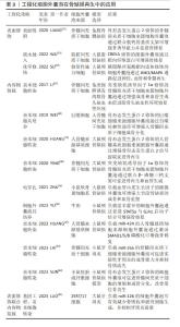
2.4 工程化细胞外囊泡在骨缺损修复中的应用 近年来,利用工程化细胞外囊泡治疗骨缺损相关疾病的研究数量增长迅速,这得益于疾病机制相关研究的完善以及药理学和材料科学的快速发展。骨形态发生蛋白2是一种强效骨诱导因子,其引入为增强细胞外囊泡成骨作用提供了一种现成的策略。间充质干细胞衍生的细胞外囊泡具有骨诱导特性,但是幼稚间充质干细胞衍生细胞外囊泡在促进骨再生方面效果甚微。为了生成具有增强成骨诱导能力的间充质干细胞衍生细胞外囊泡,HUANG等[79]将编码骨形态发生蛋白2基因的慢病毒颗粒转染人骨髓来源基质细胞,以探索骨形态发生蛋白2过表达的工程化细胞外囊泡对骨缺损的再生修复效果,结果显示这些工程化细胞外囊泡在尺寸分布、标记物表达和内吞特性方面保持了幼稚人骨髓来源基质细胞衍生细胞外囊泡的一般物理和生化特性,但是它们通过过表达骨形态发生蛋白2信号级联的miRNA,在体外增强了幼稚人骨髓来源基质细胞的成骨分化潜力[79]。此外,与骨髓来源基质细胞衍生细胞外囊泡相比,骨形态发生蛋白2过表达的细胞外囊泡在大鼠颅骨缺损模型中显示出更高的骨再生能力[79]。工程化细胞外囊泡也可以与支架材料结合来提高植入支架的生物功能。WEI等[80]从骨形态发生蛋白2刺激的巨噬细胞中分离细胞外囊泡,发现它们可以通过自噬依赖性途径增强骨髓间充质干细胞的成骨分化能力,并且骨形态发生蛋白2/巨噬细胞衍生细胞外囊泡可以用来修饰钛纳米管植入物以促进成骨。还有团队获取骨髓间充质干细胞来源细胞外囊泡,通过层层自组装的方式开发了一种骨形态发生蛋白2基因激活的脱钙骨基质支架,兔股骨髁缺损实验证明,基于细胞外囊泡的基因激活的脱钙骨基质支架不仅可以显著促进骨修复,还能促进血管生成[81]。SUN等[82]利用小鼠胚胎成纤维细胞作为工具细胞,制备了工程化骨形态发生蛋白2过表达的细胞外囊泡,并将其封装到明胶甲基丙烯酰水凝胶中,结果显示载有工程化细胞外囊泡的水凝胶显著促进了骨髓间充质干细胞的体外成骨分化,并改善了体内颅骨缺损处的原位骨再生。 考虑到miRNA内容物对细胞外囊泡功能的重要性,通过在细胞外囊泡中靶向表达选定的miRNA来推动工程化细胞外囊泡领域的发展也成为一种潜在可行的策略。为了验证这一假设,HUANG等[83]设计了工程化人骨髓来源基质细胞衍生细胞外囊泡,使其具有更高水平的miR-424,这是骨形态发生蛋白2信号级联的增强剂,结果显示,miR-424过表达的人骨髓来源基质细胞衍生细胞外囊泡通过激活SMAD1/5/8磷酸化增强了幼稚人骨髓来源基质细胞的成骨分化潜力,大鼠颅骨缺陷再生骨体积显著增加。LAI等[84]为了研究miR-26a在骨再生中的作用,用DP7-C作为转染剂转染骨髓间充质干细胞后,通过超速离心从miR-26a修饰的骨髓间充质干细胞中提取细胞外囊泡,然后在体外和体内研究中评估工程化骨髓间充质干细胞来源细胞外囊泡对成骨的影响。体外研究发现,负载miR-26a的骨髓间充质干细胞来源细胞外囊泡可增强骨髓间充质干细胞的增殖、迁移和成骨分化能力;体内研究观察到负载miR-26a的骨髓间充质干细胞来源细胞外囊泡可促进小鼠牙周炎的成骨能力并抑制骨质流失[84]。miR-126是牙周炎相关递质的转录调节因子之一,将miR-126靶向递送至巨噬细胞可能为治疗牙周炎提供有效的免疫调节策略。LUO等[85]成功构建了负载miR-126的C-X-C基序趋化因子受体4(C-X-C motif chemokine receptor 4,CXCR4)的细胞外囊泡(CXCR4-miR-126-EVs),减少了向巨噬细胞的脱靶递送,并调节巨噬细胞向抗炎表型转变。在大鼠牙周炎部位局部注射CXCR4-miR-126-EVs可有效减少骨吸收和破骨细胞生成,并抑制牙周炎进展[85]。 除了骨形态发生蛋白2和miRNA以外,研究者们也开发了其他生物活性因子来增强细胞外囊泡的骨缺损再生修复能力。缺氧诱导因子1α在骨骼发育中起着关键作用,然而缺氧诱导因子1α在常氧条件下迅速降解,仅在缺氧条件下保持稳定。为此,研究者们通过基因工程手段将缺氧诱导因子1α亚基功能区的3个氨基酸位点突变为402,564,803位的丙氨酸,成功构建了突变型缺氧诱导因子1α,突变型缺氧诱导因子1α可以在常氧条件下有效表达功能性蛋白质[86]。LI等[87]通过腺病毒转染的方式获取了突变型缺氧诱导因子1α修饰的骨髓间充质干细胞来源细胞外囊泡,并探索了其在治疗早期激素性股骨头缺血性坏死中的作用。与对照组相比,将突变型缺氧诱导因子1α修饰的骨髓间充质干细胞来源细胞外囊泡注射到坏死区域通过增强成骨和血管生成能力促进了激素性股骨头坏死修复[87]。突变型缺氧诱导因子1α修饰的骨髓间充质干细胞来源细胞外囊泡也可以负载于β-磷酸三钙支架材料上,促进临界尺寸骨缺损区域的新骨再生及新血管生成[88]。SU等[89]将磷脂酰丝氨酸靶向适体与修复性施万细胞来源细胞外囊泡偶联,构建可靶向损伤轴突和骨再生的仿生骨膜,体内外实验证明该骨膜可通过c-Jun氨基末端激酶(c-Jun N-terminal kinase 3,JNK3)/丝裂原活化蛋白激酶(mitogen-activated protein kinase,MAPK)通路靶向损伤神经,促进血管生成和骨再生。带有血管内皮生长因子质粒的软骨祖细胞衍生细胞外囊泡具有双重作用,既可作为成骨诱导基质诱导骨髓间充质干细胞成骨分化,又可以作为基因载体可控释放血管内皮生长因子基因重塑血管系统,其介导的骨支架可显著增强大段骨缺损的成骨和血管重塑能力[90]。纤维蛋白作为组织损伤部位的普遍性特征成分,是将细胞外囊泡递送到缺损骨骼的理想靶点。有团队通过纤维蛋白靶向肽半胱氨酸-精氨酸-谷氨酸-赖氨酸-丙氨酸(cysteine-arginine-glutamic acid-lysine-alanine,CREKA)修饰脂肪来源间充质干细胞衍生细胞外囊泡以增强骨修复,结果显示由于纤维蛋白结合和保留能力得到改善,其在大鼠股骨缺损模型中显著增强了骨修复能力[91]。淫羊藿苷在炎性骨缺损的治疗中起着重要作用,然而药代动力学研究表明较差的生物利用度限制了其应用[92]。在此背景下,YU等[93]分离了牛奶来源细胞外囊泡并负载了淫羊藿苷,以改善淫羊藿苷的成骨效应,结果显示负载淫羊藿苷的细胞外囊泡可以通过促进信号转导和转录激活因子5与GJA1启动子的结合,进而促进成骨细胞的增殖和分化,提高小鼠颅骨缺损的骨修复能力。不同来源的工程化细胞外囊泡在骨缺损再生中的应用见表3。 "

| [1] MAYFIELD CK, AYAD M, LECHTHOLZ-ZEY E, et al. 3D-Printing for Critical Sized Bone Defects: Current Concepts and Future Directions. Bioengineering (Basel). 2022;9(11):680. [2] STAHL A, YANG YP. Regenerative Approaches for the Treatment of Large Bone Defects. Tissue Eng Part B Rev. 2021;27(6):539-547. [3] BALDWIN P, LI DJ, AUSTON DA, et al. Autograft, Allograft, and Bone Graft Substitutes: Clinical Evidence and Indications for Use in the Setting of Orthopaedic Trauma Surgery. J Orthop Trauma. 2019;33(4):203-213. [4] SONG X, XU L, ZHANG W. Biomimetic synthesis and optimization of extracellular vesicles for bone regeneration. J Control Release. 2023; 355:18-41. [5] LIU F, SUN T, AN Y, et al. The potential therapeutic role of extracellular vesicles in critical-size bone defects: Spring of cell-free regenerative medicine is coming. Front Bioeng Biotechnol. 2023;11:1050916. [6] TAN F, LI X, WANG Z, et al. Clinical applications of stem cell-derived exosomes. Signal Transduct Target Ther. 2024;9(1):17. [7] KUMAR MA, BABA SK, SADIDA HQ, et al. Extracellular vesicles as tools and targets in therapy for diseases. Signal Transduct Target Ther. 2024;9(1):27. [8] GREGORY CD, RIMMER MP. Extracellular vesicles arising from apoptosis: forms, functions, and applications. J Pathol. 2023;260(5):592-608. [9] DIXSON AC, DAWSON TR, DI VIZIO D, et al. Context-specific regulation of extracellular vesicle biogenesis and cargo selection. Nat Rev Mol Cell Biol. 2023;24(7):454-476. [10] JEPPESEN DK, ZHANG Q, FRANKLIN JL, et al. Extracellular vesicles and nanoparticles: emerging complexities. Trends Cell Biol. 2023;33(8):667-681. [11] PAN Z, SUN W, CHEN Y, et al. Extracellular Vesicles in Tissue Engineering: Biology and Engineered Strategy. Adv Healthc Mater. 2022;11(21):e2201384. [12] LIU Q, LI D, PAN X, et al. Targeted therapy using engineered extracellular vesicles: principles and strategies for membrane modification. J Nanobiotechnology. 2023;21(1):334. [13] CHARGAFF E, WEST R. The biological significance of the thromboplastic protein of blood. J Biol Chem. 1946;166(1):189-197. [14] WOLF P. The nature and significance of platelet products in human plasma. Br J Haematol. 1967;13(3):269-288. [15] JOHNSTONE RM, ADAM M, HAMMOND JR, et al. Vesicle formation during reticulocyte maturation. Association of plasma membrane activities with released vesicles (exosomes). J Biol Chem. 1987;262(19): 9412-9420. [16] RAPOSO G, NIJMAN HW, STOORVOGEL W, et al. B lymphocytes secrete antigen-presenting vesicles. J Exp Med. 1996;183(3):1161-1172. [17] VALADI H, EKSTRÖM K, BOSSIOS A, et al. Exosome-mediated transfer of mRNAs and microRNAs is a novel mechanism of genetic exchange between cells. Nat Cell Biol. 2007;9(6):654-659. [18] LÖTVALL J, HILL AF, HOCHBERG F, et al. Minimal experimental requirements for definition of extracellular vesicles and their functions: a position statement from the International Society for Extracellular Vesicles. J Extracell Vesicles. 2014;3:26913. [19] THÉRY C, WITWER KW, AIKAWA E, et al. Minimal information for studies of extracellular vesicles 2018 (MISEV2018): a position statement of the International Society for Extracellular Vesicles and update of the MISEV2014 guidelines. J Extracell Vesicles. 2018;7(1):1535750. [20] WELSH JA, GOBERDHAN DCI, O’DRISCOLL L, et al. Minimal information for studies of extracellular vesicles (MISEV2023): From basic to advanced approaches. J Extracell Vesicles. 2024;13(2):e12404. [21] DE SOUSA KP, ROSSI I, ABDULLAHI M, et al. Isolation and characterization of extracellular vesicles and future directions in diagnosis and therapy. Wiley Interdiscip Rev Nanomed Nanobiotechnol. 2023;15(1):e1835. [22] JIA Y, YU L, MA T, et al. Small extracellular vesicles isolation and separation: Current techniques, pending questions and clinical applications. Theranostics. 2022;12(15):6548-6575. [23] PARIMON T, GARRETT NE 3RD, CHEN P, et al. Isolation of Extracellular Vesicles from Murine Bronchoalveolar Lavage Fluid Using an Ultrafiltration Centrifugation Technique. J Vis Exp. 2018;(141): 10.3791/58310. [24] FOERS AD, CHATFIELD S, DAGLEY LF, et al. Enrichment of extracellular vesicles from human synovial fluid using size exclusion chromatography. J Extracell Vesicles. 2018;7(1):1490145. [25] KOH YQ, ALMUGHLLIQ FB, VASWANI K, et al. Exosome enrichment by ultracentrifugation and size exclusion chromatography. Front Biosci (Landmark Ed). 2018;23(5):865-874. [26] LIANGSUPREE T, MULTIA E, RIEKKOLA ML. Modern isolation and separation techniques for extracellular vesicles. J Chromatogr A. 2021; 1636:461773. [27] STAM J, BARTEL S, BISCHOFF R, et al. Isolation of extracellular vesicles with combined enrichment methods. J Chromatogr B Analyt Technol Biomed Life Sci. 2021;1169:122604. [28] HASSANPOUR TAMRIN S, SANATI NEZHAD A, SEN A. Label-Free Isolation of Exosomes Using Microfluidic Technologies. ACS Nano. 2021;15(11):17047-17079. [29] PETROVA T, KALININA O, AQUINO A, et al. Topographic Distribution of miRNAs (miR-30a, miR-223, miR-let-7a, miR-let-7f, miR-451, and miR-486) in the Plasma Extracellular Vesicles. Noncoding RNA. 2024;10(1):15. [30] RIKKERT LG, NIEUWLAND R, TERSTAPPEN LWMM, et al. Quality of extracellular vesicle images by transmission electron microscopy is operator and protocol dependent. J Extracell Vesicles. 2019;8(1):1555419. [31] HÖÖG JL, LÖTVALL J. Diversity of extracellular vesicles in human ejaculates revealed by cryo-electron microscopy. J Extracell Vesicles. 2015;4:28680. [32] RIDOLFI A, BRUCALE M, MONTIS C, et al. AFM-Based High-Throughput Nanomechanical Screening of Single Extracellular Vesicles. Anal Chem. 2020;92(15):10274-10282. [33] BACHURSKI D, SCHULDNER M, NGUYEN PH, et al. Extracellular vesicle measurements with nanoparticle tracking analysis - An accuracy and repeatability comparison between NanoSight NS300 and ZetaView. J Extracell Vesicles. 2019;8(1):1596016. [34] BUSCHMANN D, MUSSACK V, BYRD JB. Separation, characterization, and standardization of extracellular vesicles for drug delivery applications. Adv Drug Deliv Rev. 2021;174:348-368. [35] MAAS SL, BROEKMAN ML, DE VRIJ J. Tunable Resistive Pulse Sensing for the Characterization of Extracellular Vesicles. Methods Mol Biol. 2017;1545:21-33. [36] ARAB T, MALLICK ER, HUANG Y, et al. Characterization of extracellular vesicles and synthetic nanoparticles with four orthogonal single-particle analysis platforms. J Extracell Vesicles. 2021;10(6):e12079. [37] DIMITRIADIS S, DOVA L, KOTSIANIDIS I, et al. Imaging Flow Cytometry: Development, Present Applications, and Future Challenges. Methods Protoc. 2024;7(2):28. [38] GOMZIKOVA MO, JAMES V, RIZVANOV AA. Therapeutic Application of Mesenchymal Stem Cells Derived Extracellular Vesicles for Immunomodulation. Front Immunol. 2019;10:2663. [39] LIN Z, XIONG Y, MENG W, et al. Exosomal PD-L1 induces osteogenic differentiation and promotes fracture healing by acting as an immunosuppressant. Bioact Mater. 2021;13:300-311. [40] VAN DEN AKKER F, VRIJSEN KR, DEDDENS JC, et al. Suppression of T cells by mesenchymal and cardiac progenitor cells is partly mediated via extracellular vesicles. Heliyon. 2018;4(6):e00642. [41] CHU C, DENG J, SUN X, et al. Collagen Membrane and Immune Response in Guided Bone Regeneration: Recent Progress and Perspectives. Tissue Eng Part B Rev. 2017;23(5):421-435. [42] REIS M, MAVIN E, NICHOLSON L, et al. Mesenchymal Stromal Cell-Derived Extracellular Vesicles Attenuate Dendritic Cell Maturation and Function. Front Immunol. 2018;9:2538. [43] GANGADARAN P, RAJENDRAN RL, OH JM, et al. Extracellular vesicles derived from macrophage promote angiogenesis In vitro and accelerate new vasculature formation In vivo. Exp Cell Res. 2020;394(2):112146. [44] HAN KY, CHANG JH, AZAR DT. MMP14-Containing Exosomes Cleave VEGFR1 and Promote VEGFA-Induced Migration and Proliferation of Vascular Endothelial Cells. Invest Ophthalmol Vis Sci. 2019;60(6):2321-2329. [45] ZHU Y, ZHANG J, HU X, et al. Extracellular vesicles derived from human adipose-derived stem cells promote the exogenous angiogenesis of fat grafts via the let-7/AGO1/VEGF signalling pathway. Sci Rep. 2020; 10(1):5313. [46] MA J, ZHAO Y, SUN L, et al. Exosomes Derived from Akt-Modified Human Umbilical Cord Mesenchymal Stem Cells Improve Cardiac Regeneration and Promote Angiogenesis via Activating Platelet-Derived Growth Factor D. Stem Cells Transl Med. 2017;6(1):51-59. [47] LIU X, LI Q, NIU X, et al. Exosomes Secreted from Human-Induced Pluripotent Stem Cell-Derived Mesenchymal Stem Cells Prevent Osteonecrosis of the Femoral Head by Promoting Angiogenesis. Int J Biol Sci. 2017;13(2):232-244. [48] ZHANG Y, HAO Z, WANG P, et al. Exosomes from human umbilical cord mesenchymal stem cells enhance fracture healing through HIF-1α-mediated promotion of angiogenesis in a rat model of stabilized fracture. Cell Prolif. 2019;52(2):e12570. [49] ZHANG Y, CAO X, LI P, et al. microRNA-935-modified bone marrow mesenchymal stem cells-derived exosomes enhance osteoblast proliferation and differentiation in osteoporotic rats. Life Sci. 2021; 272:119204. [50] HU Y, ZHANG Y, NI CY, et al. Human umbilical cord mesenchymal stromal cells-derived extracellular vesicles exert potent bone protective effects by CLEC11A-mediated regulation of bone metabolism. Theranostics. 2020;10(5):2293-2308. [51] MURPHY DE, DE JONG OG, BROUWER M, et al. Extracellular vesicle-based therapeutics: natural versus engineered targeting and trafficking. Exp Mol Med. 2019;51(3):1-12. [52] KOMURO H, AMINOVA S, LAURO K, et al. Advances of engineered extracellular vesicles-based therapeutics strategy. Sci Technol Adv Mater. 2022;23(1):655-681. [53] RICHTER M, VADER P, FUHRMANN G. Approaches to surface engineering of extracellular vesicles. Adv Drug Deliv Rev. 2021;173:416-426. [54] DOOLEY K, MCCONNELL RE, XU K, et al. A versatile platform for generating engineered extracellular vesicles with defined therapeutic properties. Mol Ther. 2021;29(5):1729-1743. [55] KOOIJMANS SAA, DE JONG OG, SCHIFFELERS RM. Exploring interactions between extracellular vesicles and cells for innovative drug delivery system design. Adv Drug Deliv Rev. 2021;173:252-278. [56] WANG M, ALTINOGLU S, TAKEDA YS, et al. Integrating Protein Engineering and Bioorthogonal Click Conjugation for Extracellular Vesicle Modulation and Intracellular Delivery. PLoS One. 2015;10(11): e0141860. [57] LEE J, LEE H, GOH U, et al. Cellular Engineering with Membrane Fusogenic Liposomes to Produce Functionalized Extracellular Vesicles. ACS Appl Mater Interfaces. 2016;8(11):6790-6795. [58] SATO YT, UMEZAKI K, SAWADA S, et al. Engineering hybrid exosomes by membrane fusion with liposomes. Sci Rep. 2016;6:21933. [59] PIFFOUX M, SILVA AKA, WILHELM C, et al. Modification of Extracellular Vesicles by Fusion with Liposomes for the Design of Personalized Biogenic Drug Delivery Systems. ACS Nano. 2018;12(7):6830-6842. [60] SZEBENI J, MOGHIMI SM. Liposome triggering of innate immune responses: a perspective on benefits and adverse reactions. J Liposome Res. 2009;19(2):85-90. [61] KOOIJMANS SAA, FLIERVOET LAL, VAN DER MEEL R, et al. PEGylated and targeted extracellular vesicles display enhanced cell specificity and circulation time. J Control Release. 2016;224:77-85. [62] MOLNAR D, LINDERS J, MAYER C, et al. Insertion stability of poly(ethylene glycol)-cholesteryl-based lipid anchors in liposome membranes. Eur J Pharm Biopharm. 2016;103:51-61. [63] GAO X, RAN N, DONG X, et al. Anchor peptide captures, targets, and loads exosomes of diverse origins for diagnostics and therapy. Sci Transl Med. 2018;10(444):eaat0195. [64] TAMURA R, UEMOTO S, TABATA Y. Augmented liver targeting of exosomes by surface modification with cationized pullulan. Acta Biomater. 2017;57:274-284. [65] CHOI ES, SONG J, KANG YY, et al. Mannose-Modified Serum Exosomes for the Elevated Uptake to Murine Dendritic Cells and Lymphatic Accumulation. Macromol Biosci. 2019;19(7):e1900042. [66] TIAN T, ZHANG HX, HE CP, et al. Surface functionalized exosomes as targeted drug delivery vehicles for cerebral ischemia therapy. Biomaterials. 2018;150:137-149. [67] LIANG Y, DUAN L, LU J, et al. Engineering exosomes for targeted drug delivery. Theranostics. 2021;11(7):3183-3195. [68] YANG Z, YANG Y, XU Y, et al. Biomimetic nerve guidance conduit containing engineered exosomes of adipose-derived stem cells promotes peripheral nerve regeneration. Stem Cell Res Ther. 2021; 12(1):442. [69] KOJIMA R, BOJAR D, RIZZI G, et al. Designer exosomes produced by implanted cells intracerebrally deliver therapeutic cargo for Parkinson’s disease treatment. Nat Commun. 2018;9(1):1305. [70] PASCUCCI L, COCCÈ V, BONOMI A, et al. Paclitaxel is incorporated by mesenchymal stromal cells and released in exosomes that inhibit in vitro tumor growth: a new approach for drug delivery. J Control Release. 2014;192:262-270. [71] BETZER O, PERETS N, ANGEL A, et al. In Vivo Neuroimaging of Exosomes Using Gold Nanoparticles. ACS Nano. 2017;11(11):10883-10893. [72] DIDIOT MC, HALL LM, COLES AH, et al. Exosome-mediated Delivery of Hydrophobically Modified siRNA for Huntingtin mRNA Silencing. Mol Ther. 2016;24(10):1836-1847. [73] ALVAREZ-ERVITI L, SEOW Y, YIN H, et al. Delivery of siRNA to the mouse brain by systemic injection of targeted exosomes. Nat Biotechnol. 2011;29(4):341-345. [74] LAMICHHANE TN, JEYARAM A, PATEL DB, et al. Oncogene Knockdown via Active Loading of Small RNAs into Extracellular Vesicles by Sonication. Cell Mol Bioeng. 2016;9(3):315-324. [75] TENCHOV R, SASSO JM, WANG X, et al. Exosomes─Nature’s Lipid Nanoparticles, a Rising Star in Drug Delivery and Diagnostics. ACS Nano. 2022;16(11):17802-17846. [76] GOH WJ, LEE CK, ZOU S, et al. Doxorubicin-loaded cell-derived nanovesicles: an alternative targeted approach for anti-tumor therapy. Int J Nanomedicine. 2017;12:2759-2767. [77] XI XM, XIA SJ, LU R. Drug loading techniques for exosome-based drug delivery systems. Pharmazie. 2021;76(2):61-67. [78] MAJNOONI MB, FAKHRI S, GHANADIAN SM, et al. Inhibiting Angiogenesis by Anti-Cancer Saponins: From Phytochemistry to Cellular Signaling Pathways. Metabolites. 2023;13(3):323. [79] HUANG CC, KANG M, LU Y, et al. Functionally engineered extracellular vesicles improve bone regeneration. Acta Biomater. 2020;109:182-194. [80] WEI F, LI M, CRAWFORD R, et al. Exosome-integrated titanium oxide nanotubes for targeted bone regeneration. Acta Biomater. 2019;86: 480-492. [81] LIANG Z, LUO Y, LV Y. Mesenchymal stem cell-derived microvesicles mediate BMP2 gene delivery and enhance bone regeneration. J Mater Chem B. 2020;8(30):6378-6389. [82] SUN J, LI G, WU S, et al. Engineering preparation and sustained delivery of bone functional exosomes-laden biodegradable hydrogel for in situ bone regeneration. Compos B Eng. 2023;261:110803. [83] HUANG CC, KANG M, LEUNG K, et al. Micro RNA based MSC EV engineering: Targeting the BMP2 cascade for bone repair. Front Cell Dev Biol. 2023;11:1127594. [84] LAI S, DENG L, LIU C, et al. Bone marrow mesenchymal stem cell-derived exosomes loaded with miR-26a through the novel immunomodulatory peptide DP7-C can promote osteogenesis. Biotechnol Lett. 2023;45(7):905-919. [85] LUO H, CHEN D, LI R, et al. Genetically engineered CXCR4-modified exosomes for delivery of miR-126 mimics to macrophages alleviate periodontitis. J Nanobiotechnology. 2023;21(1):116. [86] YANG C, LIU H, LIU D. Mutant hypoxia-inducible factor 1α modified bone marrow mesenchymal stem cells ameliorate cerebral ischemia. Int J Mol Med. 2014;34(6):1622-1628. [87] LI H, LIU D, LI C, et al. Exosomes secreted from mutant-HIF-1α-modified bone-marrow-derived mesenchymal stem cells attenuate early steroid-induced avascular necrosis of femoral head in rabbit. Cell Biol Int. 2017;41(12):1379-1390. [88] YING C, WANG R, WANG Z, et al. BMSC-Exosomes Carry Mutant HIF-1α for Improving Angiogenesis and Osteogenesis in Critical-Sized Calvarial Defects. Front Bioeng Biotechnol. 2020;8:565561. [89] SU Y, GAO Q, DENG R, et al. Aptamer engineering exosomes loaded on biomimetic periosteum to promote angiogenesis and bone regeneration by targeting injured nerves via JNK3 MAPK pathway. Mater Today Bio. 2022;16:100434. [90] ZHA Y, LI Y, LIN T, et al. Progenitor cell-derived exosomes endowed with VEGF plasmids enhance osteogenic induction and vascular remodeling in large segmental bone defects. Theranostics. 2021;11(1):397-409. [91] WU Q, FU X, LI X, et al. Modification of adipose mesenchymal stem cells-derived small extracellular vesicles with fibrin-targeting peptide CREKA for enhanced bone repair. Bioact Mater. 2022;20:208-220. [92] XU S, YU J, ZHAN J, et al. Pharmacokinetics, Tissue Distribution, and Metabolism Study of Icariin in Rat. Biomed Res Int. 2017;2017:4684962. [93] YU X, DONG M, WANG L, et al. Nanotherapy for bone repair: milk-derived small extracellular vesicles delivery of icariin. Drug Deliv. 2023; 30(1):2169414. |
| [1] | Lai Pengyu, Liang Ran, Shen Shan. Tissue engineering technology for repairing temporomandibular joint: problems and challenges [J]. Chinese Journal of Tissue Engineering Research, 2025, 29(在线): 1-9. |
| [2] | Jin Kai, Tang Ting, Li Meile, Xie Yuan. Effects of conditioned medium and exosomes of human umbilical cord mesenchymal stem cells on proliferation, migration, invasion, and apoptosis of hepatocellular carcinoma cells [J]. Chinese Journal of Tissue Engineering Research, 2025, 29(7): 1350-1355. |
| [3] | Cao Yue, Ye Xinjian, Li Biyao, Zhang Yining, Feng Jianying. Effect of extracellular vesicles for diagnosis and therapy of oral squamous cell carcinoma [J]. Chinese Journal of Tissue Engineering Research, 2025, 29(7): 1523-1530. |
| [4] | Sun Yuting, Wu Jiayuan, Zhang Jian. Physical factors and action mechanisms affecting osteogenic/odontogenic differentiation of dental pulp stem cells [J]. Chinese Journal of Tissue Engineering Research, 2025, 29(7): 1531-1540. |
| [5] | Li Shuai, Liu Hua, Shang Yonghui, Liu Yicong, Zhao Qihang, Liu Wen. Stress distribution on the maxilla when wearing the Twin-block appliance for Class II malocclusion [J]. Chinese Journal of Tissue Engineering Research, 2025, 29(5): 881-887. |
| [6] | Ding Zhili, Huang Jie, Jiang Qiang, Li Tusheng, Liu Jiang, Ding Yu. Constructing rabbit intervertebral disc degeneration models by different methods under X-ray guidance: a comparative study [J]. Chinese Journal of Tissue Engineering Research, 2025, 29(5): 995-1002. |
| [7] | Xiao Fang, Huang Lei, Wang Lin. Magnetic nanomaterials and magnetic field effects accelerate bone injury repair [J]. Chinese Journal of Tissue Engineering Research, 2025, 29(4): 827-838. |
| [8] | Wang Sifan, He Huiyu, Yang Quan, Han Xiangzhen. miRNA-378a overexpression of macrophage cell line composite collagen sponge: anti-inflammation and tissue repair promotion [J]. Chinese Journal of Tissue Engineering Research, 2025, 29(4): 789-799. |
| [9] | Li Mingzhe, Ye Xiangling, Wang Bing, Yu Xiang. Preparation and osteogenic properties of liquid crystal display light-cured polylactic acid scaffold loaded with nano-tantalum [J]. Chinese Journal of Tissue Engineering Research, 2025, 29(4): 670-677. |
| [10] | Yu Shuangqi, Ding Fan, Wan Song, Chen Wei, Zhang Xuejun, Chen Dong, Li Qiang, Lin Zuoli. Effects of polylactic acid-glycolic acid copolymer/lysine-grafted graphene oxide nanoparticle composite scaffolds on osteogenic differentiation of MC3T3 cells [J]. Chinese Journal of Tissue Engineering Research, 2025, 29(4): 707-712. |
| [11] | Dang Xiaowen, Huang Hailiang, Huang Lei, Wang Yajie . Research frontiers and hotspots of carbon nanomaterials in biomedical field over the past 10 years [J]. Chinese Journal of Tissue Engineering Research, 2025, 29(4): 752-760. |
| [12] | Ma Weibang, Xu Zhe, Yu Qiao, Ouyang Dong, Zhang Ruguo, Luo Wei, Xie Yangjiang, Liu Chen. Screening and cytological validation of cartilage degeneration-related genes in exosomes from osteoarthritis synovial fluid [J]. Chinese Journal of Tissue Engineering Research, 2025, 29(36): 7783-7789. |
| [13] | Guo Jia, Ren Yafeng, Li Bing, Huang Jing, Shang Wenya, Yang Yike, Liu Huiyao. Action mechanism of mesenchymal stem cell-derived exosomes carrying miRNAs in improving spinal cord injury [J]. Chinese Journal of Tissue Engineering Research, 2025, 29(36): 7827-7838. |
| [14] | Ge Xiao, Zhao Zhuangzhuang, Guo Shuyu, Xu Rongyao. HOXA10 gene-modified bone marrow mesenchymal stem cells promote bone regeneration [J]. Chinese Journal of Tissue Engineering Research, 2025, 29(36): 7701-7708. |
| [15] | Zhang Xiongjinfu, Chen Yida, Cheng Xinyi, Liu Daihui, Shi Qin . Exosomes derived from bone marrow mesenchymal stem cells of young rats to reverse senescence in aged rat bone marrow mesenchymal stem cells [J]. Chinese Journal of Tissue Engineering Research, 2025, 29(36): 7709-7718. |
| Viewed | ||||||
|
Full text |
|
|||||
|
Abstract |
|
|||||