Chinese Journal of Tissue Engineering Research ›› 2025, Vol. 29 ›› Issue (22): 4832-4840.doi: 10.12307/2025.442
Visual analysis of functional near-infrared spectroscopy research
Zhai Yifan1, Li Hongxia1, Tian Shuicheng2, Cheng Qiwei3, Zhen Xinyue1
- 1School of Management, 2School of Safety Science and Engineering, 3School of Materials Science and Engineering, Xi’an University of Science and Technology, Xi’an 710699, Shaanxi Province, China
-
Received:2024-04-08Accepted:2024-06-01Online:2025-08-08Published:2024-12-06 -
Contact:Li Hongxia, PhD, Professor, School of Management, Xi’an University of Science and Technology, Xi’an 710699, Shaanxi Province, China -
About author:Zhai Yifan, Master candidate, School of Management, Xi’an University of Science and Technology, Xi’an 710699, Shaanxi Province, China -
Supported by:National Social Science Fund Project, No. 20XGL025 (to LHX); National Natural Science Foundation of China, No. 51874237, U1904210 (to TSC)
CLC Number:
Cite this article
Zhai Yifan, Li Hongxia, Tian Shuicheng, Cheng Qiwei, Zhen Xinyue. Visual analysis of functional near-infrared spectroscopy research[J]. Chinese Journal of Tissue Engineering Research, 2025, 29(22): 4832-4840.
share this article
Add to citation manager EndNote|Reference Manager|ProCite|BibTeX|RefWorks
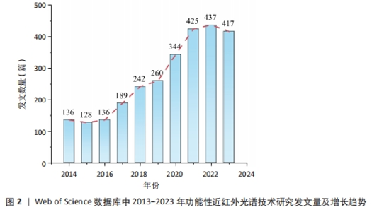
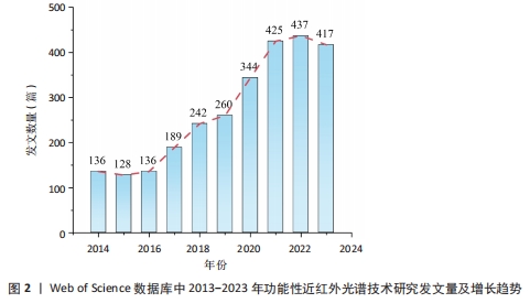
2.1 发文文献数量分析 此次研究选择了 2 714 篇文献,来自68个国家 2 400个机构的 9 171 名作者,共发表在495 种期刊上,这些文章引用了来自13 785 种期刊的79 876 篇引用文献。 图2显示了近10年来功能性近红外光谱技术研究领域论文发表的时间分布及增长趋势。由图2可以发现,近10年来功能性近红外光谱技术研究相关文献的年发文量整体态势趋于稳定,2014年发文量已达到136篇,表明该领域一直以来备受关注,已具有良好的研究基础;2016-2022年,发文量总体呈增长趋势,并在2022年达到年发文量的高峰,为437篇,说明了该领域的研究热度高涨,其研究价值受到更多认知神经学科、临床医生等相关领域科学家的关注,并且这一趋势可能会继续增长。由于时间搜索范围限制在2023-11-30,因此2023年的统计数据尚未完整。"
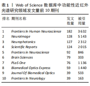
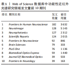
2.2 期刊文献计量分析 了解引文文献的期刊分布,有助于研究人员通过合适的期刊掌握该领域的研究热点和前沿。表1显示了功能性近红外光谱研究在Web of Science数据库中发表论文最多的前10种期刊及其被引用的次数,这些期刊的平均影响因子均大于2.5。收录功能性近红外光谱技术研究的英文文献期刊较为集中,多为生物医学、神经科学及脑科学专业期刊。前5大被引用期刊分别是《神经影像》《人类神经科学前沿》《神经光子学》《科学报告》和《生物医学光学快车》。从表1可以看出,虽然发表在《人类神经科学前沿》期刊上的论文数量排名第一,共182篇,但其引用次数少于文献数量排名第二的《神经影像》期刊。"
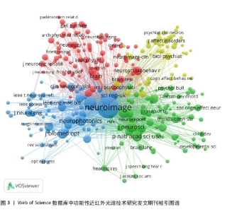
为了更直观地掌握共引期刊之间的关系,使用VOSviewer 6.1.20绘制了共引期刊分布图谱,如图3所示,其中节点的大小表示期刊文献被引用的频率。"


由图3可以发现,《神经影像》的节点最大,表明该期刊文献被引用率最高,此期刊的文章代表了该研究近年来的研究方向,其高被引文献反映了学术影响力,说明研究成果取得了重大的突破性进展与创新。结合表1来看,《神经影像》的文献被引用量为5 143,位居首位,2个分析结果一致。MORIOKA等[16]报道,脑电图-功能性近红外光谱技术混合脑机接口将空间注意力解码精度提高了8%以上,这一结果表明,基于功能性近红外光谱技术与其他技术(如脑电图、眼动追踪等技术)相结合使用的多模态成像方法,是该研究的重要方向。DING等[17]使用功能性近红外光谱技术研究了健康成人在不同复杂程度单任务和双重任务时的大脑激活和网络效率,研究结果表明网络效率降低可能是导致双重任务表现不佳的一个可能机制。这项研究的发现可能会为执行功能受损的神经系统人群提供新的治疗方法。发表在《神经影像》上的高被引文献都不同程度地代表了功能性近红外光谱技术研究当前的热点内容,并引领未来的研究方向。 2.3 国家和机构的文献计量分析 此次研究收集了来自全球68个国家的文献。图4A显示了发文数量最多的前10个国家,其中美国发表的文章数量最多,共796篇,表明美国是该领域最具影响力的国家;中国以739篇排名第二,对功能性近红外光谱技术研究的发展也做出了重要贡献。 此次研究选择的文献来自2 400家机构。图4B显示了发文数量排名前10的机构,其中美国有4个机构,中国有2个机构,英国、意大利、韩国和德国各有1个机构。结果表明,对功能性近红外光谱技术的研究成果贡献主要来自英国的伦敦大学、中国的北京师范大学及德国的图宾根大学。其中,伦敦大学在该领域贡献最多,累计发表89篇论文,研究方向主要集中在降低功能性近红外光谱技术噪声的方法、功能性近红外光谱技术在脑-计算机接口中的应用可行性研究,以及婴幼儿大脑皮质血流动力学的相关研究[18-22]。第二大贡献者是中国的北京师范大学,共发表86篇论文,研究方向侧重于静息态脑功能连接的功能性近红外光谱技术与功能性磁共振成像技术的定量比较与评估[23-24],以及利用功能性近红外光谱技术研究多动症儿童在工作记忆任务下脑区血氧含量的变化等[25-26]。图宾根大学位居第三,累计发表73篇文献,研究焦点聚集在借助功能性近红外光谱技术研究多动症、重度抑郁症及双向情感障碍患者和健康人群额叶皮层的血氧浓度的差异[27-30]。"
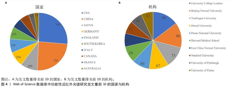
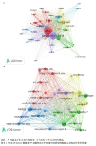
图5展示了国家和机构的合作关系可视化网络图谱,其中每个节点之间的连线代表各国家、机构之间的合作关系,连线的粗细则代表联系的紧密程度。图5A 显示美国是该网络视图的中心,与中国、英国、日本、德国等多个国家之间形成了稳定的合作团队。图5B显示了研究机构处于全球聚集状态,表明研究机构分布相对集中,形成了较大的聚集集群。然而功能性近红外光谱技术的研究机构主要来自国外高校,各机构之间的连线较细,说明各机构之间的合作发文较少,表明国内外及各机构间的合作交流较少,今后应进一步扩大国际研究合作,以推动该研究的发展和提升整体研究标准。"


2.4 作者的文献计量分析 表2展示了发文量排名前10的作者,其中Ehlis(图宾根大学)排名第一,十年来共发表50篇文章,文献被引用次数为900次,平均每篇被引用18次;其次是Fallgatter(图宾根大学),共发表43篇文章,被引用次数为856次,平均每篇被引用约20次。"
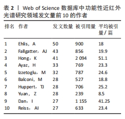

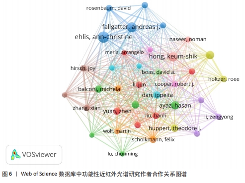
值得注意的是,尽管作者Hong (釜山国立大学)的发文数量排名第三,但其文章的被引用次数位居第一,并且平均每篇文章的被引用量最高,约为51次/篇,说明Hong在功能性近红外光谱研究领域发表的文献质量较高,例如借助功能性近红外光谱技术监测血氧浓度依赖信号的时间响应时,为了减少检测时间,提出并验证了一种基于核递归最小二乘(KRLS)算法的预测方法,结果显示在监测响应时间时可以在0.1 s内完成神经元的激活[31]。目前该方法已获得了国内外专家学者的认可。 图6展示了作者之间的合作关系,通过分析合作网络可以识别出功能性近红外光谱研究领域的代表性作者,图中的每个节点代表一位作者,节点大小表示作者的发文数量,节点连线代表作者之间的联系或者合作关系,连线的粗细和颜色分别表示联系的紧密程度及合作发表论文的频次。可以发现,Ehlis和Fallgatter的节点较大且均为蓝色,表明这2位作者之间有紧密的合作关系,形成了稳定的合作团队;此外,Ehlis和Fallgatter的发文数量也在作者排名中居于前列,这反映出团队合作对功能性近红外光谱技术研究未来的发展产生了重要影响。"
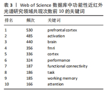
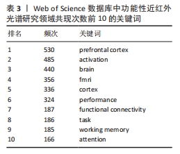
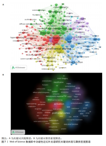
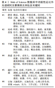
2.5 关键词分析 2.5.1 关键词共现可视化分析 关键词能够反映文章的核心内容,通过分析一个领域的高频关键词可以快速掌握该领域的研究热点和主要研究方 向[32]。 在去除与检索策略相关的检索词并合并同义关键词后,引用频次最高的10个关键词如表3所示。可以看出,该领域研究的热点关键词包括前额叶皮质、激活、大脑、脑功能核磁共振成像、行为、功能性连接、任务、工作记忆和注意力。由VOSviewer 1.6.20生成的关键词共现网络图谱如图7所示,图中每个节点代表一个关键词,节点的大小表示关键词出现的频次,节点的不同颜色表示不同的聚类,具有相似研究主题的关键词组合成一个集群,并以相同的颜色显示。从图7可以看出,生成的网络图可以大致分为6个集群,代表了6个不同的研究方向。这6个聚类分别是#1静息状态、#2运动想象、#3平衡、#4焦虑、#5超扫描和#6认知表现,主要集群及其包含的关键词如表4所示。"
"

"
2.5.2 关键词突现可视化分析 虽然VOSviewer生成的关键词共现和聚类密度可视化图谱能够直观地显示该领域的热门研究聚类,但未考虑时间因素,无法从图中获取该领域的前沿热点,因此,此次研究结合CiteSpace软件的突发性探测功能,对频次变化率高的关键词突现强度进行排序,形成关键词爆发函数。通过研究关键词爆发的强度和持续时间可以掌握该领域在不同时间段的研究热点,探索未来的研究前沿和发展趋势[33]。 图8总结了2013-2023年功能性近红外光谱技术研究中引用爆发最强的前25个关键词。图中的浅色线表示时间间隔,深色线显示每个突发时间间隔的开始、结束和持续时间。由图8可以发现,2013-2023年引用爆发强度最强的关键词是“人脑”,其强度值为9.83,其他高爆发强度的关键词包括“磁共振成像”、“光学拓扑”“可靠性”“脑血流动力学”“血液流动”“认知控制”“前额叶皮质”和“刺激”,这些关键词一直是持续的热点词,与图7关键词和聚类的图谱分析结果一致,表明使用功能性近红外光谱技术探索大脑的各种机制仍然是当前及未来一段时间的研究热点。"
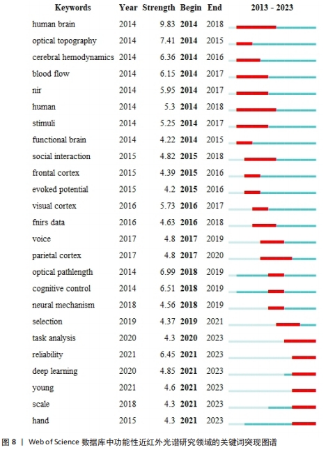

这些机制包括对不同脑区(尤其是前额叶皮质)在不同状态下的变化、个体的决策反应、认知控制和情绪调节的研究,以及对婴幼儿个体差异的检测及与其他脑功能成像技术相结合的研究。同时从图8可以看出,“任务分析(task analysis)”“可靠性(reliability)”“深度学习(deep learning)”“年轻人(young)”“尺度(scale)”和“手(hand)”是从2020年开始持续到2023年的突发关键词,表明了未来该领域的发展趋势和潜在价值会围绕以上6个方向进行研究。"

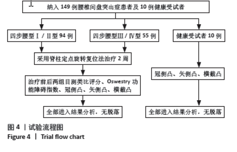
2.1 参与者数量分析 纳入149例腰椎间盘突出症患者及10例健康受试者,临床试验过程无不良事件发生,数据无脱落,全部进入结果分析。 2.2 试验流程图 见图4。"
| [1] RAHMAN MD A, SIDDIK AB, GHOSH TK, et al. A narrative review on clinical applications of fNIRS. J Digit Imaging. 2020;33(5):1167-1184. [2] ARRIDGE SR, HEBDEN JC. Optical imaging in medicine: II.modelling and reconstruction. Phys Med Biol. 1997;42(5):841-853. [3] 胡天然,于布为.功能近红外光谱成像技术在认知功能评估中的应用[J].国际麻醉学与复苏杂志,2014,35(10):932-935. [4] 熊桃,李阳,崔丽君,等.功能性近红外光谱应用于脑认知领域的可视化分析[J].中国康复,2024,39(1):46-51. [5] XU G, ZHOU M, CHEN Y, et al. Brain activation during standing balance control in dual-task paradigm and its correlation among older adults with mild cognitive impairment: a fNIRS study. BMC Geriatr. 2024;24(1):144. [6] KELES HO, KARAKULAK EZ, HANOGLU L, et al. Screening for alzheimer’s disease using prefrontal resting-state functional near-infrared spectroscopy. Front Hum Neurosci. 2022;16:1061668. [7] REN L, YIN X, WANG HY, et al. Correlation and underlying brain mechanisms between rapid eye movement sleep behavior disorder and executive functions in Parkinson’s disease: an fNIRS study. Front Aging Neurosci. 2024;15:1290108. [8] BLANCO B, LLOYD-FOX S, BEGUM-ALI J, et al. Cortical responses to social stimuli in infants at elevated likelihood of ASD and/or ADHD: a prospective cross-condition fNIRS study. Cortex. 2023;169:18-34. [9] 张宁,杨远滨,田浩林,等.功能性近红外光谱应用于康复领域的可视化分析[J].中国康复理论与实践,2023,29(10):1171-1178. [10] HAN Y, HUANG J, YIN Y, et al. From brain to worksite: the role of fNIRS in cognitive studies and worker safety. Front Public Health. 2023;11:1256895. [11] LUO H, CAI Z, HUANG Y, et al. Study on pain catastrophizing from 2010 to 2020: a bibliometric analysis via CiteSpace. Front Psychol. 2021;12:759347. [12] CHEN C. Searching for intellectual turning points: progressive knowledge domain visualization. Proc Natl Acad Sci U S A. 2004; 101(suppl_1):5303-5310. [13] 胡杰.基于科学知识图谱的我国传统与现代运动康复临床研究比较分析[D].南京:南京中医药大学,2022. [14] HUANG N. Quantitative and visual analysis of tsunami warning research: a bibliometric study using web of science and VOSviewer. Int J Disaster Risk Reduct. 2024;103:104307. [15] 任智军,朱东华,荆雷.基于可视化数据挖掘的管理科学科技文本分析研究[J].科学学与科学技术管理,2006(1):8-12. [16] MORIOKA H, KANEMURA A, MORIMOTO S, et al. Decoding spatial attention by using cortical currents estimated from electroencephalography with near-infrared spectroscopy prior information. NeuroImage. 2014;90:128-139. [17] DING Q, OU Z, YAO S, et al. Cortical activation and brain network efficiency during dual tasks: an fNIRS study. NeuroImage. 2024;289:120545. [18] CUI X, BRAY S, REISS AL. Functional near infrared spectroscopy (NIRS) signal improvement based on negative correlation between oxygenated and deoxygenated hemoglobin dynamics. NeuroImage. 2010;49(4):3039-3046. [19] BRIGADOI S, CECCHERINI L, CUTINI S, et al. Motion artifacts in functional near-infrared spectroscopy: a comparison of motion correction techniques applied to real cognitive data. NeuroImage. 2014;85:181-191. [20] LLOYD-FOX S, RICHARDS JE, BLASI A, et al. Coregistering functional near-infrared spectroscopy with underlying cortical areas in infants. Neurophotonics. 2014;1(2):025006. [21] NASEER N, HONG MJ, HONG KS. Online binary decision decoding using functional near-infrared spectroscopy for the development of brain–computer interface. Exp Brain Res. 2014;232(2):555-564. [22] ASLIN RN, SHUKLA M, EMBERSON LL. Hemodynamic correlates of cognition in human Infants. Annu Rev Psychol. 2015;66(1):349-379. [23] LU CM, ZHANG YJ, BISWAL BB, et al. Use of fNIRS to assess resting state functional connectivity. J Neurosci Methods. 2010; 186(2):242-249. [24] DUAN L, ZHANG YJ, ZHU CZ. Quantitative comparison of resting-state functional connectivity derived from fNIRS and fMRI: A simultaneous recording study. NeuroImage. 2012;60(4):2008-2018. [25] GU Y, MIAO S, HAN J, et al. Complexity analysis of fNIRS signals in ADHD children during working memory task. Sci Rep. 2017;7(1):829. [26] GU Y, MIAO S, HAN J, et al. Identifying ADHD children using hemodynamic responses during a working memory task measured by functional near-infrared spectroscopy. J Neural Eng. 2018;15(3):035005. [27] ALEKSANDROWICZ A, HAGENMULLER F, HAKER H, et al. Frontal brain activity in individuals at risk for schizophrenic psychosis and bipolar disorder during the emotional Stroop task – an fNIRS study. Neuroimage Clin, 2020;26:102232. [28] ROSENBAUM D, INT-VEEN I, KROCZEK A, et al. Amplitude of low frequency fluctuations (ALFF) of spontaneous and induced rumination in major depression: an fNIRS study. Sci Rep. 2020;10(1):21520. [29] SOMMER A, FALLGATTER AJ, PLEWNIA C. Investigating mechanisms of cognitive control training: neural signatures of PASAT performance in depressed patients. J Neural Transm. 2022;129(5-6):649-659. [30] STORCHAK H, HUDAK J, DRESLER T, et al. Monitoring processes and their neuronal correlates as the basis of auditory verbal hallucinations in a non-clinical sample. Front Psychiatry. 2021;12:644052. [31] ZAFAR A, HONG KS. Reduction of onset delay in functional near-infrared spectroscopy: prediction of HbO/HbR signals. Front Neurorobot. 2020;14:10. [32] 宋浩然,张玉强,谷娜,等.基于Citespace对人工智能在骨创伤研究的可视化分析[J].中国组织工程研究,2025,29(3):493-502. [33] HU H, DAI J, JIN Y, et al. Bibliometric analysis on desertification restoration based on CiteSpace. Arab J Geosci. 2021;14(2):72. [34] EHLIS AC, SCHNEIDER S, DRESLER T, et al. Application of functional near-infrared spectroscopy in psychiatry. NeuroImage. 2014; 85:478-488. [35] 郑权良,王庭照,史兵,等.幼儿动作发展水平与抑制控制和认知灵活性加工的差异[J].中国学校卫生,2024,45(2):258-262. [36] TAKEUCHI Y. Change in blood volume in the brain during a simulated aircraft landing task. J Occup Health. 2000;42(2):60-65. [37] YANG D, HUANG R, YOO SH, et al. Detection of mild cognitive impairment using convolutional neural network: temporal-feature maps of functional near-infrared spectroscopy. Front Aging Neurosci. 2020;12:141. [38] 李秀丽,黄富表,张通.上肢运动游戏训练对脑卒中患者注意功能障碍和日常生活活动能力的影响[J]. 中国医刊,2023,58(11): 1263-1266. [39] 辛佳炜,王岩,敖强,等.近红外功能成像观察三叉神经痛触发痛与前额叶氧合血红蛋白变化的相关性[J].中华临床医师杂志(电子版),2012,6(19):5917-5921. [40] LIM SB, YANG CL, PETERS S, et al. Phase-dependent brain activation of the frontal and parietal regions during walking after stroke - an fNIRS study. Front Neurol. 2022;13:904722. [41] SCARAPICCHIA V, BROWN C, MAYO C, et al. Functional magnetic resonance imaging and functional near-infrared spectroscopy: insights from combined recording studies. Front Hum Neurosci. 2017;11:419. [42] SU WC, DASHTESTANI H, MIGUEL HO, et al. Simultaneous multimodal fNIRS-EEG recordings reveal new insights in neural activity during motor execution,observation, and imagery. Sci Rep. 2023;13(1):5151. [43] İŞBILIR E, ÇAKIR MP, ACARTÜRK C, et al. Towards a multimodal model of cognitive workload through synchronous optical brain imaging and eye tracking measures. Front Hum Neurosci. 2019;13:375. [44] CHEN H, LIANG Q, WANG B, et al. Sports game intervention aids executive function enhancement in children with autism - an fNIRS study. Neurosci Lett. 2024;822:137647. [45] ZHAI Y, XIE H, ZHAO H, et al. Neural synchrony underlies the positive effect of shared reading on children’s language ability. Cereb Cortex. 2023;33(19):10426-10440. |
| [1] | Liang Haobo, Wang Zeyu, Ma Wenlong, Liu Hao, Liu Youwen. Hot issues in the field of joint revision: infection, rehabilitation nursing, bone defect, and prosthesis loosening [J]. Chinese Journal of Tissue Engineering Research, 2025, 29(9): 1963-1971. |
| [2] | Lyu Liting, Yu Xia, Zhang Jinmei, Gao Qiaojing, Liu Renfan, Li Meng, Wang Lu. Bibliometric analysis of research process and current situation of brain aging and exosomes [J]. Chinese Journal of Tissue Engineering Research, 2025, 29(7): 1457-1465. |
| [3] | Xie Liugang, Cui Shuke, Guo Nannan, Li Aoyu, Zhang Jingrui. Research hotspots and frontiers of stem cells for Alzheimer’s disease [J]. Chinese Journal of Tissue Engineering Research, 2025, 29(7): 1475-1485. |
| [4] | Chang Jinxia, Liu Yufei, Niu Shaohui, Wang Chang, Cao Jianchun. Visualization analysis of macrophage polarization in tissue repair process [J]. Chinese Journal of Tissue Engineering Research, 2025, 29(7): 1486-1496. |
| [5] | Li Huijun, Li Huangyan, Zhang Yeting. Physical activity and cognition in older adults: research hotspot and topic evolution [J]. Chinese Journal of Tissue Engineering Research, 2025, 29(5): 1073-1080. |
| [6] | Dang Xiaowen, Huang Hailiang, Huang Lei, Wang Yajie . Research frontiers and hotspots of carbon nanomaterials in biomedical field over the past 10 years [J]. Chinese Journal of Tissue Engineering Research, 2025, 29(4): 752-760. |
| [7] | Ma Yucong, Ouyang Zhengzheng, Liu Xiaojie, Yang Sifei. Tracking of research trends and hotspots in medical magnesium alloy materials [J]. Chinese Journal of Tissue Engineering Research, 2025, 29(34): 7470-7480. |
| [8] | Wang Yong, Li Hongyu, Liu Yuhang, Wang Fengxing. Knowledge map of surgical treatment for osteonecrosis of the femoral head: a bibliometric analysis of data from 2005 to 2024 [J]. Chinese Journal of Tissue Engineering Research, 2025, 29(33): 7250-7260. |
| [9] | Deng Qing, Wang Qingjun, Zhang Yeting. Visual analysis of dynamic evolution of research topics in the field of physical activity and hippocampal tissue [J]. Chinese Journal of Tissue Engineering Research, 2025, 29(32): 6997-7003. |
| [10] | Guo Haizhen, Cong Zidong, Zhao Yuke, Li Xiaofeng, Yu Lu, Qian Shule, Wang Runying, Du Wuxun. Development of patch clamp technology in the past 10 years: visual analysis based on CiteSpace and VOSviewer [J]. Chinese Journal of Tissue Engineering Research, 2025, 29(31): 6717-6726. |
| [11] | Zhao Xiaoxuan, Liu Shuaiyi, Xing Zheng, Li Qingwen, Chu Xiaolei, Li Qi. Research hotspots and trends in application of tissue engineering in peripheral nerve injury [J]. Chinese Journal of Tissue Engineering Research, 2025, 29(30): 6591-6600. |
| [12] | Xu Qiang, Qin Jialin, Lian Zeshuang, Wang Aoting, Li Ding, Wang Ye, Wang Junfang. Visual analysis of hot spots and trends in the study of ligamentum flavum ossification [J]. Chinese Journal of Tissue Engineering Research, 2025, 29(3): 628-636. |
| [13] | Song Haoran, Zhang Yuqiang, Gu Na, Zhi Xiaodong, Wang Wei. Visualization analysis of artificial intelligence in bone trauma research based on Citespace [J]. Chinese Journal of Tissue Engineering Research, 2025, 29(3): 493-502. |
| [14] | Zheng Xiaodong, Gao Shan, Han Wenjin, Liu Lijun, Jia Menglong, Yu Longtan. Visual analysis of treatment of adolescent idiopathic scoliosis [J]. Chinese Journal of Tissue Engineering Research, 2025, 29(3): 645-653. |
| [15] | Liu Jian, Liu Qing, Huang Ye, Cao Guanglei, Liu Yuan, Song Qingpeng. Growth factors promote knee cartilage regeneration: a bibliometric analysis of research hotspots [J]. Chinese Journal of Tissue Engineering Research, 2025, 29(29): 6351-6359. |
| Viewed | ||||||
|
Full text |
|
|||||
|
Abstract |
|
|||||