Chinese Journal of Tissue Engineering Research ›› 2025, Vol. 29 ›› Issue (22): 4679-4686.doi: 10.12307/2025.406
Previous Articles Next Articles
Finite element analysis of ultrashort implants applied to the mandibular posterior tooth area under different bone conditions
Zilalai · Julaiti, Mawulanjiang · Abudurenmu, Aikeliya · Ainiwaer, Reyila · Kuerban, Nijiati · Tuersun
- Department of Stomatology, Second Affiliated Hospital of Xinjiang Medical University, Urumqi 830063, Xinjiang Uygur Autonomous Region, China
-
Received:2024-02-04Accepted:2024-04-09Online:2025-08-08Published:2024-12-05 -
Contact:Nijiati · Tuersun, Master, Chief physician, Associate professor, Department of Stomatology, Second Affiliated Hospital of Xinjiang Medical University, Urumqi 830063, Xinjiang Uygur Autonomous Region, China -
About author:Zilalai · Julaiti, Master candidate, Department of Stomatology, Second Affiliated Hospital of Xinjiang Medical University, Urumqi 830063, Xinjiang Uygur Autonomous Region, China -
Supported by:Natural Science Foundation of Xinjiang Uygur Autonomous Region, No. 2021D01C362
CLC Number:
Cite this article
Zilalai · Julaiti, Mawulanjiang · Abudurenmu, Aikeliya · Ainiwaer, Reyila · Kuerban, Nijiati · Tuersun. Finite element analysis of ultrashort implants applied to the mandibular posterior tooth area under different bone conditions[J]. Chinese Journal of Tissue Engineering Research, 2025, 29(22): 4679-4686.
share this article
Add to citation manager EndNote|Reference Manager|ProCite|BibTeX|RefWorks

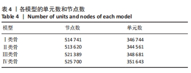
2.1 各组模型的具体单元数和节点数 表4。"
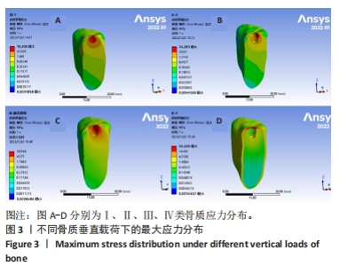
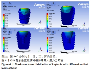
2.2 不同骨质种植修复体整体应力分布情况 2.2.1 垂直载荷 皮质骨最大应力位于种植体颈部处,且在Ⅰ、Ⅱ、Ⅲ类骨质中相对于Ⅳ类骨更趋向于稳定,力也从分散于种植体周围到Ⅳ类骨时逐渐集中至种植体颈部与皮质骨连接处,见图3A-D;Ⅰ类骨中最大应力在中央螺丝区域,Ⅱ、Ⅲ、Ⅳ类骨中最大应力则统一在种植体与基台连接处,Ⅰ-Ⅳ类骨中皮质骨受力均比松质骨大,见表5。种植体在Ⅰ、Ⅱ、Ⅲ类骨中呈现出应力逐渐增大的趋势,但到Ⅳ类骨时应力有所减小。Ⅰ、Ⅱ、Ⅲ、Ⅳ类骨质的高应力部分都集中在种植体颈部与基台连接处,但明显可以观察到在Ⅰ类和Ⅱ类骨质中,种植体颈部的应力沿植体长轴分散分布,而Ⅲ、Ⅳ类骨质的应力集中在种植体颈部与基台连接处,见图4。"
"

"

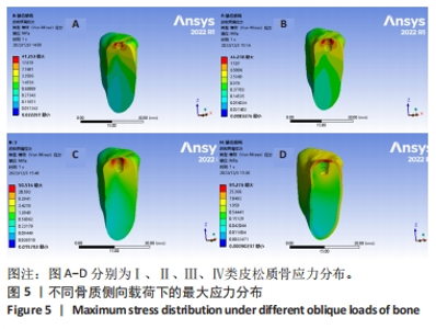
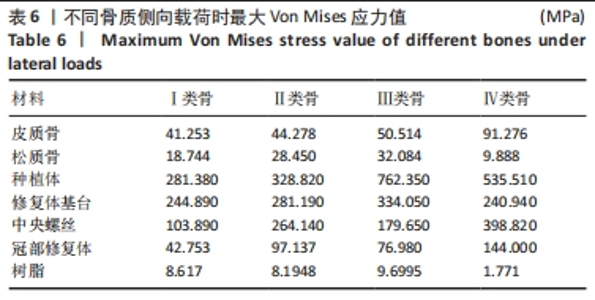
2.2.2 侧向载荷 与垂直载荷比较不难看出,侧向载荷时各类骨的最大应力峰值都明显大于垂直向载荷,且皮质骨最大应力随着骨质条件的减弱呈逐渐增大的趋势,到Ⅳ类骨时应力峰值达到最高,见图5;皮质骨应力分布在与种植体连接处,Ⅰ-Ⅲ类骨中皮质骨所受应力均匀分散在种植体周围,而在Ⅳ类骨则集中在皮质骨与种植体连接区域。皮质骨受力均比松质骨大,见表6。"
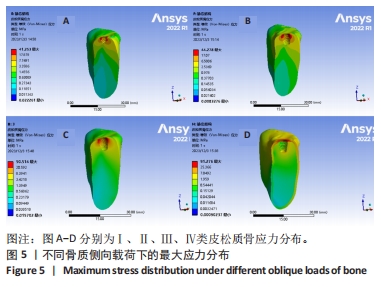
"


Ⅰ-Ⅳ类骨中整体等效应力都集中于种植体与基台连接处,其中,Ⅰ-Ⅲ类骨中种植体颈部所受应力是逐渐增大的,而到Ⅳ类骨时种植体所受应力值有所减小,但应力相对于Ⅰ、Ⅱ类骨来说更加集中于种植体的颈部,见图6。结合图6,表6发现Ⅲ、Ⅳ类骨的等效应力峰值在种植体颈部。"
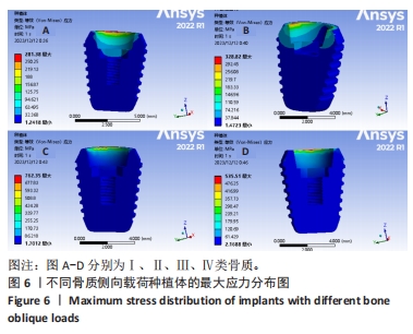
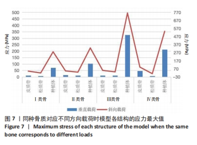
2.3 同一种骨质不同方向对种植体周围骨组织的应力分布与应力值 Ⅰ类骨在垂直向载荷时应力主要沿着种植体长轴方向向骨组织分散,应力主要集中区域占超短种植体长度的1/2,而在侧向受力时应力主要在种植体颈部颊舌侧于皮质骨连接处且应力值较垂直载荷明显增大。但种植体在不同方向中受到的应力集中区域是无明显差异的,见图4,6。 Ⅱ类骨中,当受到与牙体长轴一致的垂直向力时应力集中分布在种植体颈部颊侧与皮质骨连接处,受侧向载荷力时跟Ⅰ类骨相一致,还是集中在种植体颈部颊舌侧与皮质骨连接处。种植体的等效应力峰值则集中在植体与基台相连处,当Ⅲ类骨受不同方向载荷时,种植体周围骨质在垂直向载荷力下应力主要沿种植体长轴方向分散,分布在种植体近中颊侧与皮质骨和松质骨相连处,而侧向载荷时应力主要集中区域与Ⅱ类骨一致,而种植体在不同方向上的应力分布区域无明显差异,都在超短种植体颈部与基台相连处。Ⅳ类骨在不同载荷方向所受到的应力分布区域几乎相一致,都在皮质骨与种植体颈部相连处,种植体所受应力也表现出一致的结果,即分布区域无明显差别。在Ⅰ、Ⅱ、Ⅲ、Ⅳ这4种骨质中,各结构的等效应力峰值均为侧向载荷应力值均大于垂直向载荷应力值,见图7。"


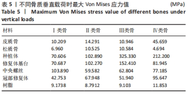
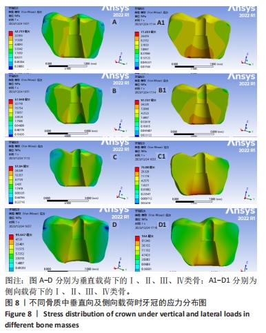
2.4 不同骨质中种植修复体各部件所受最大Von Mises应力值 见表5,表6。可以看出,牙冠在Ⅰ、Ⅱ、Ⅲ、Ⅳ类骨中,加载的方向无论是垂直向或者侧向力,牙冠所受的应力在Ⅰ、Ⅱ、Ⅲ类骨较平稳,在Ⅳ类骨时应力值最高,高应力区集中在基台与牙冠底部连接处,且当牙冠侧向受力时的应力比垂直向受力时的应力更大,见图8;修复基台在受垂直向载荷力时应力从Ⅰ-Ⅲ类骨逐渐增大,受侧向力时也同样是Ⅰ-Ⅲ类骨的应力逐渐增大,反之到Ⅳ类骨时修复体基台应力有所减小,高应力区集中在基台与种植体连接处。中央螺丝及树脂粘接材料在两种不同加载方向中及不同骨质类型下所受应力没有表现出一个明显的规律。"
| [1] YONGHUI C, ZHIYU C, DINGGUO Z, et al.Inlay osteotome sinus floor elevation with concentrated growth factor application and simultaneous short implant placement in severely atrophic maxilla.Sci Rep. 2016:6:27348. [2] 叶芷君,徐普.超短种植体在萎缩后牙区的临床研究进展[J].成都医学院学报,2021,16(5):673-675. [3] 吴越琳,刘加荣.三维有限元分析在口腔医学领域的研究进展[J].口腔医学,2021,41(7):659-663. [4] 李征,王媛,田梦婷 ,等.桩核表面制备不同形式固位槽及使用不同黏结剂对固位力的影响[J].中国组织工程研究,2020,24(34): 5508-5513. [5] 康非吾,卢军,潘可风.种植区骨皮质厚度对种植体骨界面应力分布的影响[J].同济大学学报(医学版),2006,27(1):31-33. [6] LIU C, XING Y, LI Y, et al. Bone quality effect on short implants in the edentulous mandible: a finite element study. BMC Oral Health. 2022; 22(1):139. [7] 孙江伟,王俊祥,白布加甫·叶力思,等.不同光滑颈圈种植体修复时应力分布的三维有限元分析[J].中国组织工程研究,2023,27(7): 1004-1011. [8] 李希光,郅克谦,高岭,等.基于三维有限元评价种植体不同倾斜角度在上颌后牙区骨量不足的应力分析[J].口腔医学研究,2019, 35(7):671-675. [9] 热依拉·库尔班,霞黑达·依拉尔江,陈欣,等.超短种植体不同修复方式的三维有限元分析[J].中国组织工程研究,2023,27(30): 4824-4829. [10] 施梦汝,谢伟丽,施武阁,等.柱形锥形种植体在不同种植深度的三维有限元研究[J].口腔医学,2019,39(7):577-580. [11] 鲍中波.骨质条件和种植体外形对ISRPD种植体承载能力影响的有限元分析[D].大连:大连医科大学,2021. [12] 李春.口腔种植与传统固定义齿修复牙列缺损的疗效对比[J].临床医学工程,2021,28(4):481-482. [13] NEJAT N, ÖNDER G, EMIN MK. Extra-Short Implants with Osteotome Sinus Floor Elevation: A Prospective Clinical Study.Int J Oral Maxillofac Implants. 2020;35(2):415-422. [14] LOMBARDO G, MARINCOLA M, SIGNORIELLO A, et al.Single-Crown, Short and Ultra-Short Implants, in Association with Simultaneous Internal Sinus Lift in the Atrophic Posterior Maxilla: A Three-Year Retrospective Study.Materials (Basel). 2020;13(9):2208. [15] RAMEH S, MENHALL A, YOUNES R.Key factors influencing short implant success.Oral Maxillofac Surg. 2020;24(3):263-275. [16] JOJI M, SHRUTHI E, SURESH S, et al.Clinical outcomes of ultrashort sloping shoulder implant design: A survival analysis.Clin Implant Dent Relat Res. 2018;20(4):646-652. [17] LOMBARDO G, SIGNORIELLO A, PARDO A, et al. Short and ultra-short(<6-mm)locking-taper implants supporting single crowns in posterior areas (part II): A 5-year retrospective study on periodontally healthy patients and patients with a history of periodontitis.Clin Implant Dent Relat Res. 2022;24(4):455-467. [18] CASTELLANOS-COSANO L, RODRIGUEZ-PEREZ A, SPINATO S, et al. Descriptive retrospective study analyzing relevant factors related to dental implant failure. Med Oral Patol Oral Cir Bucal. 2019;24(6): e726-e738. [19] 白布加甫·叶力思,热娜古丽·买合木提,艾孜买提江·赛依提,等.上颌中切牙联冠种植修复体在不同咬合方式中的应力分析[J].中国组织工程研究,2022,26(4):567-572. [20] NIROOMAND MR, ARABBEIKI M. Effect of the dimensions of implant body and thread on bone resorption and stability in trapezoidal threaded dental implants: a sensitivity analysis and optimization. Comput Methods Biomech Biomed Engin. 2020;23(13):1005-1013. [21] SUMRA N, DESAI S, KULSHRESTHA R, et al. Analysis of micromovements and peri-implant stresses and strains around ultra-short implants-A three-dimensional finite-element method study.J Indian Soc Periodontol. 2021;25(4):288-294. [22] LANA CZ, MARTIN F, ANDREAS R, et al.Biomechanical finite element analysis of short-implant-supported, 3-unit, fixed CAD/CAM prostheses in the posterior mandible.Int J Implant Dent. 2022;8(1):8. [23] 丁熙,朱形好,廖胜辉,等.种植牙即刻负载与延期负载的生物力学三维有限元分析[J].口腔医学,2007,27(10):519-522. [24] 许月丹,金鑫阳,赵维家,等.种植义齿的咬合设计[J].口腔医学, 2020,40(12):1124-1128. [25] LI T, KONG L, WANG Y, et al. Selection of optimal dental implant diameter and length in type IV bone: a three-dimensional finite element analysis. Int J Oral Maxillofac Surg. 2009;38(10):1077-1083. [26] LINETSKIY I, DEMENKO V, LINETSKA L, et al.Impact of annual bone loss and different bone quality on dental implant success–A finite element study.Comput Biol Med. 2017;91:318-325. [27] 王媛,张杨.不同下颌骨密度对4颗种植体支持Locator式覆盖义齿的有限元生物力学分析[J].中国组织工程研究,2022,26(22): 3492-3497. [28] HUANG HL, HSU JT, FUH LJ, et al. Biomechanical simulation of various surface roughnesses and geometric designs on an immediately loaded dental implant. Comput Biol Med. 2010;40(5):525-532. [29] BARÃO VA, DELBEN JA, LIMA J, et al. Comparison of different designs of implant-retained overdentures and fixed full-arch implant-supported prosthesis on stress distribution in edentulous mandible--a computed tomography-based three-dimensional finite element analysis. J Biomech. 2013;46(7):1312-1320. [30] BASHUTSKI JD, D’SILVA NJ, WANG HL.Implant compression necrosis: current understanding and case report.J Periodontol. 2009;80(4):700-704. [31] 陈俊良,刘旭琳,张潇月,等.短种植体修复牙槽骨严重吸收的下颌第二磨牙的三维有限元分析[J].口腔医学研究,2019,35(3):246-250. [32] YALÇıN M, KAYA B, LAÇIN N, et al. Three-Dimensional Finite Element Analysis of the Effect of Endosteal Implants with Different Macro Designs on Stress Distribution in Different Bone Qualities. Int J Oral Maxillofac Implants. 2019;34(3):e43–e50. [33] 施斌,吴涛.种植修复体机械并发症的原因、预防及处理[J].口腔疾病防治,2018,26(7):415-421. [34] 施斌,晏奇,伍昕宇.短种植体(≤6mm)的临床应用与并发症[J].口腔疾病防治,2020,28(3):137-145. |
| [1] | Zhou Jinhai, Li Jiangwei, Wang Xuquan, Zhuang Ying, Zhao Ying, Yang Yuyong, Wang Jiajia, Yang Yang, Zhou Shilian. Three-dimensional finite element analysis of anterior femoral notching during total knee arthroplasty at different bone strengths [J]. Chinese Journal of Tissue Engineering Research, 2025, 29(9): 1775-1782. |
| [2] | Li Shuai, Liu Hua, Shang Yonghui, Liu Yicong, Zhao Qihang, Liu Wen. Stress distribution on the maxilla when wearing the Twin-block appliance for Class II malocclusion [J]. Chinese Journal of Tissue Engineering Research, 2025, 29(5): 881-887. |
| [3] | Zhou Zonghao, Luo Siyang, Chen Jiawen, Chen Guangneng, Feng Hongchao. Finite element analysis of bioabsorbable plates versus miniature titanium plates in mandibular fracture fixation in different bone qualities [J]. Chinese Journal of Tissue Engineering Research, 2025, 29(4): 818-826. |
| [4] | Xiao Fang, Huang Lei, Wang Lin. Magnetic nanomaterials and magnetic field effects accelerate bone injury repair [J]. Chinese Journal of Tissue Engineering Research, 2025, 29(4): 827-838. |
| [5] | Zhang Yu, Xu Ruian, Fang Lei, Li Longfei, Liu Shuyan, Ding Lingxue, Wang Yuexi, Guo Ziyan, Tian Feng, Xue Jiajia. Gradient artificial bone repair scaffold regulates skeletal system tissue repair and regeneration [J]. Chinese Journal of Tissue Engineering Research, 2025, 29(4): 846-855. |
| [6] | Chen Yilong, Zhang Xu, Li Hong. Mechanical analysis of fiber post combined with different crown restorations for endodontically treated non-carious cervical lesions [J]. Chinese Journal of Tissue Engineering Research, 2025, 29(4): 866-871. |
| [7] | Li Mingzhe, Ye Xiangling, Wang Bing, Yu Xiang. Preparation and osteogenic properties of liquid crystal display light-cured polylactic acid scaffold loaded with nano-tantalum [J]. Chinese Journal of Tissue Engineering Research, 2025, 29(4): 670-677. |
| [8] | Ye Xiaolong, Zhang Yuxuan, Fu Rongchang, Liu Yun, Yusanjiang·Wuhuer, Escar·Aimer, Ma Yuan. A three-dimensional finite element modal analysis on adolescent idiopathic scoliosis [J]. Chinese Journal of Tissue Engineering Research, 2025, 29(33): 7072-7079. |
| [9] | Zhuang Yan, Wang Xinyu, Cao Yilin, Ding Yuanxin, Wang Jiaqi, Yu Miao, Luan Chunyang, Ding Yuansheng. Three-dimensional finite element analysis of personalized orthodontic devices for 3D printed maxillary single-rooted rotated tooth [J]. Chinese Journal of Tissue Engineering Research, 2025, 29(30): 6409-6415. |
| [10] | Akliya·Anwar, Nafisa·Gupur, Baibugafu·Yelisi, Zilalai·Gulaiti, Guzalnur·Emrayim, Nijat·Tursun. Dynamic stress analysis of maxillary sinus lifting without bone grafting and with immediate loading after bone grafting [J]. Chinese Journal of Tissue Engineering Research, 2025, 29(30): 6416-6425. |
| [11] | Deng Guanghui, Xiang Wei, Su Qifan, Chen Xiaoyu, Wang Liangwei, Wan Zhihong, Wu Jiaqi, Chen Xiaojun. Preparation of osteoporotic femoral condylar bone defect model in rabbits and its critical value [J]. Chinese Journal of Tissue Engineering Research, 2025, 29(30): 6426-6433. |
| [12] | Fang Dandan, Ma Ruijie, Huang Yi, He Kelin, Wu Lei. Three-dimensional model of swallowing musculoskeletal system based on CT image data and biomechanical characteristics analysis [J]. Chinese Journal of Tissue Engineering Research, 2025, 29(29): 6167-6163. |
| [13] | Li Shuyuan, Yang Dawen, Zeng Zhanpeng, Cai Qunbin, Zhang Jingtao, Zhou Qishi. Application of induced membrane technique for repairing critical-sized bone defects: advantages and future development [J]. Chinese Journal of Tissue Engineering Research, 2025, 29(28): 6083-6093. |
| [14] | Liu Yang, Yang Jilei, Wang Wenli, Cui Yingying, Sun Qihao, Li Yourui. Application characteristics of thermosensitive hydrogels in bone tissue engineering [J]. Chinese Journal of Tissue Engineering Research, 2025, 29(28): 6094-6100. |
| [15] | Wu Zhengmin, Li Changxu, Cui Yanwei, Chen Su. Finite element analysis of stress distribution on mandibular All-on-4 implant fixed denture with different occlusion [J]. Chinese Journal of Tissue Engineering Research, 2025, 29(28): 6020-6029. |
| Viewed | ||||||
|
Full text |
|
|||||
|
Abstract |
|
|||||