[1] PLOUMIS A, TRANSFLEDT EE, DENIS F. Degenerative lumbar scoliosis associated with spinal stenosis. Spine J. 2007;7(4):428-436.
[2] DIEBO BG, SHAH NV, BOACHIE-ADJEI O, et al. Adult spinal deformity. Lancet. 2019;394(10193):160-172.
[3] 张科峰,刘阳,张存鑫,等.退变性腰椎侧凸的认知及矫形融合固定治疗[J]. 中国组织工程研究,2019,23(32):5227-5232.
[4] TOMÉ-BERMEJO F, PIÑERA AR, ALVAREZ-GALOVICH L. Osteoporosis and the Management of Spinal Degenerative Disease (I). Arch Bone Jt Surg. 2017;5(5):272-282.
[5] KOBAYASHI T, ATSUTA Y, TAKEMITSU M, et al. A prospective study of de novo scoliosis in a community based cohort. Spine. 2006;31(2):178-182.
[6] FUJIWARA A, TAMAI K, AN HS, et al. The relationship between disc degeneration, facet joint osteoarthritis, and stability of the degenerative lumbar spine. J Spinal Disord. 2000;13(5):444-450.
[7] MURATA Y, TAKAHASHI K, HANAOKA E, et al. Changes in scoliotic curvature and lordotic angle during the early phase of degenerative lumbar scoliosis. Spine. 2002;27(20):2268-2273.
[8] AEBI M. The adult scoliosis. Eur Spine J. 2005;14(10):925-948.
[9] SCHOUTENS C, CUSHMAN DM, MCCORMICK ZL, et al. Outcomes of Nonsurgical Treatments for Symptomatic Adult Degenerative Scoliosis: A Systematic Review. Pain Med. 2020;21(6):1263-1275.
[10] HEY HWD, KIM CK, LEE WG, et al. Supra-acetabular line is better than supra-iliac line for coronal balance referencing-a study of perioperative whole spine X-rays in degenerative lumbar scoliosis and ankylosing spondylitis patients. Spine J. 2017;17(12):1837-1845.
[11] KITANAKA S, TAKATORI R, ARAI Y, et al. Facet Joint Osteoarthritis Affects Spinal Segmental Motion in Degenerative Spondylolisthesis. Clin Spine Surg. 2018;31(8):E386-E390.
[12] CHO KJ, KIM YT, SHIN SH, et al. Surgical treatment of adult degenerative scoliosis. Asian Spine J. 2014;8(3):371-381.
[13] SUN XY, KONG C, ZHANG TT, et al. Correlation between multifidus muscle atrophy, spinopelvic parameters, and severity of deformity in patients with adult degenerative scoliosis: the parallelogram effect of LMA on the diagonal through the apical vertebra. J Orthop Surg Res. 2019;14(1):276.
[14] RICHARDSON CA, JULL GA. Muscle control-pain control. What exercises would you prescribe? Man Ther. 1995;1(1):2-10.
[15] 喻学科,陈灿,李凯,等.退行性腰椎侧凸与多裂肌退变的关联性研究[J]. 陆军军医大学学报,2022,44(20):2075-2081.
[16] 苏少亭,周红海,侯召猛,等.腰椎定点旋转手法操作中拇指推力的有限元分析[J]. 中国组织工程研究,2024,28(12):1823-1828.
[17] 高飞,赵斌.腰椎关节突关节基础研究新进展[J]. 医学影像学杂志, 2009,19(7):925-927.
[18] VARLOTTA GP, LEFKOWITZ TR, SCHWEITZER M, et al. The lumbar facet joint: a review of current knowledge: part 1: anatomy, biomechanics, and grading. Skeletal Radiol. 2011;40(1):13-23.
[19] KE S, HE X, YANG M, et al. The biomechanical influence of facet joint parameters on corresponding segment in the lumbar spine: a new visualization method. Spine J. 2021;21(12):2112-2121.
[20] ROESSINGER O, HÜGLE T, WALKER UA, et al. Polg mtDNA mutator mice reveal limited involvement of vertebral bone loss in premature aging-related thoracolumbar hyperkyphosis. Bone Rep. 2022;17:101618.
[21] KITA N. Ultrastructural studies of articular cartilaginous degeneration in the facet joints in spinal scoliosis. Nihon Seikeigeka Gakkai Zasshi. 1994; 68(4):184-195.
[22] YASUDA H, MATSUMURA A, TERAI H, et al. Radiographic evaluation of segmental motion of scoliotic wedging segment in degenerative lumbar scoliosis. J Spinal Disord Tech. 2013;26(7):379-384.
[23] DE VRIES AA, MULLENDER MG, PLUYMAKERS WJ, et al. Spinal decompensation in degenerative lumbar scoliosis. Eur Spine J. 2010; 19(9):1540-1544.
[24] 郑杰,杨永宏.椎间盘和关节突关节在退变性脊柱侧凸发生发展中的作用[J]. 中国脊柱脊髓杂志,2018,28(9):826-831.
[25] 林云飞,王飞,李放.导致腰椎峡部裂的风险因素研究现状[J]. 脊柱外科杂志,2016,14(3):181-184.
[26] LAFAGE V, SCHWAB F, PATEL A, et al. Pelvic tilt and truncal inclination: two key radiographic parameters in the setting of adults with spinal deformity. Spine. 2009;34(17):E599-E606.
[27] BOURRET S, CERPA M, KELLY MP, et al. Correlation analysis of the PI-LL mismatch according to the pelvic incidence from a database of 468 asymptomatic volunteers. Eur Spine J. 2022;31(6): 1413-1420.
[28] LI J, XIAO H, JIANG S, et al. Risk Factors and Three Radiological Predictor Models for the Progression of Proximal Junctional Kyphosis in Adult Degenerative Scoliosis Following Posterior Corrective Surgery: 113 Cases With 2-years Minimum Follow-Up. Global Spine J. 2023;13(8): 2285-2295.
[29] SIMMONS ED. Surgical treatment of patients with lumbar spinal stenosis with associated scoliosis. Clin Orthop Relat Res. 2001;(384): 45-53.
[30] 丁文元,申勇,曹来震,等.腰椎退变性侧凸患者椎间盘退行性变的影像学观测[J]. 中国脊柱脊髓杂志,2010,20(8):673-676.
[31] 周仕炼,胡星新,杨曦,等.退变性腰椎侧凸六种工况运动下的生物力学的有限元分析[J]. 华西医学,2018,33(9):1099-1105.
[32] SEO JY, HA KY, HWANG TH, et al. Risk of progression of degenerative lumbar scoliosis. J Neurosurg Spine. 2011;15(5):558-566.
[33] ZHANG J, WANG Z, CHI P. Risk Factors for Immediate Postoperative Coronal Imbalance in Degenerative Lumbar Scoliosis Patients Fused to Pelvis. Global Spine J. 2021;11(5):649-655.
[34] LEWIS SJ, KESHEN SG, KATO S, et al. Risk Factors for Postoperative Coronal Balance in Adult Spinal Deformity Surgery. Global Spine J. 2018;8(7):690-697.
[35] SWAMY G, BERVEN SH, BRADFORD DS. The selection of L5 versus S1 in long fusions for adult idiopathic scoliosis. Neurosurg Clin N Am. 2007; 18(2):281-288.
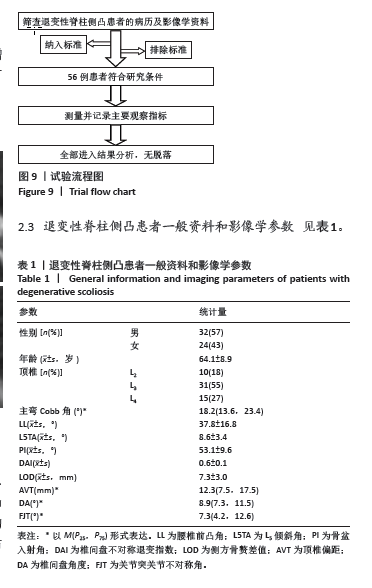
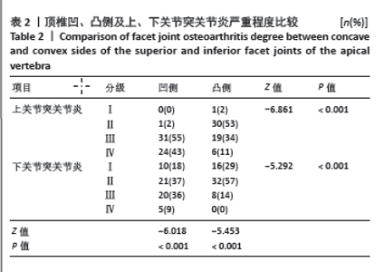
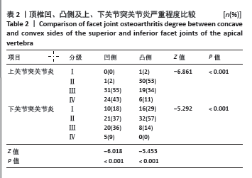
|