Chinese Journal of Tissue Engineering Research ›› 2024, Vol. 28 ›› Issue (12): 1842-1848.doi: 10.12307/2024.012
Previous Articles Next Articles
Biomechanical features of posterior “Y” osteotomy and fixation in treatment of ankylosing spondylitis based on finite element simulation analysis
Zhang Le1, 2, Cao Zhenhua3, 4, Zhang Yunfeng3, Xu Yangyang4, Jin Feng5, Su Baoke6, Wang Lidong7, Wang Xing1, Tong Ling1, Liu Qinghua1, Fang Yuan1, Sha Lirong1, Wang Haiyan1, Li Xiaohe1, Li Zhijun1
- 1Department of Human Anatomy, School of Basic Medicine, Inner Mongolia Medical University (Inner Mongolia Digital Transformation Medical Engineering Technology Research Center), Hohhot 010000, Inner Mongolia Autonomous Region, China; 2Bayannur City Hospital, Bayannur 015000, Inner Mongolia Autonomous Region, China; 3Department of Imaging, Second Affiliated Hospital of Inner Mongolia Medical University, Hohhot 010000, Inner Mongolia Autonomous Region, China;
-
Received:2022-12-01Accepted:2023-03-09Online:2024-04-28Published:2023-08-22 -
Contact:Zhang Yunfeng, Associate chief physician, Department of Imaging, Second Affiliated Hospital of Inner Mongolia Medical University, Hohhot 010000, Inner Mongolia Autonomous Region, China Li Xiaohe, Professor, Doctoral supervisor, Department of Human Anatomy, School of Basic Medicine, Inner Mongolia Medical University (Inner Mongolia Digital Transformation Medical Engineering Technology Research Center), Hohhot 010000, Inner Mongolia Autonomous Region, China -
About author:Zhang Le, Master candidate, Department of Human Anatomy, School of Basic Medicine, Inner Mongolia Medical University (Inner Mongolia Digital Transformation Medical Engineering Technology Research Center), Hohhot 010000, Inner Mongolia Autonomous Region, China; Bayannur City Hospital, Bayannur 015000, Inner Mongolia Autonomous Region, China Cao Zhenhua, MD, Associate chief physician, Department of Imaging, Second Affiliated Hospital of Inner Mongolia Medical University, Hohhot 010000, Inner Mongolia Autonomous Region, China; School of Biological and Medical Engineering, Beihang University, Beijing 100083, China -
Supported by:Natural Science Foundation of Inner Mongolia Autonomous Region, No. 2021MS08086 (to WHY); 2022 Inner Mongolia Autonomous Region Health Science and Technology Plan Project, No. 202201188 (to WHY); “Achievement Transformation” Project of Inner Mongolia Medical University in 2020, No. YKD2020CGZH009 (to WHY); Inner Mongolia Natural Science Foundation, No. 2020MS08124 (to LXH); “Grassland Talents” Project Youth Innovation and Entrepreneurship Talent Program of Inner Mongolia Autonomous Region, No. 2020 (to LXH); Follow-Up Scientific Research Project of Inner Mongolia Medical University, No. 2020 (to LXH); Inner Mongolia Science and Technology Plan Project, No. 2019GG115 (to LZJ); 2021 Mongolian Medicine Collaborative Innovation Center Scientific Research Project of Inner Mongolia Autonomous Region (to LXH); 2021 Zhiyuan Talent Project of Inner Mongolia Medical University (to LXH); 2021 School-Level Key Scientific Research Project of Inner Mongolia Medical University, No. YKD2021ZD001 (to LXH); Higher Education Innovation Team Development Plan of Inner Mongolia Education Department, No. NMGIRT2227 (to LXH)
CLC Number:
Cite this article
Zhang Le, Cao Zhenhua, Zhang Yunfeng, Xu Yangyang, Jin Feng, Su Baoke, Wang Lidong, Wang Xing, Tong Ling, Liu Qinghua, Fang Yuan, Sha Lirong, Wang Haiyan, Li Xiaohe, Li Zhijun. Biomechanical features of posterior “Y” osteotomy and fixation in treatment of ankylosing spondylitis based on finite element simulation analysis[J]. Chinese Journal of Tissue Engineering Research, 2024, 28(12): 1842-1848.
share this article
Add to citation manager EndNote|Reference Manager|ProCite|BibTeX|RefWorks

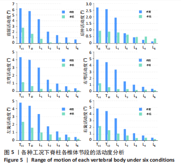
2.1 脊柱各椎体节段的活动度分析 有限元仿真得到脊柱各椎体节段的活动度,如图5所示。术前模型在6种工况下的最大活动度分别为6.16°(前屈)、2.67°(后伸)、4.32°(左弯)、4.78°(右弯)、4.74°(左旋)和4.80°(右旋),术后模型在6种工况下的最大活动度分别为2.70°(前屈)、0.92°(后伸)、1.17°(左弯)、1.21°(右弯)、2.32°(左旋)和2.14°(右旋)。术前模型在6种工况下的活动度多数大于术后模型,且椎体节段越靠下活动度越小。"

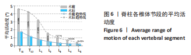
将前屈、后伸、左弯、右弯、左旋和右旋工况下的平均活动度作为评价脊柱运动的能力,以区分运动能力在椎体节段之间的差异,如图6所示。术前模型运动能力在T11-L5椎体节段分别为4.58°,4.22°,3.18°,1.54°,1.02°,0.69°和0.40°,术后模型运动能力在T11-L5椎体节段分别为1.74°,1.14°,0.70°,0.48°,0.27°,0.30°和0.18°,均呈现逐渐递减趋势。根据术前术后差异计算活动度损失比例,分别为T11(61.88%)、T12(72.83%)、L1(77.95%)、L2(68.7%)、L3(72.77%)、L4(55.54%)和L5(54.52%)。"
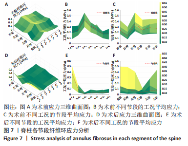
2.2 脊柱各椎间盘的应力分析 为了评估脊柱手术对腰椎的力学影响,需要了解手术前后椎间盘的力学变化,如图7所示,得到脊柱各节段纤维环应力图。图7A为术前6种工况下各节段的纤维环应力图,该三维曲面在左弯工况和L1-L2节段交界处达到峰值(0.55 MPa),为了更直观地了解应力趋势,将各工况应力及各节段应力平均以观测应力在单方面的变化趋势;图7B的平均应力表现为先上升后下降的趋势,在L1-L2节段应力最大(0.42 MPa);图7C的平均应力表现为起伏状,分别在后伸工况下达到最低(0.15 MPa),在左弯工况下达到最高(0.34 MPa);图7D为术后6种工况下各节段的纤维环应力图,该三维曲面在前屈工况和T11-T12节段交界处达到峰值(0.50 MPa);图7E的平均应力表现为逐渐下降的趋势,在T11-T12节段应力最大(0.37 MPa);图7F的平均应力表现为平缓的趋势,其中右弯工况下的应力最小(0.06 MPa)。"

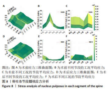
脊柱各节段的髓核应力情况如图8所示,其中图8A为术前6种工况下各节段的髓核应力图,该三维曲面在左旋工况和L1-L2节段的交界处达到峰值(0.052 MPa);图8B的平均应力表现为先上升后下降的趋势,在L1-L2节段应力最大(0.040 MPa);图8C的平均应力表现为起伏状,分别在右弯工况下达到最低(0.01 MPa),在左旋工况下达到最高(0.021 MPa);图8D为术后6种工况下各节段的髓核应力图,该三维曲面在前屈工况和T11-T12节段交界处达到峰值(0.015 MPa);图8E的平均应力表现为先减小后增大的趋势,在T11-T12节段应力最大(0.012 MPa);图8F的平均应力表现为平缓的趋势,无较大差异。"

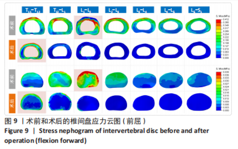
为了直观地看出应力在椎间盘上的表现,得到术前及术后的椎间盘云图,如图9所示,以前屈工况下的椎间盘应力为例,其中最大应力所在的椎间盘和髓核均以红色阴影示出。与图7,8结果一致,术前模型在L1-L2椎间盘表现出最大应力,术后模型在T11-T12椎间盘表现出最大应力。"

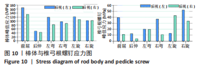
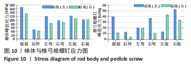
2.3 钉棒系统应力 仿真得到棒体和椎弓根螺钉的应力值,如图10所示。6个工况下,左侧棒体的峰值应力均大于右侧棒体,其中6种工况下最大的峰值应力分别是前屈(166.67 MPa)、后伸(49.02 MPa)、左弯(118.13 MPa)、右弯(96.05 MPa)、左旋(120.88 MPa)和右旋(102.34 MPa);椎弓根螺钉的峰值应力在左右两侧并没有表现出一致的趋势,其中6种工况的最大峰值应力分别为前屈(39.46 MPa)、后伸(12.59 MPa)、左弯(20.11 MPa)、右弯(36.87 MPa)、左旋(42.78 MPa)和右旋(52.05 MPa)。棒体的应力远大于椎弓根螺钉应力。"
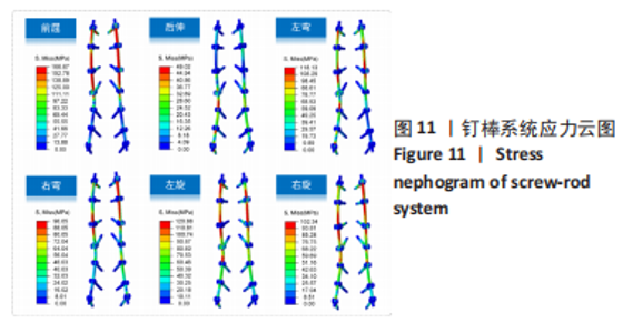
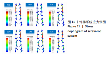
为了更直观地了解钉棒系统的应力情况,得到了钉棒系统的应力云图,如图11所示。前屈工况下,峰值应力主要集中于棒体的上半部分,且最上端螺钉应力大于其他螺钉;后伸工况下,峰值应力主要集中于棒体中段偏下部分,峰值应力小于前屈工况;在左弯和右弯工况下,明显表现出一侧棒体应力明显,另一侧棒体底端应力较小,最大应力出现在棒体的上半部分;在左旋和右旋工况下,两侧棒体均表现出相似的应力趋势,最大应力集中在棒体的上半部分。6种工况下,棒体的应力集中主要出现在上端,椎弓根螺钉的应力集中均出现在根部,这些地方应为断钉断棒的易发生区域。"
| [1] CARON T, BRANSFORD R, NGUYEN Q, et al. Spine fractures in patients with ankylosing spinal disorders. Spine (Phila Pa 1976). 2010;35(11):458-464. [2] TAN S, YAO JH, FLYNN JA, et al. Zygapophyseal Joint Fusion in Ankylosing Spondylitis Assessed by Computed Tomography: Associations with Syndesmophytes and Spinal Motion. J Rheumatol. 2017;44(7):1004-1010. [3] MARTINDALE J, SHUKLA R, GOODACRE J. The impact of ankylosing spondylitis/axial spondyloarthritis on work productivity. Best Pract Res Clin Rheumatol. 2015;29(3): 512-523. [4] PRAY C, FEROZ NI, HAROON NN. Bone Mineral Density and Fracture Risk in Ankylosing Spondylitis: A Meta-Analysis. Calcif Tissue Int. 2017;101(2):182-192. [5] MIN Y, HUI-YUN G, HOU-CHENG Z, et al. The surgical treatment strategies for thoracolumbar spine fractures with ankylosing spondylitis: a case report. BMC Surg. 2019;19(1):99. [6] REINHOLD M, KNOP C, KNEITZ C, et al. Spine Fractures in Ankylosing Diseases: Recommendations of the Spine Section of the German Society for Orthopaedics and Trauma (DGOU). Global Spine J. 2018;8(2 Suppl):56-68. [7] SHEN FH, SAMARTZIS D. Surgical management of lower cervical spine fracture in ankylosing spondylitis. J Trauma. 2006;61(4):1005-1009. [8] LINDTNER RA, KAMMERLANDER C, GOETZEN M, et al. Fracture reduction by postoperative mobilisation for the treatment of hyperextension injuries of the thoracolumbar spine in patients with ankylosing spinal disorders. Arch Orthop Trauma Surg. 2017;137(4):531-541. [9] RUSTAGI T, DRAZIN D, ONER C, et al. Fractures in Spinal Ankylosing Disorders: A Narrative Review of Disease and Injury Types, Treatment Techniques, and Outcomes. J Orthop Trauma. 2017;31(Suppl 4):57-74. [10] DEBARGE R, DEMEY G, ROUSSOULY P. Radiological analysis of ankylosing spondylitis patients with severe kyphosis before and after pedicle subtraction osteotomy. Eur Spine J. 2010;19(1):65-70. [11] QIAN BP, WANG XH, QIU Y, et al. The influence of closing-opening wedge osteotomy on sagittal balance in thoracolumbar kyphosis secondary to ankylosing spondylitis: a comparison with closing wedge osteotomy. Spine (Phila Pa 1976). 2012;37(16): 1415-1423. [12] DEBARGE R, DEMEY G. ROUSSOULY P. Sagittal balance analysis after pedicle subtraction osteotomy in ankylosing spondylitis. Eur Spine J. 2011;20(Suppl 5):619-625. [13] BESS S, HARRIS JE, TURNER AW, et al. The effect of posterior polyester tethers on the biomechanics of proximal junctional kyphosis: a finite element analysis. J Neurosurg Spine. 2017;26(1):125-133. [14] CAHILL PJ, WANG W, ASGHAR J, et al. The Use of a Transition Rod May Prevent Proximal Junctional Kyphosis in the Thoracic Spine Following Scoliosis Surgery: A Finite Element Analysis. Spine (Phila Pa 1976). 2012;37(12):687-695. [15] PEI B, LU D, WU X, et al. Effects of Growing Rod Technique with Different Surgical Modes and Growth Phases on the Treatment Outcome of Early Onset Scoliosis: A 3-D Finite Element Analysis. Int J Environ Res Public Health. 2022;19(4):2057. [16] PEI B, LU D, WU X, et al. Kinematic and biomechanical responses of the spine to distraction surgery in children with early onset scoliosis: A 3-D finite element analysis. Front Bioeng Biotechnol. 2017;10:933341. [17] LIU Y, WANG Z, LIU M, et al. Biomechanical influence of the surgical approaches, implant length and density in stabilizing ankylosing spondylitis cervical spine fracture. Sci Rep. 2021;11(1):6023. [18] MUHEREMU A, LI H, MA JY, et al. Establishment of a three-dimensional finite element model of severe kyphotic deformity secondary to ankylosing spondylitis. J Int Med Res. 2017;45(2):639-646. [19] ZHANG TY, WANG YH, ZHANG PX, et al. Different fixation pattern for thoracolumbar fracture of ankylosing spondylitis: A finite element analysis. PloS One. 2021;16(4): e0250009. [20] ROBINSON Y, LISON ALMKVIST V, OLERUD C, et al. Finite Element Analysis of Long Posterior Transpedicular Instrumentation for Cervicothoracic Fractures Related to Ankylosing Spondylitis. Global Spine J. 2018;8(6):570-578. [21] 马涌, 斯刊达尔斯依提, 欧勇, 等. 不同截骨方式修复强直性脊柱炎后凸畸形的有限元分析[J]. 中国组织工程研究,2017,21(19):3038-3043. [22] 李辉, 张鹏, 尚俊, 等. 去松质骨截骨术与全脊柱截骨术治疗强直性脊柱炎后凸畸形有限元分析[J]. 山西医科大学学报,2019,50(3):368-372. [23] TONG Z, XIAO B, YAN K, et al. Biomechanical Evaluation of the Transcortical and Transpedicular Trajectories for Pedicle Screw Insertion in Thoracolumbar Fracture Fixation for Ankylosing Spondylitis. Front Surg. 2021;8:706597. [24] NIGIL HAROON N, SZABO E, RABOUD JM, et al. Alterations of bone mineral density, bone microarchitecture and strength in patients with ankylosing spondylitis: a cross-sectional study using high-resolution peripheral quantitative computerized tomography and finite element analysis. Arthritis Res Ther. 2015;17:37. [25] FINLEY SM, BRODKE DS, SPINA NT, et al. FEBio finite element models of the human lumbar spine. Comput. Methods Biomech Biomed Eng. 2018;21(6):444-452. [26] WANG JL, PARNIANPOUR M, SHIRAZI-ADL A, et al. Viscoelastic finite-element analysis of a lumbar motion segment in combined compression and sagittal flexion. Spine (Phila Pa 1976). 2000;25(3):310-318. [27] BURSTEIN AH, REILLY DT, MARTENS M. Aging of bone tissue: mechanical properties. J Bone Joint Surg Am. 1976;58(1):82-86. [28] SHIRAZI-ADL SA, SHRIVASTAVA SC, AHMED AM. Stress Analysis of the Lumbar Disc-Body Unit in Compression A Three-Dimensional Nonlinear Finite Element Study. Spine (Phila Pa 1976). 1984;9(2):120-134. [29] KURUTZ M, OROSZVÁRY L. Finite element analysis of weightbath hydrotraction treatment of degenerated lumbar spine segments in elastic phase J. Biomech. 2010;43(3):433-441. [30] CAI XY, SANG DC, YUCHI CX, et al. Using finite element analysis to determine effects of the motion loading method on facet joint forces after cervical disc degeneration. Comput Biol Med. 2020;116:103519. [31] AGAWAL A, ZAKERI A, AGARWAL AK, et al. Distraction magnitude and frequency affects the outcome in juvenile idiopathic patients with growing rods: Finite element study using a representative scoliotic spine model. Spine J. 2015;15(8):1848-1855. [32] WHITE AAI, PANJABI MM. Clinical Biomechanics of the Spine. Lippincott-Raven,1992. [33] MATSUKAWA K, YATO Y, IMABAYASHI H, et al. Biomechanical evaluation of the fixation strength of lumbar pedicle screws using cortical bone trajectory: a finite element study. J Neurosurg Spine. 2015;23(4):471-478. [34] WERNER BC, SAMARTZIS D, SHEN FH. Spinal fractures in patients with ankylosing spondylitis: etiology, diagnosis, and management. J Am Acad Orthop Surg. 2016; 24(4):241-249. [35] LEONE A, MARINO M, DELLATTI C, et al. Spinal fractures in patients with ankylosing spondylitis. Rheumatol Int. 2016;36(10):1335-1346. [36] CHARLES YP, BUY X, GANGI A, et al Fracture in ankylosing spondylitis after minor trauma: radiological pitfalls and treatment by percutaneous instrumentation. A case report. Orthop Traumatol Surg Res. 2013;99(1):115-119. [37] WESTERVELD LA, VERLAAN JJ, ONER FC. Spinal fractures in patients with ankylosing spinal disorders: a systematic review of the literature on treatment, neurological status and complications. Eur Spine J. 2009;18(2):145-156. [38] 李民, 周灵, 汪桂珍, 等. 不同腰椎生理曲度下牵引的三维有限元分析[J]. 中国组织工程研究,2020,24(20):3162-3167. [39] YAMAMOTO I, PANJABI M, CRISCO T, et al. Three-dimensional movements of the whole lumbar spine. Spine. 1989;14(1):1256-1260. [40] XIAO Z, WANG L, GONG H, et al. Biomechanical evaluation of three surgical scenarios of posterior lumbar interbody fusion by finite element analysis. Biomed Eng Online. 2012;11(1):31. [41] Agarwal A, Kodigudla M, Kelkar A, et al. Towards a validated patient-specific computational modeling framework to identify failure regions in traditional growing rods in patients with early onset scoliosis. N Am Spine Soc J. 2021;5(5):100043. [42] 李辉. 强直性脊柱炎后凸矫形生物力学研究和临床疗效分析[D]. 乌鲁木齐: 新疆医科大学.2017. |
| [1] | Kang Zhijie, Cao Zhenhua, Xu Yangyang, Zhang Yunfeng, Jin Feng, Su Baoke, Wang Lidong, Tong Ling, Liu Qinghua, Fang Yuan, Sha Lirong, Liang Liang, Li Mengmeng, Du Yifei, Lin Lin, Wang Haiyan, Li Xiaohe, Li Zhijun. Finite element model establishment and stress analysis of lumbar-sacral intervertebral disc in ankylosing spondylitis [J]. Chinese Journal of Tissue Engineering Research, 2024, 28(6): 840-846. |
| [2] | Leng Siyi, Pu Rui, Chen Ziyang, Yang Qihang, Song Yongjing, Liu Hui. Roles of exercise intervention in intestinal flora in autoimmune diseases [J]. Chinese Journal of Tissue Engineering Research, 2023, 27(32): 5219-5226. |
| [3] | Jia Hongsheng, Li Xianlin, Cai Lei, Zhang Chongfeng, Chen Zuchuang, Zhang Ye. Network meta-analysis of different drugs for the treatment of ankylosing spondylitis [J]. Chinese Journal of Tissue Engineering Research, 2021, 25(33): 5404-5412. |
| [4] | Xie Jiang, Dai Jie, Li Hui, Zhu Xu. Biomechanical analysis of sagittal balance restoration of ankylosing kyphosis based on pelvic sagittal parameters [J]. Chinese Journal of Tissue Engineering Research, 2021, 25(30): 4762-4766. |
| [5] | Wang Zhuo, Shi Zhongfeng, Wang Fengyun, Li Weidong, Han Liang. Mechanism of Hanshi Bi granules in the treatment of ankylosing spondylitis based on network pharmacology [J]. Chinese Journal of Tissue Engineering Research, 2020, 24(11): 1738-1744. |
| [6] | Zhou Jin, Fu Lin, Zhou Zhen, Li Qiurong. One-year evaluation of recombinant human tumor necrosis factor-alpha receptor II decrement combined with thalidomide increment in the treatment of active ankylosing spondylitis [J]. Chinese Journal of Tissue Engineering Research, 2019, 23(35): 5664-5669. |
| [7] | Xiong Chunxiang, , Wei Xiaochun, Yin Dong, Huang Yu, Du Chang, Mo Bingfeng. Susceptibility of ankylosing spondylitis early-onset hip ankylosis and human leukocyte antigen-B27 subtypes [J]. Chinese Journal of Tissue Engineering Research, 2019, 23(23): 3710-3715. |
| [8] | Shen Cheng-kai, Lü Cheng-yu, Wang Ying-zhen, Zhang Hai-ning, Sui Ai-hua, Meng Fei . Association between interleukin-10 promoter regions gene polymorphisms and susceptibility of ankylosing spondylitis [J]. Chinese Journal of Tissue Engineering Research, 2013, 17(20): 3793-3800. |
| [9] | Li Bei-bei, Ren Cui-ping, Li Ying, Shi Xiao-ying, Ren Xian. Magnetic resonance diffusion weighted imaging sequence shows the best b value of sacroiliac joint imaging of healthy volunteers [J]. Chinese Journal of Tissue Engineering Research, 2013, 17(17): 3124-3131. |
| Viewed | ||||||
|
Full text |
|
|||||
|
Abstract |
|
|||||