Chinese Journal of Tissue Engineering Research ›› 2015, Vol. 19 ›› Issue (49): 7885-7889.doi: 10.3969/j.issn.2095-4344.2015.49.003
Previous Articles Next Articles
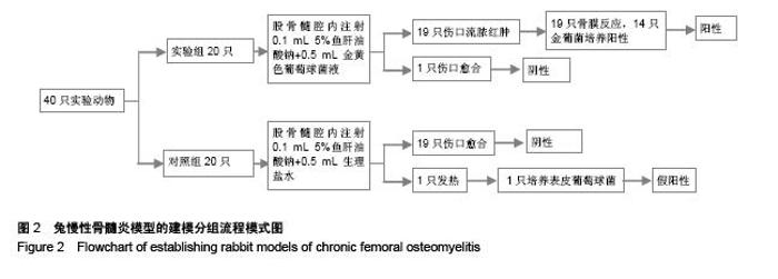
Establishment of rabbit models of chronic femoral osteomyelitis
Chen Jin-shui1, Liao Zhao-shan2, Lin Song-qing1, Lu Qiao-ping2, Wang Ben-hai1, Huang Wen-qi2, Lin Yong-yang2, Zhang Hui-hao2
- 1First Department of Orthopedics, Fuzhou General Hospital of Nanjing Military Command, Fuzhou 350025, Fujian Province, China; 2College of Osteopathy, Fujian University of Traditional Chinese Medicine, Fuzhou 350108, Fujian Province, China
-
Received:2015-09-07Online:2015-11-30Published:2015-11-30 -
Contact:Lin Song-qing, Master, Associate chief physician, First Department of Orthopedics, Fuzhou General Hospital of Nanjing Military Command, Fuzhou 350025, Fujian Province, China -
About author:Chen Jin-shui, M.D., Attending physician, First Department of Orthopedics, Fuzhou General Hospital of Nanjing Military Command, Fuzhou 350025, Fujian Province, China -
Supported by:Medical and Health Scientific Research Foundation of Nanjing Military Area Command in 2012, China, No. 12MA103; the Natural Science Foundation of Fujian Province of China, No. 2013J01344
CLC Number:
Cite this article
Chen Jin-shui, Liao Zhao-shan, Lin Song-qing, Lu Qiao-ping, Wang Ben-hai, Huang Wen-qi, Lin Yong-yang, Zhang Hui-hao. Establishment of rabbit models of chronic femoral osteomyelitis[J]. Chinese Journal of Tissue Engineering Research, 2015, 19(49): 7885-7889.
share this article
"

"


"

"
| [1] 侯喜君,王春华,廉红星,等. β-磷酸三钙煅烧骨负载抗生素治疗慢性骨髓炎的实验研究[J]中国组织工程研究,2012,16(51): 9518-9522.[2] Winters R,Tatum SA 3rd. Chronic nonbacterial osteomyelitis. Curr Opin Otolaryngol Head Neck Surg. 2014;22(4):332-335.[3] Conterno LO, da Silva Filho CR. Antibiotics for treating chronic osteomyelitis in adults.Cochrane Database Syst Rev. 2009;(3): CD004439.[4] Jae JJ, Ho SL, Young RC, et al. Surgical treatment of non -diabetic chronic osteomyelitis involving the foot and ankle. Foot & AnkleInter.2012;33(2):128-132.[5] 易远历,杨茂伟,王凯,等.VSD联合骨移植替代材料植入治疗跟骨慢性骨髓炎疗效观察[J].中国骨与关节损伤杂志,2013, 28(12): 1210-1211.[6] Roderick MR, Ramanan AV. Chronic recurrent multifocal osteomyelitis. Adv Exp Med Biol.2013;764:99-107.[7] Marais LC, Ferreira N. Bone transport through an induced membrane in the management of tibial bone defects resulting from chronic osteomyelitis. Strategies Trauma Limb Reconstr. 2015;10(1):27-33.[8] Spellberg B, Lipsky BA. Systemic antibiotic therapy for chronic osteomyelitis in adults. Clin Infect Dis. 2012;54(3): 393-407. [9] Panteli M,Puttaswamaiah R,Lowenberg DW,et al.Malignant Transformationbin Chronic Osteomyelitis: Recognition and Principles of Management. J Am Acad Orthop Surg. 2014; 22(9):586-594. [10] Senneville E, Nguyen S.Current pharmacotherapy options for osteomyelitis: convergences, divergences and lessons to be drawn.Expert Opin Pharmacother. 2013;14(6):723-734. [11] Inanmaz ME, Uslu M, Isik C, et al.Extracorporeal shockwave increases the effectiveness of systemic antibiotic treatment in implant-related chronic osteomyelitis: experimental study in a rat model.J Orthop Res. 2014;32(6):752-756. [12] Vergidis P, Schmidt-Malan SM, Mandrekar JN, et al. Comparative activities of vancomycin, tigecycline and rifampin in a rat model of methicillin-resistant Staphylococcus aureus osteomyelitis.J Infect. 2015;70(6):609-615. [13] Gahukamble AD, McDowell A, Post V, et al.Propionibacterium acnes and Staphylococcus lugdunensis cause pyogenic osteomyelitis in an intramedullary nail model in rabbits.J Clin Microbiol. 2014;52(5):1595-1606. [14] 闻一新,周田华,徐永清,等.兔胫骨金黄色葡萄球菌慢性骨髓炎模型的制备[J].实用骨科杂志,2013,19(11):1001-1003.[15] Ozturan KE, Yucel I, Kocoglu E, et al.Efficacy of moxifloxacin compared to teicoplanin in the treatment of implant-related chronic osteomyelitis in rats.J Orthop Res. 2010;28(10): 1368-1372.[16] Norden CW, Shinners E, Niederriter K.Clindamycin treatment of experimental chronic osteomyelitis due to Staphylococcus aureus.J Infect Dis. 1986;153(5):956-959. [17] 黄金亮,唐辉,范新宇.等.慢性难治性骨髓炎动物模型制备与鉴定[J]. 西南国防医药,2012,22(3):236-239. [18] Odekerken JC,Arts JJ,Surtel DA,et al. A rabbit osteomyelitis model for the longitudinal assessment of early post -operative implant infections. J Orthop Surg Res.2013;4(8):38.[19] 陈孝平.外科常用实验方法及动物模型的建立[M].北京:人民卫生出版社,2003:380.[20] 杜颖,徐文瑜,任珺,等.热原检查用家兔正常体温的统计分析[J]. 实验动物科学与管理,2003,20(1):17-19.[21] Kaarsemaker S,Walenkamp GH,vd Bogaard AE.New model for chronic osteomyelitis with staphylococcus aureus in sheep. Clin Orthop Relat Res.1997;339:246 -252.[22] Petty W,Spanier S,Shuster JJ,et al.The influence of skeletal implants on incidence of infection. Experiments in a canine model.J Bone Joint Surg Am.1985;67(8):1236-1244.[23] 刘立峰,邹林,蔡锦方.创伤性骨髓炎[J].中国矫形外科杂志, 2009, 17(10):766-768.[24] Horn J, Schlegel U, Krettek C,et al.Infection resistance of unreamed solid, hollow slotted and cannulated intramedullary nails: An in-vivo experimental comparison.J Orthop Res.2005; 23(4):810-815.[25] 高翠芳,李晓泉. 5%鱼肝油酸钠对慢性骨髓炎造模的影响[J].中医正骨,2002,14(9):12-13.[26] Helbig L,Simank HG,Lorenz H,et al,Establishment of a new methicillin resistant Staphylococcus aureus animal model of osteomyelitis. Int Orthop.2014;38(4):891-897. |
| [1] | Chen Ziyang, Pu Rui, Deng Shuang, Yuan Lingyan. Regulatory effect of exosomes on exercise-mediated insulin resistance diseases [J]. Chinese Journal of Tissue Engineering Research, 2021, 25(25): 4089-4094. |
| [2] | Chen Yang, Huang Denggao, Gao Yuanhui, Wang Shunlan, Cao Hui, Zheng Linlin, He Haowei, Luo Siqin, Xiao Jingchuan, Zhang Yingai, Zhang Shufang. Low-intensity pulsed ultrasound promotes the proliferation and adhesion of human adipose-derived mesenchymal stem cells [J]. Chinese Journal of Tissue Engineering Research, 2021, 25(25): 3949-3955. |
| [3] | Yang Junhui, Luo Jinli, Yuan Xiaoping. Effects of human growth hormone on proliferation and osteogenic differentiation of human periodontal ligament stem cells [J]. Chinese Journal of Tissue Engineering Research, 2021, 25(25): 3956-3961. |
| [4] | Sun Jianwei, Yang Xinming, Zhang Ying. Effect of montelukast combined with bone marrow mesenchymal stem cell transplantation on spinal cord injury in rat models [J]. Chinese Journal of Tissue Engineering Research, 2021, 25(25): 3962-3969. |
| [5] | Gao Shan, Huang Dongjing, Hong Haiman, Jia Jingqiao, Meng Fei. Comparison on the curative effect of human placenta-derived mesenchymal stem cells and induced islet-like cells in gestational diabetes mellitus rats [J]. Chinese Journal of Tissue Engineering Research, 2021, 25(25): 3981-3987. |
| [6] | Hao Xiaona, Zhang Yingjie, Li Yuyun, Xu Tao. Bone marrow mesenchymal stem cells overexpressing prolyl oligopeptidase on the repair of liver fibrosis in rat models [J]. Chinese Journal of Tissue Engineering Research, 2021, 25(25): 3988-3993. |
| [7] | Liu Jianyou, Jia Zhongwei, Niu Jiawei, Cao Xinjie, Zhang Dong, Wei Jie. A new method for measuring the anteversion angle of the femoral neck by constructing the three-dimensional digital model of the femur [J]. Chinese Journal of Tissue Engineering Research, 2021, 25(24): 3779-3783. |
| [8] | Meng Lingjie, Qian Hui, Sheng Xiaolei, Lu Jianfeng, Huang Jianping, Qi Liangang, Liu Zongbao. Application of three-dimensional printing technology combined with bone cement in minimally invasive treatment of the collapsed Sanders III type of calcaneal fractures [J]. Chinese Journal of Tissue Engineering Research, 2021, 25(24): 3784-3789. |
| [9] | Qian Xuankun, Huang Hefei, Wu Chengcong, Liu Keting, Ou Hua, Zhang Jinpeng, Ren Jing, Wan Jianshan. Computer-assisted navigation combined with minimally invasive transforaminal lumbar interbody fusion for lumbar spondylolisthesis [J]. Chinese Journal of Tissue Engineering Research, 2021, 25(24): 3790-3795. |
| [10] | Hu Jing, Xiang Yang, Ye Chuan, Han Ziji. Three-dimensional printing assisted screw placement and freehand pedicle screw fixation in the treatment of thoracolumbar fractures: 1-year follow-up [J]. Chinese Journal of Tissue Engineering Research, 2021, 25(24): 3804-3809. |
| [11] | Shu Qihang, Liao Yijia, Xue Jingbo, Yan Yiguo, Wang Cheng. Three-dimensional finite element analysis of a new three-dimensional printed porous fusion cage for cervical vertebra [J]. Chinese Journal of Tissue Engineering Research, 2021, 25(24): 3810-3815. |
| [12] | Wang Yihan, Li Yang, Zhang Ling, Zhang Rui, Xu Ruida, Han Xiaofeng, Cheng Guangqi, Wang Weil. Application of three-dimensional visualization technology for digital orthopedics in the reduction and fixation of intertrochanteric fracture [J]. Chinese Journal of Tissue Engineering Research, 2021, 25(24): 3816-3820. |
| [13] | Sun Maji, Wang Qiuan, Zhang Xingchen, Guo Chong, Yuan Feng, Guo Kaijin. Development and biomechanical analysis of a new anterior cervical pedicle screw fixation system [J]. Chinese Journal of Tissue Engineering Research, 2021, 25(24): 3821-3825. |
| [14] | Lin Wang, Wang Yingying, Guo Weizhong, Yuan Cuihua, Xu Shenggui, Zhang Shenshen, Lin Chengshou. Adopting expanded lateral approach to enhance the mechanical stability and knee function for treating posterolateral column fracture of tibial plateau [J]. Chinese Journal of Tissue Engineering Research, 2021, 25(24): 3826-3827. |
| [15] | Zhu Yun, Chen Yu, Qiu Hao, Liu Dun, Jin Guorong, Chen Shimou, Weng Zheng. Finite element analysis for treatment of osteoporotic femoral fracture with far cortical locking screw [J]. Chinese Journal of Tissue Engineering Research, 2021, 25(24): 3832-3837. |
| Viewed | ||||||
|
Full text |
|
|||||
|
Abstract |
|
|||||