Chinese Journal of Tissue Engineering Research ›› 2015, Vol. 19 ›› Issue (48): 7747-7751.doi: 10.3969/j.issn.2095-4344.2015.48.007
Previous Articles Next Articles
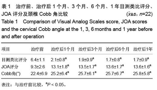
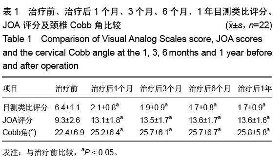
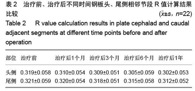
The clinical application of zero notch anterior cervical fusion plate (Zero-P) on anterior cervical decompression and bone fusion
Cheng Jun-jie, Dai Jie, Ma Yuan, Tian Hui-zhong
- First Department of Spine Surgery, the Sixth Affiliated Hospital of Xinjiang Medical University, Urumqi 830002, Xinjiang Uygur Autonomous Region, China
-
Received:2015-09-13Online:2015-11-26Published:2015-11-26 -
Contact:Ma Yuan, Master, Chief physician, First Department of Spine Surgery, the Sixth Affiliated Hospital of Xinjiang Medical University, Urumqi 830002, Xinjiang Uygur Autonomous Region, China -
About author:Cheng Jun-jie, Attending physician, First Department of Spine Surgery, the Sixth Affiliated Hospital of Xinjiang Medical University, Urumqi 830002, Xinjiang Uygur Autonomous Region, China
CLC Number:
Cite this article
Cheng Jun-jie, Dai Jie, Ma Yuan, Tian Hui-zhong. The clinical application of zero notch anterior cervical fusion plate (Zero-P) on anterior cervical decompression and bone fusion[J]. Chinese Journal of Tissue Engineering Research, 2015, 19(48): 7747-7751.
share this article
"
"


"
| [1] Park Y, Maeda T, Cho W, et al. Comparison of anterior cervical fusion after two-level discectomy or single-level corpectomy: sagittal alignment, cervical lordosis, graft collapse, and adjacent-level ossification. Spine J. 2010;10(3): 193-199.
[2] Yorimitsu E, Chiba K, Toyama Y, et al. Long-term outcomes of standard discectomy for lumbar disc herniation: a follow-up study of more than 10 years. Spine (Phila Pa 1976). 2001; 26(6):652-657.
[3] 冯虎,马志兵,齐祥如,等.颈前路钢板固定位置与相邻节段退变之间关系的研究[J].中国骨与关节损伤杂志,2011,26(1):1-4.
[4] 金明熙,谢林.经口咽入路行上位颈椎手术[J].中国脊柱脊髓杂志, 2001,11(1):12-15.
[5] 金大地,王健,瞿东滨.颈椎前路手术早期并发症原因分析及对策[J].中华骨科杂志, 2005,25(2):102-106.
[6] 陈建明,张成程,庄颖,等.经颈椎前方联合胸骨柄劈开术治疗颈胸段椎体结核[J].西南国防医药,2013,23(8):875-876.
[7] 陈智,黄轩,李凤宁,等.颈椎前路术后吞咽困难的相关因素分析[J]. 中国脊柱脊髓杂志,2013,22(11):979-983.
[8] 江兵,朱成润,曹燕庆,等.颈椎前路手术早期并发症及防范措施[J]. 疑难病杂志, 2014,13(1): 67-69.
[9] 孙韶华,方剑利,马维虎,等.经口咽入路钢板内固定治疗不稳定性寰椎骨折[J].中国骨伤,2013,26(1):81-84.
[10] 张秋航,严波,郭宏川,等.内镜经口入路齿状突切除治疗颅底凹陷[J].中国微侵袭神经外科杂志,2014,19(1):13-17.
[11] 陈超君,汪四花,马姚静. 6例经口咽松解后路融合治疗陈旧性 Ⅱ 型齿状突骨折的护理[J].中华护理杂志, 2013,48(12):1121-1122.
[12] 王达义,常巍,尚晖,等.经口咽前路控制性松解复位+后路枕颈融合内固定术治疗颅底凹陷症[J].骨科,2015,6(4):177-182.
[13] 谢恩,郝定均,吴起,等.经前路部分切除胸骨上端入路手术治疗上胸椎病变[J].中国脊柱脊髓杂志,2013,23(2):135-139.
[14] 谢恩, 郝定均.前路胸骨上端部分潜行切除入路治疗上胸椎病变的手术技巧研究[J].美中国际创伤杂志,2013,11(5):3-4.
[15] 王子璋,梁秦龙,李继锋,等.胸骨前入路与锁骨下入路腔镜辅助下甲状腺切除术对比分析[J].现代肿瘤医学,2013,21(4): 743-746.
[16] 江兵,朱成润,曹燕庆,等.颈椎前路手术早期并发症及防范措施[J]. 疑难病杂志,2014,13(1):67-69.
[17] 李强,陈向阳,冯虎,等.颈椎前路手术早期并发症防治探讨[J]. 颈腰痛杂志, 2013,34(2): 124-126.
[18] 魏鲁青,曾昭勇,张健平,等. Zero-P颈椎前路融合治疗颈椎病 8 例临床观察[J].当代医学,2013,19(34):84-85.
[19] 陈波,金格勒.颈椎前路手术术后吞咽困难的研究进展[J].中国脊柱脊髓杂志,2015,25(1):80-84.
[20] 王宵光,王淙清.颈椎前路钢板手术后期并发症的预防[J].中国骨与关节损伤杂志, 2013, 28(1): 41-42.
[21] 缪锦浩,匡勇,陈德玉,等.颈前路减压零切迹椎间植骨融合内固定系统治疗颈椎病的早期疗效分析[J].中国脊柱脊髓杂志, 2012, 22(6):536-540.
[22] 陈凯,陈超,朱晓东,等. Zero-P用于颈椎前路手术的早期临床疗效分析[J].脊柱外科杂志,2012,10(3):155-158.
[23] 程彩霞,卢旭华,谢宁,等. Zero-P椎间融合术在治疗颈前路融合术后相邻节段退变性疾病中的应用[J].脊柱外科杂志,2012, 10(3):152-154.
[24] 王彬彬,倪斌,谢宁,等.单节段颈椎前路减压术中应用不同内置物的疗效分析[J].脊柱外科杂志,2014,12(4):239-244.
[25] 刘上楼, 顾德毅,徐饶,等.应用Zero-p颈椎融合器cage治疗颈椎外伤合并脊髓型颈椎病[J].中国骨与关节损伤杂志, 2012, 27(1): 85.
[26] 何国雄,欧光信,李文锐,等.颈椎前路潜行减压植骨融合术治疗多节段脊髓型颈椎病32例[J].海南医学,2013,24(4):551-553.
[27] 王刚,吴涛,黄鑫鹏,等. Zero-P椎间融合器颈椎前路置入内固定治疗颈椎病的优势[J].中国组织工程研究, 2014,18(31): 4980-4985.
[28] 庄超,周栋,汤雪明,等.颈前路零切迹椎间融合内固定系统治疗脊髓型颈椎病的疗效分析[J].中国修复重建外科杂志,2015,29(6): 751-755.
[29] 祁敏,梁磊,王新伟,等.颈前路多节段融合术后吞咽困难的原因分析[J].中华骨科杂志,2013, 33(5):467-472.
[30] 于从,谢小华,陈雪萍,等.两种颈椎前路手术致吞咽困难的比较研究及护理[J].中国临床解剖学杂志, 2014,32(4):480-483.
[31] 陈波,金格勒.颈椎前路手术术后吞咽困难的研究进展[J].中国脊柱脊髓杂志,2015, 25(1):80-84. |
| [1] | Chen Ziyang, Pu Rui, Deng Shuang, Yuan Lingyan. Regulatory effect of exosomes on exercise-mediated insulin resistance diseases [J]. Chinese Journal of Tissue Engineering Research, 2021, 25(25): 4089-4094. |
| [2] | Chen Yang, Huang Denggao, Gao Yuanhui, Wang Shunlan, Cao Hui, Zheng Linlin, He Haowei, Luo Siqin, Xiao Jingchuan, Zhang Yingai, Zhang Shufang. Low-intensity pulsed ultrasound promotes the proliferation and adhesion of human adipose-derived mesenchymal stem cells [J]. Chinese Journal of Tissue Engineering Research, 2021, 25(25): 3949-3955. |
| [3] | Yang Junhui, Luo Jinli, Yuan Xiaoping. Effects of human growth hormone on proliferation and osteogenic differentiation of human periodontal ligament stem cells [J]. Chinese Journal of Tissue Engineering Research, 2021, 25(25): 3956-3961. |
| [4] | Sun Jianwei, Yang Xinming, Zhang Ying. Effect of montelukast combined with bone marrow mesenchymal stem cell transplantation on spinal cord injury in rat models [J]. Chinese Journal of Tissue Engineering Research, 2021, 25(25): 3962-3969. |
| [5] | Gao Shan, Huang Dongjing, Hong Haiman, Jia Jingqiao, Meng Fei. Comparison on the curative effect of human placenta-derived mesenchymal stem cells and induced islet-like cells in gestational diabetes mellitus rats [J]. Chinese Journal of Tissue Engineering Research, 2021, 25(25): 3981-3987. |
| [6] | Hao Xiaona, Zhang Yingjie, Li Yuyun, Xu Tao. Bone marrow mesenchymal stem cells overexpressing prolyl oligopeptidase on the repair of liver fibrosis in rat models [J]. Chinese Journal of Tissue Engineering Research, 2021, 25(25): 3988-3993. |
| [7] | Liu Jianyou, Jia Zhongwei, Niu Jiawei, Cao Xinjie, Zhang Dong, Wei Jie. A new method for measuring the anteversion angle of the femoral neck by constructing the three-dimensional digital model of the femur [J]. Chinese Journal of Tissue Engineering Research, 2021, 25(24): 3779-3783. |
| [8] | Meng Lingjie, Qian Hui, Sheng Xiaolei, Lu Jianfeng, Huang Jianping, Qi Liangang, Liu Zongbao. Application of three-dimensional printing technology combined with bone cement in minimally invasive treatment of the collapsed Sanders III type of calcaneal fractures [J]. Chinese Journal of Tissue Engineering Research, 2021, 25(24): 3784-3789. |
| [9] | Qian Xuankun, Huang Hefei, Wu Chengcong, Liu Keting, Ou Hua, Zhang Jinpeng, Ren Jing, Wan Jianshan. Computer-assisted navigation combined with minimally invasive transforaminal lumbar interbody fusion for lumbar spondylolisthesis [J]. Chinese Journal of Tissue Engineering Research, 2021, 25(24): 3790-3795. |
| [10] | Hu Jing, Xiang Yang, Ye Chuan, Han Ziji. Three-dimensional printing assisted screw placement and freehand pedicle screw fixation in the treatment of thoracolumbar fractures: 1-year follow-up [J]. Chinese Journal of Tissue Engineering Research, 2021, 25(24): 3804-3809. |
| [11] | Shu Qihang, Liao Yijia, Xue Jingbo, Yan Yiguo, Wang Cheng. Three-dimensional finite element analysis of a new three-dimensional printed porous fusion cage for cervical vertebra [J]. Chinese Journal of Tissue Engineering Research, 2021, 25(24): 3810-3815. |
| [12] | Wang Yihan, Li Yang, Zhang Ling, Zhang Rui, Xu Ruida, Han Xiaofeng, Cheng Guangqi, Wang Weil. Application of three-dimensional visualization technology for digital orthopedics in the reduction and fixation of intertrochanteric fracture [J]. Chinese Journal of Tissue Engineering Research, 2021, 25(24): 3816-3820. |
| [13] | Sun Maji, Wang Qiuan, Zhang Xingchen, Guo Chong, Yuan Feng, Guo Kaijin. Development and biomechanical analysis of a new anterior cervical pedicle screw fixation system [J]. Chinese Journal of Tissue Engineering Research, 2021, 25(24): 3821-3825. |
| [14] | Lin Wang, Wang Yingying, Guo Weizhong, Yuan Cuihua, Xu Shenggui, Zhang Shenshen, Lin Chengshou. Adopting expanded lateral approach to enhance the mechanical stability and knee function for treating posterolateral column fracture of tibial plateau [J]. Chinese Journal of Tissue Engineering Research, 2021, 25(24): 3826-3827. |
| [15] | Zhu Yun, Chen Yu, Qiu Hao, Liu Dun, Jin Guorong, Chen Shimou, Weng Zheng. Finite element analysis for treatment of osteoporotic femoral fracture with far cortical locking screw [J]. Chinese Journal of Tissue Engineering Research, 2021, 25(24): 3832-3837. |
| Viewed | ||||||
|
Full text |
|
|||||
|
Abstract |
|
|||||