Chinese Journal of Tissue Engineering Research ›› 2014, Vol. 18 ›› Issue (24): 3840-3844.doi: 10.3969/j.issn.2095-4344.2014.24.012
Previous Articles Next Articles
Geometric accuracy of magnetic resonance imaging displaying the inferior alveolar nerve
Deng Wei1, Huang Dai-ying2, Chen Song-ling2, Chen Jing-xin1, Liao Tian-an1
- 1Department of Oral and Maxillofacial Surgery, Hainan Provincial People’s Hospital, Haikou 570001, Hainan Province, China
2Department of Oral and Maxillofacial Surgery, the First Affiliated Hospital, Sun Yat-sen University, Guangzhou 510080, Guangdong Province, China
-
Revised:2014-05-16Online:2014-06-11Published:2014-06-11 -
Contact:Chen Song-ling, M.D., Professor, Department of Oral and Maxillofacial Surgery, the First Affiliated Hospital, Sun Yat-sen University, Guangzhou 510080, Guangdong Province, China -
About author:Deng Wei, M.D., Attending physician, Department of Oral and Maxillofacial Surgery, Hainan Provincial People’s Hospital, Haikou 570001, Hainan Province, China -
Supported by:the National Natural Science Foundation of China, No. 81360172
CLC Number:
Cite this article
Deng Wei, Huang Dai-ying, Chen Song-ling, Chen Jing-xin, Liao Tian-an . Geometric accuracy of magnetic resonance imaging displaying the inferior alveolar nerve[J]. Chinese Journal of Tissue Engineering Research, 2014, 18(24): 3840-3844.
share this article
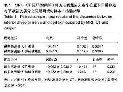
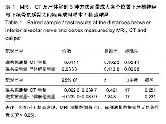
不同方法测得的各位置下颌牙槽神经与皮质骨之间的距离:应用MRI、CT及尸体解剖3种方法测量得到各个位置下牙槽神经与下颌骨皮质骨之间距离的平均值分别为(4.894±1.692),(4.906±1.715),(4.861±1.718) mm。经配对t 检验发现,MRI测量数据与CT、解剖测量数据差异无显著性意义(P > 0.05),见表1。"
| [1] Libersa P, Savignat M, Tonnel A. Neurosensory disturbances of the inferior alveolar nerve: a retrospective study of complaints in a 10-year period. J Oral Maxillofac Surg. 2007; 65(8):1486-1489. [2] Hillerup S. Iatrogenic injury to the inferior alveolar nerve: etiology, signs and symptoms, and observations on recovery. Int J Oral Maxillofac Surg.2008;37(8):704-709. [3] Jeries W, Swinson B, Moles DR, et al. Permanent sensory nerve impairment following third molar surgery: a prospective study. Oral Surg Oral Med Oral Pathol Oral Radiol Endod. 2006;102(4):e1-e7. [4] Seeberger R, Asi Y, Thiele OC, et al. Neurosensory alterations and function of the temporomandibular joint after high oblique sagittal split osteotomy: an alternative technique in orthognathic surgery. Br J Oral Maxillofac Surg. 2013;51(6):536-540. [5] Lin MH, Mau LP, Cochran DL, et al. Risk assessment of inferior alveolar nerve injury for immediate implant placement in the posterior mandible: a virtual implant placement study. J Dent. 2014;42(3):263-270. [6] Selvi F, Dodson TB, Nattestad A, et al. Factors that are associated with injury to the inferior alveolar nerve in high-risk patients after removal of third molars.Br J Oral Maxillofac Surg. 2013;51(8):868-873. [7] Bede SY, Ismael WK, Al-Assaf DA, et al. Inferior alveolar nerve injuries associated with mandibular fractures. J Craniofac Surg. 2012;23(6):1776-1778. [8] Takazakura D, Ueki K, Nakagawa K, et al. A comparison of postoperative hypoesthesia between two types of sagittal split ramus osteotomyand intraoral vertical ramus osteotomy, using the trigeminal somatosensory-evoked potential method. Int J Oral Maxillofac Surg. 2007;36(1):11-14. [9] Shiratori K, Nakamori K, Ueda M, et al. Assessment of the shape of the inferior alveolar canal as a marker for increased risk of injury to the inferior alveolar nerve at third molar surgery: a prospective study. J Oral Maxillofac Surg. 2013; 71(12):2012-2019. [10] Dula K, Mini R, Van der Stelt PF, et al. The radiographic assessment of implant patients: decision-making criteria. Int J Oral Maxillofacial Impl. 2001:16:80-89. [11] Katakam SK, Shankar U, Thakur D, et al. Comparison of orthopantomography and computed tomography image for assessing the relationship between impacted mandibular third molar and mandibular canal. J Contemp Dent Pract. 2012; 13(6): 819-823. [12] Guerrero ME, Nackaerts O, Beinsberger J,et al. Inferior alveolar nerve sensory disturbance after impacted mandibular third molar evaluation using cone beam computed tomography and panoramic radiography: a pilot study. J Oral Maxillofac Surg. 2012;70(10):2264-2270. [13] 王虎,张静,牛玉明,等.牙种植数字化全景X线片失真率的临床评价[J].中国口腔种植学杂志,2006,11(2):73-76. [14] Lam EW, Ruprecht A, Yang J. Comparison of two-dimensional orthoradially reformatted computed tomography and panoramic radiography for dental implant treatment planning. J Prosthet Dent. 1995;74(1):42-46. [15] Bou Serhal C, van Steenberghe D, Quirynen M, et al. Localisation of the mandibular canal using conventional spiral tomography: a human cadaver study. Clin Oral Impl Res 2001; 12:230-236. [16] Gaia BF, Pinheiro LR, Umetsubo OS, et al. Accuracy and reliability of linear measurements using 3-dimensional computed tomographic imaging software for Le Fort I Osteotomy. Br J Oral Maxillofac Surg. 2014; 52(3):258-263. [17] Frei C, Buser D, Dula K. Study on the necessity for cross-section imaging of the posterior mandible for treatment planning of standard cases in implant dentistry. Clin Oral Impl Res.2004; 15(4):490-497. [18] Hanazawa T, Sano T, Seki K, et al. Radiologic measurements of the mandible: a comparison between CT-reformatted and conventional tomographic images. Clin Oral Impl Res.2004; 15(2):226-232. [19] Wanschitz F, Birkfellner W, Watzinger F, et al. Evaluation of accuracy of computer-aided intraoperative positioning of endosseous oral implants in the edentulous mandible. Clin Oral Impl Res.2002; 13(1):59-64. [20] Bou Serhal C, van Steenberghe D, Bosmans H, et al. Organ radiation dose assessment for conventional spiral tomography: a human cadaver study. Clin Oral Implants Res 2001:12(1): 85-90. [21] Cassetta M, Barchetti F, Pranno N, et al. High resolution 3-T MR imaging in the evaluation of the facial nerve course. G Chir. 2014;35(1-2):15-19. [22] Poma R, Chambers H, da Costa RC, et al. MRI measurement of the canine auditory pathways and relationship with brainstem auditory evoked responses. Vet Comp Orthop Traumatol. 2008; 21(3):238-242. [23] 鲜军舫,王振堂,满凤媛,等.正常成人活体视神经的MRI研究[J]. 中国医学影像技术,2003,19(4):405-407. [24] Lutz AM, Gold G, Beaulieu C.MR imaging of the brachial plexus. Neuroimaging Clin N Am. 2014;24(1):91-108. [25] Deng W, Chen SL, Zhang ZW, et al. High-resolution magnetic resonance imaging of the inferior alveolar nerve using 3-dimensional magnetization-prepared rapid gradient-echo sequence at 3.0T. J Oral Maxillofac Surg. 2008; 66(12): 2621-2626. [26] Deng W, Chen SL, Huang DY. Traumatic neuroma of mental nerve following chin augmentation. Int J Oral Maxillofac Surg. 2009;38(12):1324-1326. [27] 邓伟,陈松龄,张中伟,等.磁共振图像重建显示下牙槽神经及其在下颌骨检查的应用[J].中山大学学报:医学科学版,2008,29(4): 501-503. [28] 黄代营,邓伟,陈松龄,等.磁共振成像显示犬下牙槽神经的实验研究[J].中华口腔医学研究杂志:电子版,2008,2(4):336-339. [29] Nasel C, Gahleitner A, Breitenseher M, et al. Dental MR Tomography of the Mandible. J Comput Assist Tomogr.1998; 22(3): 498-502. [30] Nasel C, Gahleitner A, Breitenseher M, et al. Localization of the mandibular neurovascular bundle using dental magnetic resonance imaging. Dentomaxillofacial Radiology.1998; 27(5):305-7. [31] Kress B, Nissen S, Gottschalk A, et al. High-resolution MR technique allowing visualization of the course of the inferior alveolar nerve along cystic precesses. Eur Radiol 2003;13(7): 1612-1614. [32] Kress B, Gottschalk A, Anders L, et al. High-resolution dental magnetic resonance imaging of inferior alveolar nerve responses to the extraction of third molars. Eur Radiol 2004; 14(8):1416-1420. [33] Kress B, Gottschalk A, Stippich C, et al. MR imaging of traumatic lesions of the inferior alveolar nerve in patients with fractures of the mandible. AJNR Am J Neuroradiol. 2003; 24(8):1635-1638. [34] Gray CF, Redpath TW, Smith FW. Magnetic resonance imaging: a useful tool for evaluation of bone prior to implant surgery. British Dental Journal. 1998;184(12):603-607. [35] Gray CF, Redpath TW, Smith FW. Pre-surgical dental implant assessment by magnetic resonance imaging. J Oral Implantol. 1996;22(2):147-153. [36] Salvolini E, De Florio L, Regnicolo L, et al. Magnetic resonance applications in dental implantology: technical notes and preliminary results. Radio Med(Torino) 2002; 103 (5-6):526-529. [37] Imamura H, Sato H, Matsuura T, IshikawaM, et al. A comparative study of computed tomography and magnetic resonance imaging for the detection of mandibular canals and cross-sectional areas in diagnosis prior to dental implant treatment. Clin Implant Dent Relat Res.2004;6(2):75-81. [38] Eggers G, Rieker M, Fiebach J, et al.Geometric accuracy of magnetic resonance imaging of the mandibular nerve.Dentomaxillofac Radiol.2005;34(5):285-291. [39] Besimo CE, Lambrecht JT, Guindy JS. Accuracy of implant treatment planning utilizing template-guided reformatted computed tomography. Dentomaxillofac Radiol. 2000:29(1): 46-51. [40] Mangione F, Meleo D, Talocco M, et al. Comparative evaluation of the accuracy of linear measurements between cone beam computed tomography and 3D microtomography. Ann Ist Super Sanita. 2013;49(3):261-265. |
| [1] | Min Youjiang, Yao Haihua, Sun Jie, Zhou Xuan, Yu Hang, Sun Qianpu, Hong Ensi. Effect of “three-tong acupuncture” on brain function of patients with spinal cord injury based on magnetic resonance technology [J]. Chinese Journal of Tissue Engineering Research, 2021, 25(在线): 1-8. |
| [2] | Yi Meizhi, Luo Guanghua, Xiao Yawen, Hu Rong, Chen Xiaolong, Zhao Heng. MRI findings of anatomical variations of the talus [J]. Chinese Journal of Tissue Engineering Research, 2021, 25(24): 3888-3893. |
| [3] | Chen Xiaolong, Zhao Heng, Hu Rong, Luo Guanghua, Liu Jincai . Correlation of infrapatellar fat pad edema with trochlear and patellofemoral joint morphology: MRI evaluation [J]. Chinese Journal of Tissue Engineering Research, 2021, 25(15): 2410-2415. |
| [4] | Li Ying, Guan Hantian, Zhou Yu. Semantic memory impairment and neuroregulation in patients with mild cognitive impairment [J]. Chinese Journal of Tissue Engineering Research, 2020, 24(32): 5236-5242. |
| [5] | Li Wei, Chu Zhanfei, Yu Zechen, Yu Jinghong, Jia Yanbo, Wang Zongbo. T2-mapping quantitative imaging technique based on sequence optimization in the ankle talus osteochondral injury [J]. Chinese Journal of Tissue Engineering Research, 2020, 24(27): 4333-4337. |
| [6] | Song Yancheng, Kang Liqing, Shen Canghai, Liu Fenghai, Feng Yongjian. Application of task-state fMRI in evaluating disease severity and prognosis of cervical spondylotic myelopathy [J]. Chinese Journal of Tissue Engineering Research, 2020, 24(21): 3341-3346. |
| [7] | Shen Canghai, Feng Yongjian, Song Yancheng, Liu Gang, Liu Zhiwei, Wang Ling, Dai Haiyang. Value of quantitative MRI T2WI parameters in predicting surgical outcome of thoracic ossification of the ligamentum flavum [J]. Chinese Journal of Tissue Engineering Research, 2020, 24(18): 2893-2899. |
| [8] |
Huang Xuejie, Chang Xiaodan, Zhao Dewei.
Focused research of dynamic contrast-enhanced magnetic
resonance imaging in bone and joint |
| [9] | Xie Yuchen, Chen Wendong, Ma Li . Anatomical differences in three-dimensional finite element model of difficult airway patients [J]. Chinese Journal of Tissue Engineering Research, 2019, 23(4): 562-566. |
| [10] | Liu Bin, Liu Xiangyang, Wang Guoping, Shen Xiongjie, Chang Lei, Peng Shuai, Zhang Mingyan. Measurement of paravertebral muscle indicators on MRI in the patients with osteoporotic vertebral fracture and its clinical significance [J]. Chinese Journal of Tissue Engineering Research, 2019, 23(4): 578-583. |
| [11] | Zong Qiang, Xu Yanan, Qu Tianyi, Li Lijun, Hai Miti·Abuduaini, Ni Dongkui. Magnetic verapamil nanoparticles promote peripheral nerve regeneration [J]. Chinese Journal of Tissue Engineering Research, 2019, 23(34): 5425-5429. |
| [12] | Cao Jingli1, 2, Marina S. Ferguson3, Sun Jie3, Zhang Dong1, Liu Li1, Li Zirui1, Wang Yajie1, Sui Binbin1, Shen Mi1, Gao Peiyi1, 4, Thomas S. Hatsukami3, Zhao Xihai5, Yuan Chun1, 3, 5. An en bloc paraffin embedding method for serial sectioning of carotid atherosclerotic plaque [J]. Chinese Journal of Tissue Engineering Research, 2019, 23(31): 5041-5045. |
| [13] | Wang Jian, Zhang Xiaodong. Magnetic resonance imaging-based 3D printing technology: advantages and prospects in clinical application [J]. Chinese Journal of Tissue Engineering Research, 2019, 23(30): 4897-4904. |
| [14] | Zhao Heng, Hu Rong, Liu Jincai, Luo Guanghua, Qing Weipeng, Peng Zhaojie. MR anatomical variation of sacroiliac joints [J]. Chinese Journal of Tissue Engineering Research, 2019, 23(28): 4516-4521. |
| [15] |
Guo Juan, Qian Lixia, Wang Xiaodong.
Magnetic resonance imaging in the assessment of hallux valgus deformity: changes in the structure and position of the metatarsophalangeal joints
[J]. Chinese Journal of Tissue Engineering Research, 2019, 23(24): 3857-3861.
|
| Viewed | ||||||
|
Full text |
|
|||||
|
Abstract |
|
|||||