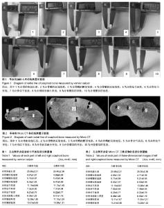
| [1] 胥少汀,葛宝丰,徐印坎.实用骨科学[M]. 3版.北京:人民卫生出版社,2005.
[2] Herbert TJ. Fisher WE.Management of the fractured scaphoid using a new bone screw. J Bone Joi nt Surg Br.1986;66(1): 114-117.
[3] Herbert TJ, Fischer WE.Management of the fratured scaphoid usi ng a new bone screw. J Bone Joint Surg.1984;66: 114-123.
[4] Reting AC. Manage ment of acut e scaphoid fractures. Hand Clin.2000;16(3):381-395.
[5] Hambidge JE,Desai VV,Schranz PJ,et al.Acute fractures of the scaphoid.Treatment by cast immobilisation with the wrist in flexion or extension. J Bone Joint Surg Br.1999;81(1): 91-92.
[6] Geiss ler WB.Carpal fractures in athletes. Clin Sports Med. 2001;20(1):167-188.
[7] McAdams TR, Spisak S, Beaulieu CF, et al. The effect of pronation and supination on the minimally displaced scaphoid fracture.Clin Orthop Relat Res.2003;(411):255-259.
[8] McAdams TR, Srivas tava S. Arthroscopic evaluation of scaphoid waist fracture stability and the role of the radioscaphocapitate ligament.Arthroscopy.2004; 20(2): 152-157.
[9] Jeon IH, Oh CW, Park BC, et al. Minimal invasive percutaneous Herbert screw fixation in acute unstable scaphoid fracture.Hand Surg.2003;8(2):213-218.
[10] Cooney WP 3rd. Scaphoid fractures: current treatments and techniques. Instr Course Lect.2003;52:197-208.
[11] Bhandari M, Hanson BP. Acute nondisplaced fractures of the scaphoid. J Orthop Trauma.2004;18(4):253-255.
[12] Shin AY. Percutaneous fixation of stable scaphoid fractures. Tech HandUp Extrem Surg.2004;8(2):87-94.
[13] Shih JT,Lee HM, Hou YT, et al.Results of arthroscopic reduction and percutaneous fixation for acute displaced s caphoid fractures.Arthroscopy.2005; 21(5):620-626.
[14] Kong WY, Xu YQ, Wang YF, et al.Anatomic measurement of scaphoid and its clinical significance. Chin J Traumatol. 2009; 12(1):41-44.
[15] 章莹,尹庆水,许家军,等. 手舟骨微创内固定解剖学基础[J].中国临床解剖学杂志,2004,22(2):176-178.
[16] 韩利军.手舟月骨间韧带和月三角骨间韧带的解剖学特点[J].中国组织工程研究与临床康复,2009,13(37):7318-7321.
[17] 曾立军,徐永清,骆华松,等.舟骨、大、小多角骨的临床解剖学研究[J].中国临床解剖学杂志,2008,26(1):29-31.
[18] 李民.腕关节镜治疗手舟骨骨折解剖学的研究[J].医学信息:下旬刊,2009,1(5):64-65.
[19] 王毅,徐永清,陈东源,等.可吸收手舟骨螺钉的研制和生物力学研究[J].中国临床解剖学杂志,2009,27(3):329-332.
[20] 覃励明,徐永清,吴农欣,等.头状骨、月骨、三角骨、钩骨测量及四角融合器的设计[J].实用手外科杂志,2009,23(1):11-13.
[21] 唐亮,卢弘栩,丁健,等.舟骨-大-小多角骨新型融合器的稳定性[J].中国组织工程研究与临床康复,2009,13(39):7651-7656.
[22] 孔维云,徐永清,王宇飞,等.镍钛记忆合金腕舟骨内固定器的研制及生物力学测试[J].中国修复重建外科杂志,2008,22(1): 48-52.
[23] von Stechow D, Balto K, Stashenko P, et al. Three- dimensional quantitation of periradicular bone destruction by micro-computed tomography.J Endod.2003;29:252-256.
[24] Fan B,Cheung GS,Fan M,et al. C-shaped canal system in mandibular second molars: part II-Radiographic features.J Endod.2004;30:904-908.
[25] Park CH,Abramson ZR,Taba MJ,et al.Three-dimensional microCT imajing of a alveolar bone in experimental bone loss or repair. J Periodontology.2007;78(3):273-281.
[26] Sone T,Tamada T,Jo Y,et al.Analysis of three dimensional microarchitecture and degree of mineralization in bone metastases from prostate cancer using synchrotron CT.Bone. 2004;35:432-438.
[27] Hanson NA,Bagi CM.Alternative approach to assessment of bone quality using micro-computed tomography. Bone. 2004; 35:326-333.
[28] Gribel BF, Gribel MN, Frazão DC, et al. Accuracy and reliability of craniometric measurements on lateral cephalometry and 3D measurements on CBCT scans.Angle Orthod.2011;81(1):26-35.
[29] 宋薇,刘琛,何惠明,等. Micro CT法建立新生儿单侧完全性腭裂三维模型的研究[J].实用口腔医学杂志,2013,29(1):36-39.
[30] 徐永清,钟世镇,朱青安,等.正常腕关节运动学的实验研究[J].中国临床解剖学杂志,2003, 21(2):173-175.
[31] 沈华,蔡嬿娴,王永春,等.加压骑缝钉克氏针治疗经舟骨月骨周围骨折脱位[J].组织工程与重建外科杂志,2010,6(1):35-37.
[32] 常青,黄迅悟,关长勇,等.应用Herbert螺钉内固定治疗腕舟骨骨折[J].中华手外科杂志,2002,18(4):217-218.
[33] Dodds SD, Panjabi MM, Slade JF 3rd, et al. Screw fixation of scaphoid fracture: a biomechanical assessment of screw length and screw augmentation.J Hand Surg(Am).2006;31(3):405-413.
[34] Bohringer G, Schadel-Hopfner M, Lemke T, et al. Arthroscopically controlled minimal invasive screw fixation of scaphoid fractures.A pilot study. Unfallchirurg. 2000; 103(12): 1086-1092.
[35] Daecke W, Wieloch P, Vergetis P, et al. Occurrence of carpal osteoarthritis after treatment of scaphoid nonunion with bone graft and Herbert screw: a long-term follow-up study. J Hand Surg(Am).2005;30(5):923-931.
[36] 陈振兵,洪光祥,王发斌,等.加压螺钉治疗舟骨骨折的临床疗效[J].中华手外科杂志,2005, 21(1):28-30. |

