Chinese Journal of Tissue Engineering Research ›› 2014, Vol. 18 ›› Issue (22): 3510-3516.doi: 10.3969/j.issn.2095-4344.2014.22.012
Previous Articles Next Articles
Finite element analysis of posterolateral fracture of tibial plateau using three types of internal fixation
Fan Xin-bin1, Zhang Yan1, Yang Tie-yi1, Luo Cong-feng1, Gong Lu-lu2, Liang Xu1, Liu Shu-yi1, Wu Liang1, Liu Yue1
- 1Department of Orthopedics, Shanghai Pudong New Area Gongli Hospital, Shanghai 200135, China; 2School of Life Sciences and Technology, Tongji University, Shanghai 200092, China
-
Revised:2014-03-04Online:2014-05-28Published:2014-05-28 -
Contact:Zhang Yan, M.D., Associate chief physician, Department of Orthopedics, Shanghai Pudong New Area Gongli Hospital Shanghai 200135, China -
About author:Fan Xin-bin, Master, Physician, Department of Orthopedics, Shanghai Pudong New Area Gongli Hospital, Shanghai 200135, China -
Supported by:the Shanghai Pudong District Science and Technology Development Innovation Project, No. PKJ2011-Y9; the Academic Leaders of Health System Project of Shanghai Pudong District, No. PWRd2012-16
CLC Number:
Cite this article
Fan Xin-bin, Zhang Yan, Yang Tie-yi, Luo Cong-feng, Gong Lu-lu, Liang Xu, Liu Shu-yi, Wu Liang, Liu Yue. Finite element analysis of posterolateral fracture of tibial plateau using three types of internal fixation[J]. Chinese Journal of Tissue Engineering Research, 2014, 18(22): 3510-3516.
share this article
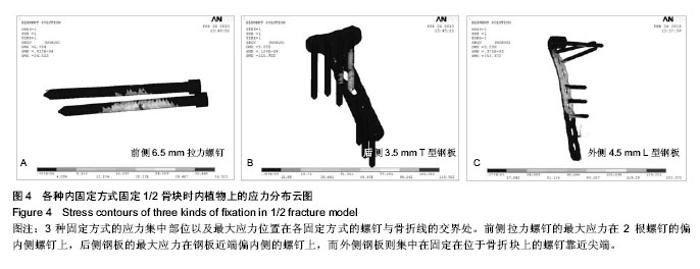
2.1 骨折模型的位移变化 在胫骨平台后外侧1/2和1/4骨块模型上分别施加500 N应力载荷后,测得3种内固定方式在X、Y、Z轴方向上的最大位移见表2。由结果得知,在1/2骨块模型中,前侧拉力螺钉与后侧钢板在各个方向上的位移较小,提供较为坚固的稳定性,且两者之间差距较小,而外侧钢板的位移较其他两种固定方式大,稳定性欠佳。同时相比X、Y轴,骨干纵轴(Z轴)方向上,外侧钢板固定模型移位更为明显;而在1/4骨块模型中,前侧拉力螺钉在各方向的位移优势更为明显,后侧钢板的位移居次,外侧钢板的位移最大。 2.2 骨折模型的应力分布 对加载后的模型进行应力的分析,得到各模型的应力分布情况,明确应力集中部位。从结果中得知,3种内固定方式的应力集中部位以及最大应力位置在各内固定方式的螺钉与骨折线的交界处,分别测得1/2骨块和1/4骨块上3种内固定方式的最大应力(图3)。 1/2骨块上前侧拉力螺钉的最大应力为36.523 MPa,外侧钢板为153.372 MPa,后侧钢板为115.922 MPa;而在骨块上的最大应力分别为:前侧拉力螺钉模型4.309 MPa,外侧钢板 4.37 MPa,后侧钢板3.124 MPa。 1/4骨块上前侧拉力螺钉的最大应力为36.803 MPa,外侧钢板为153.336 MPa,后侧钢板为104.234 MPa;而在骨块上的最大应力分别为:前侧拉力螺钉模型1.195 MPa,外侧钢板0.827 MPa,后侧钢板1.196 MPa。 3种内固定方式的应力集中部位以及最大应力点在各固定方式的螺钉与骨折线的交界处(图4)。"

通过对胫骨平台后外侧骨折多种固定方式的有限元分析,前侧拉力螺钉能够承担更大的应力,并在受到应力后位移变化较小,提供较稳定的支持;而后侧钢板在骨块较大(1/2骨块)时,能够提供较强的稳定性,与拉力螺钉相仿,而在骨块较小(1/4骨块)时,稳定性不如前侧拉力螺钉;外侧钢板在固定胫骨平台后外侧骨折时,稳定性较差,不如前侧拉力螺钉和后侧钢板。 需要说明的是,由于骨折位置的特殊性,特别是胫骨平台后外侧1/4骨块骨折时,外侧钢板仅能固定一两个螺钉,且螺钉方向与骨折线方向不相垂直,且有较大角度,固定困难,这也是实验中外侧钢板组的力学性能较其他两组差的原因之一。"

| [1] Schatzker J, McBroom R, Bruce D. The tibial plateau fracture:the Toronto experience 1968-1975. Clin Orthop. 1979;138:94-104.
[2] Papagelopoulos PJ, Partsinevelos AA, Themistocleous GS, et al. Complications after tibia plateau fracture surgery.Injury. 2006;37(6):475-484.
[3] Stauffer KD, Tuttle TA, Elkins AD, et al. Complications associated with 696 tibial plateau leveling osteotomies (2001-2003). J Am Anim Hosp Assoc. 2006;42(1):44-50.
[4] Cross WW 3rd, Levy BA, Morgan JA, et al. Periarticular raft constructs and fracture stability in split-depression tibial plateau fractures. Injury. 2013;44(6):796-801.
[5] Heikkilä JT, Kukkonen J, Aho AJ, et al. Bioactive glass granules: a suitable bone substitute material in the operative treatment of depressed lateral tibialplateau fractures: a prospective, randomized 1 year follow-up study. J Mater Sci Mater Med. 2011;22(4):1073-1080.
[6] Hsieh CH, Huang HT, Liu PC,et al.Anterior approach for posteromedial tibial plateau fractures. Kaohsiung J Med Sci. 2010;26(3):130-135.
[7] Helfet DL, Haas NP, Schatzker J,et al.AO philosophy and principles of fracture management-its evolution and evaluation. J Bone Joint Surg Am. 2003;85-A(6):1156-1160.
[8] Khan RM,Khan SH,Ahmad AJ,et al.Tibial plateau fractures.A new classification scheme. Clin Orthop Relat Res. 2000;375: 231-242.
[9] Gicquel T, Najihi N, Vendeuvre T,et al. Tibial plateau fractures: reproducibility of three classifications (Schatzker, AO, Duparc) and a revised Duparc classification. Orthop Traumatol Surg Res. 2013;99(7):805-816.
[10] Luo CF, Sun H, Zhang B. Three-column fixation for complex tibial plateau fractures. J Orthop Trauma. 2010;24(11):683-692.
[11] Carlson DA. Posterior bicondylar tibial plateau fractures. J Orthop Trauma. 2005;19:73-78.
[12] Ren WF,Zhang NN,Zhu YY,et al. Medial plus anterolateral approaches for the treatment of tibial plateau fractures involving three columns. Zhongguo Gu Shang. 2013;26(9): 768-771.
[13] Berber R,Lewis CP, Copas D,et al.Postero-medial approach for complex tibial plateau injuries with a postero-medial or postero-lateral shear fragment.Injury. 2013. pii: S0020-1383 (13)00563-9.
[14] Chiu CH, Cheng CY, Tsai MC, et al. Arthroscopy-assisted reduction of posteromedial tibial plateau fractures with buttress plate and cannulated screw construct.Arthroscopy. 2013;29(8):1346-1354.
[15] Yushkevich PA, Piven J, Hazlett HC, et al. User-guided 3D active contour segmentation of anatomical structures: Significantly improved efficiency and reliability. Neuroimage. 2006; 31(3):1116-1128.
[16] Sonoda N, Chosa E, Totoribe K,et al. Biomechanical analysis for stress fractures of the anterior middle third of the tibia in athletes:nonlinear analysis using a three-dimensional finite element method. J Orthop Sci. 2003;8:505-513.
[17] Lacroix D, Prendergast PJ. Three-dimensional Simulation of Fracture Repair in the Human Tibia. Comput Methods Biomech Biomed Engin. 2002;5(5):369-376.
[18] 罗从风,胡承方,高洪,等. 基于CT的胫骨平台骨折的三柱分型[J].中华创伤骨科杂志,2009,11(3):201-206.
[19] Patange Subba Rao SP, Lewis J, Haddad Z,et al. Three-column classification and Schatzker classification: a three- and two-dimensional computed tomography characterisation and analysis of tibial plateau fractures. Eur J Orthop Surg Traumatol. 2013. [Epub ahead of print]
[20] Wismans J, Veldpaus F, Janssen J, et al. A three-dimensional mathematical model of the knee-joint. J Biomech. 1980;13(8): 677-685.
[21] Howard NE, Phaff M, Aird J,et al. Does human immunodeficiency virus status affect early wound healing in open surgically stabilised tibial fractures?: A prospective study. Bone Joint J. 2013;95-B(12):1703-1737.
[22] Uchiyama S, Itsubo T, Nakamura K,et al. Effect of early administration of alendronate after surgery for distal radial fragility fracture on radiological fracture healing time.Bone Joint J. 2013;95-B(11):1544-1550.
[23] Matsunaga FT, Tamaoki MJ, Matsumoto MH, et al. Treatment of the humeral shaft fractures--minimally invasive osteosynthesis with bridge plate versus conservative treatment with functional brace: study protocol for a randomised controlled trial. Trials. 2013;14:246.
[24] Urita A, Iwasaki N, Kondo M, et al. Effect of low-intensity pulsed ultrasound on bone healing at osteotomy sites after forearm bone shortening. J Hand Surg Am. 2013;38(3): 498-503.
[25] Epari DR, Kassi JP, Schell H, et al. Timely fracture-healing requires optimization of axial fixation stability. J Bone Joint Surg Am. 2007;89(7):1575-1585.
[26] Lin CL, Chang YH, Pai CA. Evaluation of failure risks in ceramic restorations for endodontically treated premolar with MOD preparation. Dent Mater. 2011;27(5):431-438.
[27] Chevalier Y, Quek E, Borah B,et al. Biomechanical effects of teriparatide in women with osteoporosis treated previously with alendronate and risedronate: results from quantitative computed tomography-based finite element analysis of the vertebral body. Bone. 2010;46(1):41-48.
[28] Burstein A,Reilly D.Aging of bone tissue:mechanica properties. J Bone Joint Surg Am. 1976;58:82-86.
[29] 孙荣鑫,张玉新,白靖平,等.Sanders II型跟骨骨折内固定治疗的有限元分析[J].中国矫形外科杂志,2012,8(20):1409-1412.
[30] Gilula L, Persenaire M.Subsequent fractures post-vertebral augmentation: analysis of a prospective randomized trial in osteoporotic vertebral compression fractures. AJNR Am J Neuroradiol. 2013;34(1):221-227.
[31] Henderson CE, Lujan TJ, Kuhl LL, et al. 2010 Mid-America Orthopaedic Association Physician in Training Award: Healing Complications Are Common After Locked Plating for Distal Femur Fractures. Clin Orthop Relat Res. 2011;469(6):1757- 1765.
[32] Herrera A, Ibarz E, Cegoñino J, et al. Applications of finite element simulation in orthopedic and trauma surgery. World J Orthop. 2012; 3(4): 25-41.
[33] Liu XS, Wang J, Zhou B,et al. Fast trabecular bone strength predictions of HR-pQCT and individual trabeculae segmentation-based plate and rod finite element model discriminate postmenopausal vertebral fractures. J Bone Miner Res. 2013;28(7):1666-1678.
[34] Varga P, Schefzig P, Unger E, et al. Finite element based estimation of contact areas and pressures of the human scaphoid in various functional positions of the hand. J Biomech. 2013;46(5):984-990.
[35] Nishiyama KK, Macdonald HM, Hanley DA, et al. Women with previous fragility fractures can be classified based on bone microarchitecture and finite element analysis measured with HR-pQCT. Osteoporos Int. 2013;24(5):1733-1740.
[36] Meng L, Zhang Y, Lu Y. Three-dimensional finite element analysis of mini-external fixation and Kirschner wire internal fixation in Bennettfracture treatment. Orthop Traumatol Surg Res. 2013;99(1):21-29.
[37] Kantardzi? I, Vasiljevi? D, Blazi? L,et al. Influence of cavity design preparation on stress values in maxillary premolar: a finite element analysis. Croat Med J. 2012;53(6):568-576.
[38] Jia YW, Cheng LM, Yu GR, et al. A finite element analysis of the pelvic reconstruction using fibular transplantation fixed with four different rod-screw systems after type I resection. Chin Med J. 2008;121(4):321-326.
[39] Graeff C, Marin F, Petto H,et al.High resolution quantitative computed tomography-based assessment of trabecular microstructure and strength estimates by finite-element analysis of the spine, but not DXA, reflects vertebral fracture status in men with glucocorticoid-induced osteoporosis. Bone. 2013;52(2):568-577.
[40] Wang X, Sanyal A, Cawthon PM,et al. Prediction of new clinical vertebral fractures in elderly men using finite element analysis of CT scans. J Bone Miner Res. 2012;27(4):808-816. |
| [1] | Chen Qun-qun, Qiao Rong-qin, Duan Rui-qi, Hu Nian-hong, Li Zhao, Shao Min. Acu-Loc®2 volar distal radius bone plate system for repairing type C fracture of distal radius [J]. Chinese Journal of Tissue Engineering Research, 2017, 21(7): 1025-1030. |
| [2] | Xie Qiang. Three-dimensional finite element model for biomechanical analysis of stress in knee inversion and external rotation after posterior cruciate ligament rupture [J]. Chinese Journal of Tissue Engineering Research, 2017, 21(7): 1036-1040. |
| [3] | He Ze-dong, Zhao Jing, Chen Liang-yu, Li Ke, Weng Jie. Multilevel finite element analysis on the biological tribology damage of water on bone tissue [J]. Chinese Journal of Tissue Engineering Research, 2017, 21(7): 1041-1045. |
| [4] | Li Hui, Ma Jun-yi, Ma Yuan, Zhu Xu . Establishment of a three-dimensional finite element model of ankylosing spondylitis kyphosis [J]. Chinese Journal of Tissue Engineering Research, 2017, 21(7): 1069-1073. |
| [5] | Zou Wei, Xiao Jie, Long Hao, Zhang Yang, Wu Chen, Du Yu-hui, Feng Ming-xing, Zhou Chang-jun. Screw placement selection of minimally invasive percutaneous pedicle screw fixation for thoracolumbar fractures [J]. Chinese Journal of Tissue Engineering Research, 2017, 21(3): 356-361. |
| [6] | Jia Yan-bo, Liang Zi-hong, Ren Yi-zhong, Han Chang-xu, Kong Ling-yue, Eerduntu. Tibial avulsion fractures of anterior cruciate ligament repaired with Arthrex sutures passing through combining free knotting technique [J]. Chinese Journal of Tissue Engineering Research, 2017, 21(3): 367-372. |
| [7] | Chen Lu-yao, Hu Shi-qiang, Wang Xiao-ping, Wu Wei-wei, Wei Zhan-tu, Huang Jian. Accuracy of digital orthopedic three-dimensional reconstruction for thoracolumbar pedicle screw placement [J]. Chinese Journal of Tissue Engineering Research, 2017, 21(3): 373-377. |
| [8] | Du Shi-yao, Zhou Feng-jin, Ni Bin, Chen Bo, Chen Jin-shui. Finite-element analysis of a novel posterior atlantoaxial restricted non-fusion fixation system [J]. Chinese Journal of Tissue Engineering Research, 2017, 21(3): 383-389. |
| [9] | Liu Jun, Liao Su-ping. Three-dimensional finite element analysis of Kirschner nails and external fixation for Bennett fracture [J]. Chinese Journal of Tissue Engineering Research, 2017, 21(3): 390-395. |
| [10] | Zhang Li-chao, Zhang Li-min, Lv Yong-ming, Wang Zhi-hui, Yang Yang, Xu Fei, Dai Hai-feng, Li Jia, Cao Xiang-yu, Wu Li-zhu. Finite element analysis of knee flexion and extension movement [J]. Chinese Journal of Tissue Engineering Research, 2017, 21(3): 396-400. |
| [11] | Jia Jin-ling, Dong Yu-zhen. Finite element analysis of prosthesis position during hip arthroplasty [J]. Chinese Journal of Tissue Engineering Research, 2017, 21(3): 401-405. |
| [12] | Sheng Xiao-lei, Yuan Feng, Li Zhi-duo, Yang Yu-ming, Lu Hai-tao, Zhang Jun-wei. Comparison of the accuracy of lower cervical anterior transpedicular screws between three-dimensional printing assembly navigation template and free hand placement [J]. Chinese Journal of Tissue Engineering Research, 2017, 21(3): 406-411. |
| [13] | Wu Min-hao, Sun Wen-chao, Yan Fei-fei, Xie Yuan-long, Hou Zhi-qiang, Feng Fan, Cai Lin . Treatment research and new progress of early-onset scoliosis [J]. Chinese Journal of Tissue Engineering Research, 2017, 21(3): 433-439. |
| [14] | Wang Lei, Wang Feng-feng, Ma Yan-hui, Zhang Jie, Hu Fang, Ma Gai-ping, Liu Mei-mei, Ma Zhang-wen. Meta analysis of clinical outcome of intramedullary nails versus locking plates for two-part proximal humerus fracture [J]. Chinese Journal of Tissue Engineering Research, 2017, 21(3): 478-484. |
| [15] | Hu Jun, Zhang De-qiang, Tang Xin. Postoperative quality of life of internal fixation versus hemiarthroplasty for femoral neck fractures in the elderly [J]. Chinese Journal of Tissue Engineering Research, 2017, 21(19): 2953-2960. |
| Viewed | ||||||
|
Full text |
|
|||||
|
Abstract |
|
|||||