Chinese Journal of Tissue Engineering Research ›› 2013, Vol. 17 ›› Issue (53): 9196-9202.doi: 10.3969/j.issn.2095-4344.2013.53.016
Previous Articles Next Articles
Regeneration and structural changes of mouse cirrhotic liver after partial hepatectomy
Zheng Xiao-jun, Luo Qian-xin, Chen Qian-qi, Xiong Xi-feng, Liu Yu-rong, Ma Ning-fang
- Department of Histology and Embryology, School of Basic Sciences, Guangzhou Medical University, Guangzhou 510182,Guangdong Province, China
-
Revised:2013-09-19Online:2013-12-31Published:2013-12-31 -
Contact:Ma Ning-fang, M.D., Professor, Department of Histology and Embryology, School of Basic Sciences, Guangzhou Medical University, Guangzhou 510182, Guangdong Province, China nfma@gzhmc.edu.cn -
About author:Zheng Xiao-jun, Department of Histology and Embryology, School of Basic Sciences, Guangzhou Medical University, Guangzhou 510182, Guangdong Province, China 383271008@qq.com -
Supported by:the College Students’ Innovative Experimental Projects of Guangdong Province (2011)*
CLC Number:
Cite this article
Zheng Xiao-jun, Luo Qian-xin, Chen Qian-qi, Xiong Xi-feng, Liu Yu-rong, Ma Ning-fang. Regeneration and structural changes of mouse cirrhotic liver after partial hepatectomy[J]. Chinese Journal of Tissue Engineering Research, 2013, 17(53): 9196-9202.
share this article

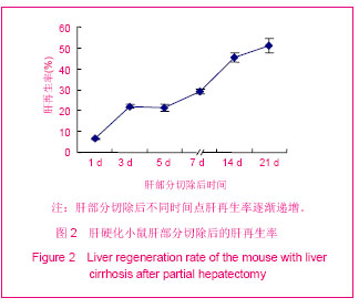
2.1 实验动物数量分析 实验共选用雄性小鼠60只制作肝硬化模型,其中11只在造模过程中死亡,存活 49只,选取5只小鼠用于组织病理学鉴定,证明造模成功率为82%。剩余的44只早期肝硬化小鼠进一步实施肝大部切除,死亡4只,术后存活率88%。肝切除后第1,3天分别取5只小鼠,其余时间点各取6只小鼠进行指标观察。 2.2 肝部分切除后的肝再生率 肝硬化小鼠肝部分切除后第3天肝质量明显回升,见图1。"

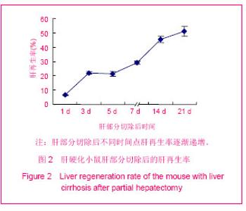
肝部分切除后第1,3,5,7,10,14,21天肝再生率分别为6.58%,22.03%,21.39%,29.05%,45.22%,50.98%,见图2。"
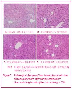
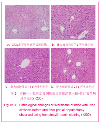
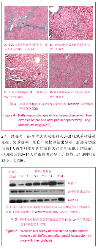
2.3 肝部分切除前后肝组织的结构变化 见图3,4。 苏木精-伊红染色结果显示,CCl4皮下注射8周后小鼠肝小叶结构破坏,肝细胞有空泡样变及脂肪变,Massson染色显示肝小叶间胶原纤维增生并伸入肝小叶内形成间隔,与周围增生的纤维间隔互相连接、包绕分割原来的肝小叶形成假小叶,呈现出早期肝硬化改变。对肝硬化小鼠实施肝部分切除后第1-5天肝组织结构与造模前肝织相比结构无明显变化;肝部分切除后第7天再生肝组织结构出现改善,随术后时间推移肝细胞脂肪变现象逐渐减少,肝小叶间增生的胶原纤维减少或消失,至肝部分切除后21 d肝小叶结构基本恢复正常。"
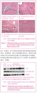
"
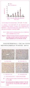
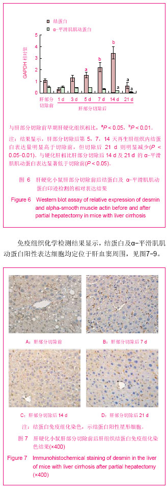
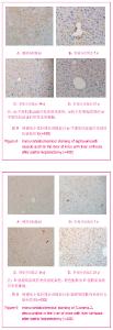
与硬化肝相比差异均有显著性意义(P < 0.05或P < 0.01);肝部分切除后各时间点α-平滑肌肌动蛋白表达比切除前明显减少,肝再生过程中α-平滑肌肌动蛋白表达依时递减,与硬化肝相比肝部分切除后14 d及21 d的α-平滑肌肌动蛋白表达差异均有显著性意义(P < 0.05),见图6。"
"
| [1] 范婷婷,谢渭芬.肝纤维化和肝硬化治疗进展[J].国际消化病杂志, 2010,30(6): 349-351.[2] Schuppan D, Kim YO. Evolving therapies for liver fibrosis. J Clin Invest. 2013;123(5):1887-1901.[3] 汤钊猷.肝癌研究进展[J].中国肿瘤,2001,10(1): 37-40.[4] 吴孟超,陈汉,沈锋. 原发性肝癌的外科治疗一附5524例报告[J].中华外科杂志,2001,39(1):25-28.[5] Nagasue N, Kohno H, Chang YC, et al. Liver resection for hepatocelluar carcinoma. Ann Surg. 1993; 217(4): 375-384.[6] Moser MA,Kneteman NM,Minuk GY. Research towards safer resection of the cirrhotic liver. HBP Surg. 2000;11: 285-297.[7] Bravo E, D'Amore E, Ciaffoni F, et al. Evaluation of the spontaneous reversibility of carbon tetrachloride-induced liver cirrhosis in rabbits. Lab Anim. 2012;46(2):122-128.[8] Mederacke I.Liver fibrosis-mouse models and relevance in human liver diseases. Z Gastroenterol. 2013;51(1):55-62.[9] Higgins GM, Anderson RM. Experimental pathology of the liver. I. Restoration of the liver of the white rat following partial surgical removal. Arch Pathol Lab Med. 1931;12:186-202.[10] Michalopoulos GK. Liver regeneration.J Cell Physiol. 2007; 213(2):286-300.[11] 陈栋,吴力群,曹景玉,等.大鼠肝切除术后肝损伤程度与肝再生状态的动态对比研究[J].中华肝胆外科杂志,2002,8(6):354-357.[12] Jones LD, Nielsen MK, Britton RA. Genetic variation in liver mass, body mass, and liver: body mass in mice. J Anim Sci. 1992;70: 2999-3006.[13] Tan X, Behari J, Cieply B, et al. Conditional deletion of beta-catenin reveals its role in liver growth and regeneration. Gastroenterology. 2006; 131: 1561-1572. [14] Inderbitzin D, Gass M, Beldi G, et al. Magnetic resonance imaging provides accurate and precise volume determination of the regenerating mouse liver. J Gastrointest Surg. 2004; 8: 806-811.[15] Kumar S, Zou Y, Bao Q, et al. Proteomic analysis of immediate-early response plasma proteins after 70% and 90% partial hepatectomy. Hepatol Res. 2012. doi: 10.1111/hepr.12030. [Epub ahead of print] [16] Hayashi H, Nagaki M, Imose M, et al. Normal liver regeneration and liver cell apoptosis after partial hepatectomy in tumor necrosis factor-alpha-deficient mice. Liver Int. 2005; 25:162-170.[17] Fujita J, Marino MW, Wada H, et al. Effect of TNF gene depletion on liver regeneration after partial hepatectomy in mice. Surgery. 2001;129: 48-54.[18] 方芳,徐丽萍,董沛杰,等.不同性别小鼠肝大部切除术后肝再生能力的比较[J].中国组织工程研究与临床康复,2010,14(53): 9909-9912.[19] Selzner M, Clavian PA. Failure of regeneration of the steatotic rat liver: disruption at two different levels in the regeneration pathway. Hepatology. 2000;31: 35-42.[20] Martins PN, Theruvath TP, Neuhaus P. Rodent models of partial hepatectomies. Liver Int.2008;28:3-11.[21] Safadi R,Friedman SL.Hepatic fibrosis-role of hepatic stellate cell activation.Med Gen Med. 2002;4(3):27-32.[22] Benyon RC,Iredale JP. Is liver fibrosis reversible? Gut. 2000; 46(4):443-446.[23] Reeves HL,Friedman SL. Activation of hepatic stellate cells- a key issue in liver fibrosis. Front Biosci. 2002;7:808-826.[24] 熊喜峰,何伟业,谢湘梅,等.β-catenin在四氯化碳诱导的小鼠肝纤维化过程中的定位表达及意义[J].解剖学报,2012,43(1): 97-102.[25] Hohme S, Hengstler JG, Brulport M. Mathematical modelling of liver regeneration after intoxication with CCl(4). Chem Biol Int. 2007;168(1):74-93.[26] Hora C, Romanque P, Dufour JF. Effect of sorafenib on murine liver regeneration. Hepatology. 2011;53(2):577-56.[27] Safadi R,Friedman SL. Hepatic fibrosis-role of hepatic stellate cell activation. Med Gen Med. 2002;4(3):27-32.[28] Kocabayoglu P, Friedman SL.Cellular basis of hepatic fibrosis and its role in inflammation and cancer. Front Biosci (Schol Ed). 2013;5:217-230.[29] Berg T, DeLanghe S, Al Alam D, et al. β-catenin regulates mesenchymal progenitor cell differentiation during hepatogenesis. J Surg Res. 2010;164(2):276-285. [30] Asahina K, Tsai SY, Li P, et al. Mesenchymal origin of hepatic stellate cells, submesothelial cells, and perivascular mesenchymal cells during mouse liver development. Hepatology. 2009;49(3):998-1011.[31] Van Rossen E, Vander Borght S, van Grunsven LA, et al. Vinculin and cellular retinol-binding protein-1 are markers for quiescent and activated hepatic stellate cells in formalin-fixed paraffin embedded human liver. Histochem Cell Biol. 2009; 131(3):313-325.[32] Zhao L, Burt AD. The diffuse stellate cell system.J Mol Histol. 2007;38(1):53-64. [33] Kalinichenko VV, Bhattacharyya D, Zhou Y, et al. Foxfl+/-mice exhibit defective stellate cell activation and abnormal liver regeneration following CCl4 injury. Hepatology 2003;37: 107-117.[34] Thomas RJ, Bhandari R, Barrett DA, et al. The effect of three dimensional co-culture of hepatocytes and hepatic stellate cells on key hepatocyte functions in vitro. Cells Tissues Organs. 2005;181:67-79.[35] Gratzner HG. Monoclonal antibody to 5-bromo and 5-iododeoxyuridine:a new reagent for detection of DNA replication. Science. 1982;218:474-479. |
| [1] | Zhu Ning, Yang Xinming, Ruan Jianwei. Triptolide improves spinal cord injury recovery via upregulation of autophagy and inhibition of apoptosis in Thy-yfp transgenic mice [J]. Chinese Journal of Tissue Engineering Research, 2020, 24(在线): 1-. |
| [2] | Liao Yuan, Qu Mengjian, Liu Jing, Zhong Peirui Zeng Yahua, Wang Ting, Xiao Hao, Li Lan, Liu Danni, Yang Lu, Zhou Jun. Effects of ultrashort wave on inflammatory response in rats with acute lung injury [J]. Chinese Journal of Tissue Engineering Research, 2020, 24(在线): 2-. |
| [3] | Kong Desheng, He Jingjing, Feng Baofeng, Guo Ruiyun, Asiamah Ernest Amponsah, Lü Fei, Zhang Shuhan, Zhang Xiaolin, Ma Jun, Cui Huixian. Efficacy of mesenchymal stem cells in the spinal cord injury of large animal models: a meta-analysis [J]. Chinese Journal of Tissue Engineering Research, 2020, 24(在线): 3-. |
| [4] | Li Xiaoqun, Xu Kaihang, Ji Fang. Corylin inhibits osteoclastogenesis and attenuates postmenopausal osteoporosis in mice [J]. Chinese Journal of Tissue Engineering Research, 2020, 24(在线): 4-. |
| [5] | Zhang Shuang, Xu Xiaomei, Zeng Yang, Yuan Xiaoping, Lin Fuwei. Rev-erbα’s effect on osteoblastogenesis of mouse bone marrow mesenchymal stem cells [J]. Chinese Journal of Tissue Engineering Research, 2020, 24(31): 4921-4926. |
| [6] | Xun Chong, Wang Qiang, Li Changzhou, Liu Xiaofeng. Potential molecular targets and therapeutic mechanisms underlying transplantation of autologous bone marrow stem cells for the treatment of spinal cord injury based on bioinformatics [J]. Chinese Journal of Tissue Engineering Research, 2020, 24(31): 4927-4933. |
| [7] | Chen Jia, Yang Yiqiang, Hu Chen, Chen Qi, Zhao Tian, Yong Min, Ma Dongyang, Ren Liling. Fabrication of prevascularized osteogenic differentiated cell sheet based on human bone marrow mesenchymal stem cells and human umbilical vein endothelial cells [J]. Chinese Journal of Tissue Engineering Research, 2020, 24(31): 4934-4940. |
| [8] | Liao Jian, Huang Xiaolin, Zhou Qian, Huo Hua, Qi Yuhan, Wu Chao, Shi Qianhui, Yang Tongjing, Liao Yunmao, Liang Xing. Calcined bone/chitosan composite promotes osteogenic differentiation of bone marrow mesenchymal stem cells in Sprague-Dawley rats [J]. Chinese Journal of Tissue Engineering Research, 2020, 24(31): 4941-4947. |
| [9] | Wang Guoliang, Li Yanlin, Xiang Yaoyu, Jia Di, Li Canzhang, He Lu. MicroRNA expression profiles of chondrocytes in osteoarthritis induced by stromal cell derived factor 1 [J]. Chinese Journal of Tissue Engineering Research, 2020, 24(31): 4948-4953. |
| [10] | Li Yuanqi, Lin Hai, Luo Hongrong, Zhang Xingdong. Relationship between mitochondrial autophagy and chondrogenesis of bone marrow mesenchymal stem cells [J]. Chinese Journal of Tissue Engineering Research, 2020, 24(31): 4954-4960. |
| [11] | Liu Hao, Liu Jun, Rui Yongjun, Tang Fenglin, Lu Miao, Ding Tao. Co-transfection by Nell-1 and Noggin-shRNA promotes osteoblast differentiation of adipose derived mesenchymal stem cells [J]. Chinese Journal of Tissue Engineering Research, 2020, 24(31): 4966-4970. |
| [12] | Wei Renyue, Li Xuechun, Li Yan, Yu Yang, Zhang Yu, Liu Zhonghua. A serum-free monolayer method for differentiation of porcine induced pluripotent stem cells into vascular endothelial cells [J]. Chinese Journal of Tissue Engineering Research, 2020, 24(31): 4971-4978. |
| [13] | Gu Yubo, Zhang Dongliang, Yuan Wei, , Song Lujie. Exosomes from adipose-derived stem cells can promote cavernous nerve regeneration in rats [J]. Chinese Journal of Tissue Engineering Research, 2020, 24(31): 4979-4985. |
| [14] |
Zheng Haijun, Jin Hui, Cui Hongling, Zhu Yakun, Zeng Hui, Han Fengjie, Qiu Cuiting, Liu Jing.
Safety of drug-coated balloon versus drug-eluting stents in the treatment of type 2 diabetes mellitus complicated by coronary artery small vessel disease in older adult patients [J]. Chinese Journal of Tissue Engineering Research, 2020, 24(28): 4573-4579. |
| [15] |
Ma Chao, Wang Huishan, Han Jinsong, Yin Zongtao, Zhang Xiling.
Cardiac valve prosthesis implantation and surgical maze ablation for the treatment of valvular disease with atrial fibrillation [J]. Chinese Journal of Tissue Engineering Research, 2020, 24(28): 4580-4587. |
| Viewed | ||||||
|
Full text |
|
|||||
|
Abstract |
|
|||||