Chinese Journal of Tissue Engineering Research ›› 2013, Vol. 17 ›› Issue (20): 3618-3625.doi: 10.3969/j.issn.2095-4344.2013.20.002
Previous Articles Next Articles
Expression prolife of mitogen-activated protein kinase pathway genes in vascular calcification associated with osteogenic differentiation of smooth muscle cells
Jin Bo1, Yin Heng-chong2, Wang Ling-qing1, Bao Li-wen1, Li Yan-lin1, Zhu Jun1, Shi Hai-ming1
- 1 Department of Cardiology, Huashan Hospital, Fudan University, Shanghai 200040, China
2 Department of Cardiology, Ruyang People’s Hospital, Luoyang 471200, Henan Province, China
-
Received:2012-10-18Revised:2012-11-20Online:2013-05-14Published:2013-05-14 -
Contact:Shi Hai-ming, M.D., Chief physician, Professor, Doctoral supervisor, Department of Cardiology, Huashan Hospital, Fudan University, Shanghai 200040, China shihmhs@yahoo.com.cn -
About author:Jin Bo☆, M.D., Attending physician, Department of Cardiology, Huashan Hospital, Fudan University, Shanghai 200040, China jinbo7711@yahoo.com.cn -
Supported by:the National Natural Science Foundation of China, No. 81100157
CLC Number:
Cite this article
Jin Bo, Yin Heng-chong, Wang Ling-qing, Bao Li-wen, Li Yan-lin, Zhu Jun, Shi Hai-ming. Expression prolife of mitogen-activated protein kinase pathway genes in vascular calcification associated with osteogenic differentiation of smooth muscle cells[J]. Chinese Journal of Tissue Engineering Research, 2013, 17(20): 3618-3625.
share this article

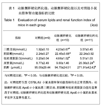
2.1 实验动物数量分析 动脉粥样硬化钙化组共20只SPF级ApoE-/-小鼠接受二步外科手术5/6肾脏切除法建立尿毒症模型,造模手术操作直接导致6只小鼠死亡,主要与创面出血密切相关,高脂饲料喂养过程中3只小鼠死亡;动脉粥样硬化组行假手术操作无小鼠死亡;对照组假手术过程中1只小鼠死亡,与麻醉药过量有关。 2.2 血脂和肾功能测定 对照组野生型C57BL/6J小鼠的血脂和肾功能指标均在正常范围内;动脉粥样硬化组ApoE-/-小鼠血清三酰甘油、胆固醇和低密度脂蛋白值均显著高于对照组;动脉粥样硬化钙化组和动脉粥样硬化组ApoE-/-小鼠的血脂水平差异无显著性意义;动脉粥样硬化钙化组小鼠血清尿素氮和肌酐值显著高于动脉粥样硬化组(P < 0.05),见表1。"

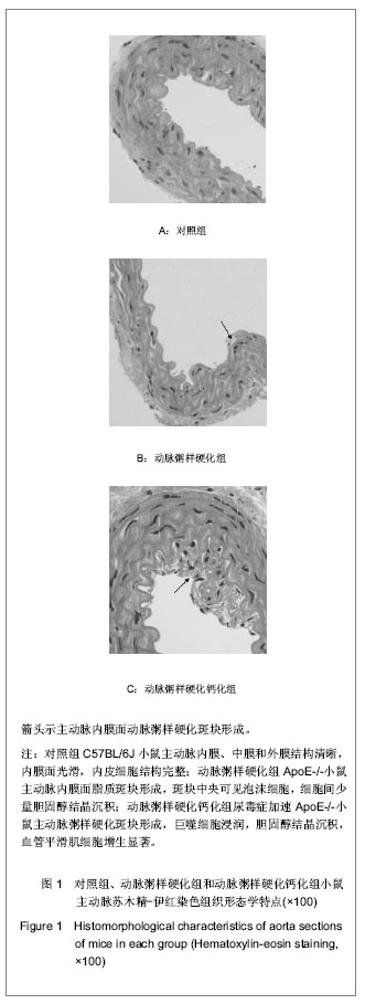
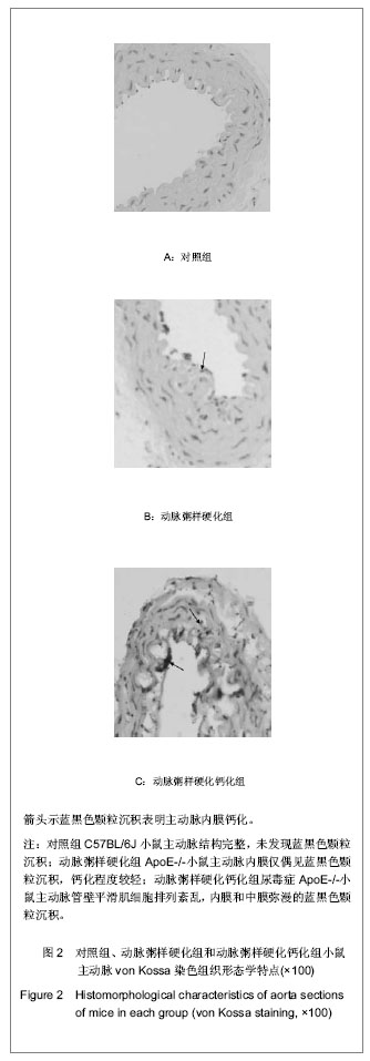
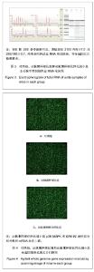
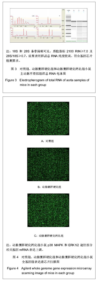
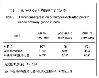
2.3 主动脉组织形态学特点 苏木精-伊红染色:对照组C57BL/6J小鼠主动脉内膜、中膜和外膜结构清晰,内膜面光滑,内皮细胞结构完整,见图1A;动脉粥样硬化组ApoE-/-小鼠主动脉内膜面脂质斑块形成,斑块中央可见泡沫细胞,细胞间少量胆固醇结晶沉积,见图1B;动脉粥样硬化钙化组尿毒症加速ApoE-/-小鼠主动脉粥样硬化斑块形成,巨噬细胞浸润,胆固醇结晶沉积,血管平滑肌细胞增生显著,见图1C。 von Kossa染色:对照组C57BL/6J小鼠主动脉结构完整,未发现蓝黑色颗粒沉积,见图2A;动脉粥样硬化组ApoE-/-小鼠主动脉内膜仅偶见蓝黑色颗粒沉积,钙化程度较轻,见图2B;动脉粥样硬化钙化组尿毒症ApoE-/-小鼠主动脉管壁平滑肌细胞排列紊乱,内膜和中膜弥漫的蓝黑色颗粒沉积,血管钙化显著,见图2C。 2.4 基因芯片样品的质量鉴定 用Trizol试剂一步法抽提3组小鼠主动脉平滑肌组织总RNA,QIAGEN RNAeasy mini kit 纯化RNA,应用Agilent 2100 Bioanalyzer测定RNA浓度和纯度,对样品进行变性琼脂糖凝胶电泳,18S和28S条带清晰可见,见图3,质检指标2100 RIN≥7.0且28S/18S≥0.7,结果表明样品总RNA纯度较高,RNA无明显降解,符合基因芯片检测要求。 2.5 基因芯片检测结果 应用Agilent小鼠全基因组表达谱芯片检测3组小鼠主动脉平滑肌组织mRNA表达,见图4。"

"
"

通过对基因芯片结果中差异表达的基因进行GO分析找出与丝裂原活化蛋白激酶信号通路相关的基因,初步结果提示,尿毒症ApoE-/-小鼠p38 丝裂原活化蛋白激酶和ERK1/2途径部分相关基因mRNA表达上调,见表2。"
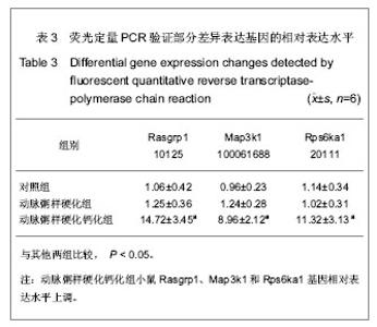
2.6实时荧光定量PCR结果 选择丝裂原活化蛋白激酶通路中3个差异表达的关键基因,应用实时荧光定量PCR法验证Rasgrp1、Map3k1和Rps6ka1基因的相对表达水平,见表3,应用Spearman相关分析,RT-PCR检测结果与基因芯片检测结果相吻合。"

| [1] Johnson RC, Leopold JA, Loscalzo J. Vascular calcification: pathobiological mechanisms and clinical implications. Circ Res. 2006;99(10):1044-1059. [2] Lee MJ, Shin DH, Kim SJ, et al. Progression of aortic arch calcification over 1 year is an independent predictor of mortality in incident peritoneal dialysis patients. PLos One. 2012;7(11):e48793. [3] Iyemere VP, Proudfoot D, Weissberg PL, et al.Vascular smooth muscle cell phenotypic plasticity and the regulation of vascular calcification. J Intern Med. 2006;260(3):192-210. [4] Zhu D, Mackenzie NC, Millán JL, et al. The appearance and modulation of osteocyte marker expression during calcification of vascular smooth muscle cells. PLos One. 2011;6(5):e19595.[5] Tanaka T, Sato H, Doi H, et al. Runx2 represses myocardin-mediated differentiation and facilitates osteogenic conversion of vascular smooth muscle cells. Mol Cell Biol. 2008;28(3):1147-1160.[6] Speer MY, Li X, Hiremath PG, et al. Runx2/Cbfa1, but not loss of myocardin, is required for smooth muscle cell lineage reprogramming toward osteochondrogenesis. J Cell Biochem. 2010;110(4):935-947. [7] Gagnon RF, Gallimore B. Characterization of a mouse model of chronic uremia. Urol Res. 1988;16(2):119-126.[8] Duan XH, Qi YF, Tang CS. Zhongguo Dongmai Yinghua Zazhi. 2008;16(2):153-157.段晓辉,齐永芬,唐朝枢. 血管钙化动物模型的研究进展[J]. 中国动脉硬化杂志,2008,16(2):153-157.[9] Cheng XZ,Huang B, Zhong H. Shengwu Jishu Tongxun. 2010; 21(1):112-115.程孝中,黄蓓,钟辉.动脉钙化发生机制研究进展[J].生物技术通讯, 2010,21(1):112-115.[10] Neven E, D'Haese PC. Vascular calcification in chronic renal failure: what have we learned from animal studies? Circ Res. 2011;108(2):249-264.[11] Moe SM, Chen NX. Pathophysiology of vascular calcification in chronic kidney disease. Circ Res. 2004;95(6):560-567. [12] You HZ, Ding F. Guoji Miniao Xitong Zazhi. 2007;27(2): 244-247.游怀舟,丁峰. 慢性肾脏病血管钙化的研究进展[J]. 国际泌尿系统杂志,2007,27(2): 244-247.[13] Yao Y, Bennett BJ, Wang X, et al. Inhibition of bone morphogenetic proteins protects against atherosclerosis and vascular calcification. Circ Res. 2010;107(4):485-494. [14] Boström KI, Jumabay M, Matveyenko A, et al. Activation of vascular bone morphogenetic protein signaling in diabetes mellitus. Circ Res. 2011;108(4):446-457. [15] Nakagami H, Osako MK, Morishita R. New concept of vascular calcification and metabolism. Curr Vasc Pharmacol. 2011;9(1):124-127. [16] Lai LQ, Yuan YS, Hao J, et al. 2010;32(10):1043-1050.赖林泉,袁运生,郜尽,等. MAPK信号通路基因在小鼠肝再生中的表达谱变化[J]. 遗传, 2010,32(10):1043-1050.[17] Miersalijiang•Yasen, Guo CJ, Fei QM, et al. Zhongguo Zuzhi Gongcheng Yanjiu yu Linchuang Kangfu. 2011;15(27): 4975-4978.米尔萨力江•亚森,郭常军,费琴明,等. 基因芯片分析成骨生长肽对OPG-/-小鼠骨髓间充质干细胞的作用[J]. 中国组织工程研究与临床康复,2011,15(27):4975-4978.[18] Mebratu Y, Tesfaigzi Y. How ERK1/2 activation controls cell proliferation and cell death: Is subcellular localization the answer? Cell Cycle. 2009;8(8):1168-1175. [19] Meloche S, Pouysségur J. The ERK1/2 mitogen-activated protein kinase pathway as a master regulator of the G1- to S-phase transition. Oncogene. 2007;26(22):3227-3239. [20] Oeztuerk-Winder F, Ventura JJ. The many faces of p38 mitogen-activated protein kinase in progenitor/stem cell differentiation. Biochem J. 2012;445(1):1-10.[21] del Barco Barrantes I, Nebreda AR. Roles of p38 MAPKs in invasion and metastasis. Biochem Soc Trans. 2012;40(1): 79-84. [22] Ding HT, Wang CG, Zhang TL, et al. Fibronectin enhances in vitro vascular calcification by promoting osteoblastic differentiation of vascular smooth muscle cells via ERK pathway. J Cell Biochem. 2006;99(5):1343-1352. [23] You H, Yang H, Zhu Q, et al. Advanced oxidation protein products induce vascular calcification by promoting osteoblastic trans-differentiation of smooth muscle cells via oxidative stress and ERK pathway. Ren Fail. 2009;31(4): 313-319. [24] Panizo S, Cardus A, Encinas M, et al. RANKL increases vascular smooth muscle cell calcification through a RANK-BMP4-dependent pathway. Circ Res. 2009;104(9): 1041-1048. [25] Tseng W, Graham LS, Geng Y, et al. PKA-induced receptor activator of NF-kappaB ligand (RANKL) expression in vascular cells mediates osteoclastogenesis but not matrix calcification. J Biol Chem. 2010;285(39):29925-29931.[26] Di Bartolo BA, Schoppet M, Mattar MZ, et al. Calcium and osteoprotegerin regulate IGF1R expression to inhibit vascular calcification. Cardiovascu Res. 2011;91(3):537-545. [27] Zheng MT, Xue J, You HZ, et al. Zhongguo Xueye Jinghua. 2008;7(2):81-84.郑曼韬,薛骏,游怀舟,等.他汀类药物对高磷介导血管平滑肌细胞成骨细胞转分化和钙化的影响[J]. 中国血液净化,2008,7(2): 81-84.[28] Kavurma MM, Schoppet M, Bobryshev YV, et al. TRAIL stimulates proliferation of vascular smooth muscle cells via activation of NF-kappaB and induction of insulin-like growth factor-1 receptor. J Biol Chem. 2008;283(12):7754-7762.[29] Chan J, Prado-Lourenco L, Khachigian LM, et al. TRAIL promotes VSMC proliferation and neointima formation in a FGF-2-, Sp1 phosphorylation-, and NFkappaB-dependent manner. Circ Res. 2010;106(6):1061-1071.[30] Li J, Li P, Zhang Y, et al. c-Ski inhibits the proliferation of vascular smooth muscle cells via suppressing Smad3 signaling but stimulating p38 pathway. Cell Signal. 2012;25(1): 159-167.[31] Bironaite D, Brunk U, Venalis A. Protective induction of Hsp70 in heat-stressed primary myoblasts. Involvement of MAPKs. J Cell Biochem. 2013 Mar 28. [32] Iwayama H, Ueda N. Role of mitochondrial Bax, caspases, and MAPKs for ceramide-induced apoptosis in renal proximal tubular cells. Mol Cell Biochem. 2013 Mar 31. [33] Wei ZF, Tong B, Xia YF, et al. Norisoboldine Suppresses Osteoclast Differentiation through Preventing the Accumulation of TRAF6-TAK1 Complexes and Activation of MAPKs/NF-κB/c-Fos/NFATc1 Pathways. PLoS One. 2013; 8(3):e59171. [34] Ma X, You X, Zeng Y, et al. Mycoplasma MALP-2 induces HO-1 expression via MAPKs and Nrf2 pathways to modulate COX-2 expression in human monocytes. Clin Vaccine Immunol. 2013 Mar 27. [35] Long X, Cowan SL, Miano JM. Mitogen-activated protein kinase 14 is a novel negative regulatory switch for the vascular smooth muscle cell contractile gene program. Arterioscler Thromb Vasc Biol. 2013;33(2):378-386. [36] Kim M, Kim S, Lim JH, et al. Laminar flow activation of ERK5 protein in vascular endothelium leads to atheroprotective effect via NF-E2-related factor 2 (Nrf2) activation. J Biol Chem. 2012;287(48):40722-40731. |
| [1] | Liu Zhichao, Zhang Fan, Sun Qi, Kang Xiaole, Yuan Qiaomei, Liu Genzhe, Chen Jiang. Morphology and activity of human nucleus pulposus cells under different hydrostatic pressures [J]. Chinese Journal of Tissue Engineering Research, 2021, 25(8): 1172-1176. |
| [2] | Guan Qian, Luan Zuo, Ye Dou, Yang Yinxiang, Wang Zhaoyan, Wang Qian, Yao Ruiqin. Morphological changes in human oligodendrocyte progenitor cells during passage [J]. Chinese Journal of Tissue Engineering Research, 2021, 25(7): 1045-1049. |
| [3] | Li Ping, Lin Yu, Chen Xiang, Liu Zhentao, Xiao Lili, Lin Xueyi, Hua Peng . Characteristics of bone remodeling in female ovariectomized rat models of osteoporosis undergoing Erzhi Pill extract intervention [J]. Chinese Journal of Tissue Engineering Research, 2021, 25(2): 191-195. |
| [4] | Shen Fu, Kuang Gaoyan, Yang Zhuo, Wen Meng, Zhu Kaimin, Yu Guizhi, Xu Wuji, Deng Bo . Immune infiltration mechanism of differential expression genes in rheumatoid arthritis and potential therapeutic prediction of Chinese herbs [J]. Chinese Journal of Tissue Engineering Research, 2021, 25(14): 2183-2191. |
| [5] | Pei Tianlong, Wang Yongkang, Wu Wenjie, Zhao Hong, Wang Longsheng . Anatomical and morphological characteristics of knee joint in hemophilic arthritis patients with three-dimensional CT and X-ray films [J]. Chinese Journal of Tissue Engineering Research, 2021, 25(12): 1886-1890. |
| [6] | Ye Dou, , Ma Xuexia , Guan Qian, , Luan Zuo , Yang Yinxiang , Wang Zhaoyan , Wang Qian , He Ying , Yao Ruiqin. Proportion and morphological characteristics of human oligodendrocyte precursor cells in different cell culture vessels [J]. Chinese Journal of Tissue Engineering Research, 2021, 25(1): 44-49. |
| [7] | Zhao Chuntao, Qing Mingsong, Yu Langbo, Peng Jiachen . Meta-analysis of total knee arthroplasty guided by kinematic alignment and mechanical alignment [J]. Chinese Journal of Tissue Engineering Research, 2020, 24(9): 1435-1442. |
| [8] |
Zhang Cong, Zhao Yan, Du Xiaoyu, Du Xinrui, Pang Tingjuan, Fu Yining, Zhang Hao, Zhang Buzhou, Li Xiaohe, Wang Lidong.
Biomechanical analysis of the lumbar spine and pelvis in adolescent
idiopathic scoliosis with lumbar major curve |
| [9] | Xu Guofeng, Li Xuebin, Tang Yifan, Zhao Yin, Zhou Shengyuan, Chen Xiongsheng, Jia Lianshun. The role of autophagy in ossification of the human ligamentum flavum [J]. Chinese Journal of Tissue Engineering Research, 2020, 24(8): 1174-1181. |
| [10] |
Cen Yanhui, Xia Meng, Jia Wei, Luo Weisheng, Lin Jiang, Chen Songlin, Chen Wei, Liu Peng, Li Mingxing, Li Jingyun, Li Manli, Ai Dingding, Jiang Yunxia.
Baicalein inhibits the biological behavior of hepatocellular
carcinoma stem cells by downregulation of Decoy receptor 3 expression |
| [11] | He Yujie, Wang Haiyan, Li Zhijun, Li Xiaohe, Cai Yongqiang, Dai Lina, Xu Yangyang, Wang Yidan, Xu Xuebin. Digital measurements of the anatomical parameters of pedicle-rib unit screw fixation in thoracic vertebrae of preschoolers [J]. Chinese Journal of Tissue Engineering Research, 2020, 24(6): 869-876. |
| [12] | Yan Shu, Lu Yan, Ouyang Zhaolian. Analysis of programs on tissue engineering funded by the National Natural Science Foundation of China between 2013 and 2018 [J]. Chinese Journal of Tissue Engineering Research, 2020, 24(5): 731-735. |
| [13] | Wei Gang, Gao Shangyuan, Zhang Ying, Huang Weiyi. Immunostimulation combined with liquid nitrogen freezing to construct a rat model of atherosclerotic vulnerable plaque [J]. Chinese Journal of Tissue Engineering Research, 2020, 24(35): 5656-5661. |
| [14] | Gong Heng, Huang Bin, Fu Ligong, Liu Huawei, Chen Lianxu. Comparison of knee geometry between Chinese Han people and American Caucasians [J]. Chinese Journal of Tissue Engineering Research, 2020, 24(33): 5366-5370. |
| [15] | Wang Guoliang, Li Yanlin, Xiang Yaoyu, Jia Di, Li Canzhang, He Lu. MicroRNA expression profiles of chondrocytes in osteoarthritis induced by stromal cell derived factor 1 [J]. Chinese Journal of Tissue Engineering Research, 2020, 24(31): 4948-4953. |
| Viewed | ||||||
|
Full text |
|
|||||
|
Abstract |
|
|||||