Chinese Journal of Tissue Engineering Research ›› 2012, Vol. 16 ›› Issue (49): 9232-9235.doi: 10.3969/j.issn.2095-4344.2012.49.020
Previous Articles Next Articles
Naringin induces bone marrow mesenchymal stem cells to repair femoral head necrosis in rabbits
Yang Yuan1, Li Xiao-feng1, Luo Dao-ming2, Wen Chao-hai2
- 1Guangxi Orthopedic Traumatology Hospital, Nanning 530012, Guangxi Zhuang Autonomous Region, China; 2Guangxi Traditional Chinese Medical University, Nanning 530000, Guangxi Zhuang Autonomous Region, China
-
Online:2012-12-02Published:2013-01-16 -
Contact:Li Xiao-feng, Master, Guangxi Orthopedic Traumatology Hospital, Nanning 530012, Guangxi Zhuang Autonomous Region, China lxfeng2000@126.com E-mail: lxfeng2000@126.com -
Supported by:a grant from Department of Science and Technology of Guangxi Zhuang Autonomous Region of China, No. Gui-ke-gong0816004-15*; a grant from Health Department of Guangxi Zhuang Autonomous Region of China, No. Zhong200863*
CLC Number:
Cite this article
Yang Yuan, Li Xiao-feng, Luo Dao-ming, Wen Chao-hai. Naringin induces bone marrow mesenchymal stem cells to repair femoral head necrosis in rabbits[J]. Chinese Journal of Tissue Engineering Research, 2012, 16(49): 9232-9235.
share this article

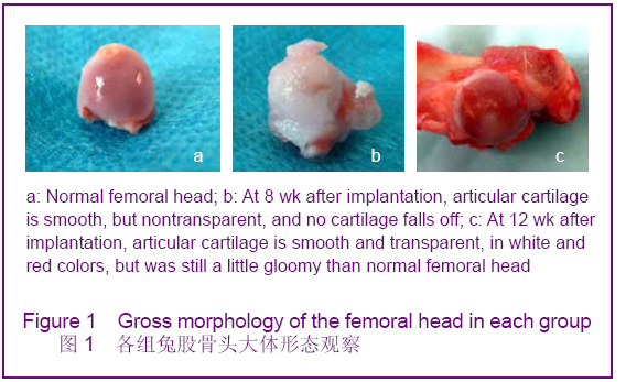
2.1 动物基本状况 动物麻醉苏醒后,即可自由活动,饮食正常。所有动物全部成活,皮毛光滑。3只对照侧术后髋关节脱位,为病灶区较广,刮除坏死组织后发生应力性骨折而导致。 2.2 股骨头大体形态观察 正常股骨头见图1a;植入后4周实验侧股骨头软骨有纤维组织隆起,关节软骨面毛糙,不透亮。植入后8周实验侧股骨头软骨较光滑,红白相间,但不透亮,见图1b。植入后12周实验侧股骨头软骨较光滑,红白相间,透亮,形态正常,但较正常股骨头灰暗,见图1c。"

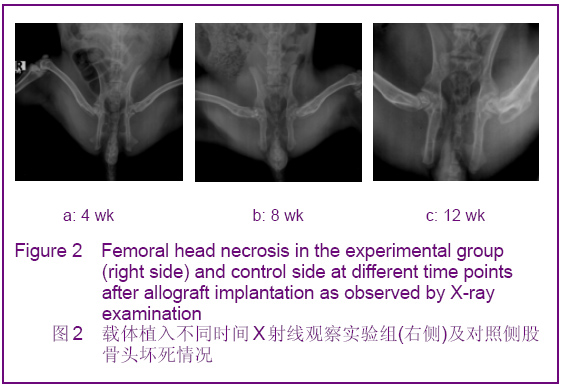
2.3 X射线影像学观察 植入后第4周实验组(右侧)病灶区内模糊,与周围组织界限清楚,对照侧见明显的骨缺损,见图2a。植入后第8周X射线观察实验组(右侧)病灶区内出现散在的密度增高影,较周围组织稍低,对照侧骨缺损区内模糊,与周围组织界限清楚,依然可见缺损轮廓,见图2b。植入后第12周X射线观察实验组(右侧)病灶区内密度增高影,较周围组织稍高,对照侧骨缺损区内有点状骨密度增高影,缺损轮廓依然可见,见图2c。"

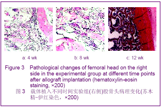
2.4 病理学观察 实验侧(骨髓间充质干细胞复合松质骨载体)4周,充填区周围有肉芽组织形成,间充质细胞大量增生,新生组织开始呈网状长入填充物,周边有明显的成骨反应,有较多的新骨形成,纤维肉芽样组织向载体内长入,植入物表面有纤维骨痂形成,与骨端形成纤维性连接,界限不明显,见图3a;8周,周边新骨带明显增宽,成骨反应向中心部推进,植入物表面有大量骨痂形成,与骨端连接紧密,少部分标本形成连续性骨桥,见图3b。12周,部分充填区为幼稚的骨小梁所占据,表面有较多的成骨细胞,部分充填区广布相对较成熟的骨小梁,但较正常骨小梁粗、排列不够规则,骨痂形成,可见植入物完全骨化,缺损区完全被新骨代替,见图3c。 对照组(单纯载体植入):4周,植入物体积无变化,纤维组织包裹植入物,其表面有骨样组织形成;8周,植入物两端有少量新骨形成,材料中部有纤维骨样组织;12周,植入物两端有新骨包绕,与骨端连接紧密,植入物部分被新骨代替,骨痂未包绕。对照组左侧,12周时缺损区仍无骨组织形成,仅为软组织充填。"
| [1] Yu XZ, Chen XL, Wang YZ, et al. Zhongguo Jiaoxing Waike Zazhi. 2002;9(4):374-377.于学忠,陈晓亮,王英振,等.股骨头缺血性坏死骨髓活性的研究[J].中国矫形外科杂志,2002,9(4):374-377.[2] Hernigou P, Beaujean F, Lambotte JC. Decrease in the mesenchymal stem-cell pool in the proximal femur in corticosteroid-induced osteonecrosis. J Bone Joint Surg Br. 1999;81(2):349-355.[3] Shahdadfar A, Frønsdal K, Haug T, et al. In vitro expansion of human mesenchymal stem cells: choice of serum is a determinant of cell proliferation, differentiation, gene expression, and transcriptome stability. Stem Cells. 2005; 23(9):1357-1366.[4] Yu Y, Zhao G, Xu K, et al. Zhongguo Zuzhi Gongcheng Yanjiu yu Linchuang Kangfu. 2008;12(8):1449-1452.于音,赵刚,许侃,等.兔骨髓间充质干细胞体外培养向成骨和成脂方向的诱导分化[J].中国组织工程研究与临床康复,2008,12(8):1449-1452.[5] Xu ZW, Zhang JX, Li J, et al. Zhongyi Zhenggu. 2005;17(4): 1-3.徐展望,张建新,李军,等.骨碎补提取液对兔骨髓基质细胞增殖的影响[J].中医正骨,2005,17(4):1-3.[6] Tan GQ. Shandong Zhongyiyao Daxue. 2005.谭国庆.骨碎补提取液对体外培养兔骨髓基质细胞向成骨细胞分化影响的实验研究[D].山东中医药大学,2005.[7] Yang ZL, Wei YJ, He ZJ, et al. Zhongguo Zhongyao Zazhi. 2001;26(10):683-684.杨中林,韦英杰,何执静,等.骨碎补不同炮制品中总黄酮及柚皮苷含量测定[J].中国中药杂志,2001,26(10):683-684.[8] Wang YS, Li JP. Zhongguo Zhongyao Zazhi. 1998;23(11): 685-686.王跃生,李计萍.骨碎补中柚皮甙的薄层定性定量方法的研究[J].中国中药杂志,1998,23(11):685-686.[9] Gronthos S, Zannettino AC, Hay SJ, et al. Molecular and cellular characterisation of highly purified stromal stem cells derived from human bone marrow. J Cell Sci. 2003;116(Pt 9):1827-1835.[10] Zhu XQ, Ma XF, He YL, et al. Zhongguo Zuzhi Gongcheng Yanjiu yu Linchuang Kangfu. 2008;12(27):5226-5229.朱肖奇,马雪峰,贺用礼,等.生物衍生骨复合成骨诱导骨髓间充质干细胞构建组织工程化骨修复兔桡骨-骨膜缺损的血管化进程[J].中国组织工程研究与临床康复,2008,12(27):5226-5229.[11] Mauney JR, Kaplan DL, Volloch V. Matrix-mediated retention of osteogenic differentiation potential by human adult bone marrow stromal cells during ex vivo expansion. Biomaterials. 2004;25(16):3233-3243.[12] Iwata H, Sakano S, Itoh T, et al. Demineralized bone matrix and native bone morphogenetic protein in orthopaedic surgery. Clin Orthop Relat Res. 2002;(395):99-109.[13] Barry FP, Murphy JM. Mesenchymal stem cells: clinical applications and biological characterization. Int J Biochem Cell Biol. 2004;36(4):568-584. |
| [1] | Lyu Ruyue, Gu Lulu, Liu Qian, Zhou Siyi, Li Beibei, Xue Letian, Sun Peng. Regulatory mechanisms of exosome secretion and its application prospects in biomedicine [J]. Chinese Journal of Tissue Engineering Research, 2026, 30(1): 184-193. |
| [2] | Xu Canli, He Wenxing, Wang Yuping, Ba Yinying, Chi Li, Wang Wenjuan, Wang Jiajia. Research context and trend of TBK1 in autoimmunity, signaling pathways, gene expression, tumor prevention and treatment [J]. Chinese Journal of Tissue Engineering Research, 2026, 30(在线): 1-11. |
| [3] | Liu Xun, Ouyang Hougan, Pan Rongbin, Wang Zi, Yang Fen, Tian Jiaxuan . Optimal parameters for physical interventions in bone marrow mesenchymal stem cell differentiation [J]. Chinese Journal of Tissue Engineering Research, 2025, 29(31): 6727-6732. |
| [4] | Hu Enxi, He Wenying, Tao Xiang, Du Peijing, Wang Libin. Regulation of THZ1, an inhibitor of cyclin-dependent kinase 7, on stemness of glioma stem cells and its mechanism [J]. Chinese Journal of Tissue Engineering Research, 2025, 29(25): 5374-5381. |
| [5] | Lin Meiyu, Zhao Xilong, Gao Jing, Zhao Jing, Ruan Guangping. Action mechanism and progress of stem cells against ovarian granulosa cell senescence [J]. Chinese Journal of Tissue Engineering Research, 2025, 29(25): 5414-5421. |
| [6] | Tian Zhenli, Zhang Xiaoxu, Fang Xingyan, Xie Tingting. Effects of sodium arsenite on lipid metabolism in human hepatocytes and regulatory factors [J]. Chinese Journal of Tissue Engineering Research, 2025, 29(23): 4956-4964. |
| [7] | Han Fang, Shu Qing, Jia Shaohui, Tian Jun. Electrotactic migration and mechanisms of stem cells [J]. Chinese Journal of Tissue Engineering Research, 2025, 29(23): 4984-4992. |
| [8] | Hu Chen, Jiang Ying, Chen Jia, Qiao Guangwei, Dong Wen, Ma Jian. Preparation and characterization of alendronate/chitosan/polyvinyl alcohol composite hydrogel films [J]. Chinese Journal of Tissue Engineering Research, 2025, 29(22): 4720-4730. |
| [9] | Yang Chao, Luo Zongping. Small molecule drug TD-198946 enhances osteogenic differentiation of rat bone marrow mesenchymal stem cells [J]. Chinese Journal of Tissue Engineering Research, 2025, 29(13): 2648-2654. |
| [10] | Li Xiaofeng, Zhao Duo, Ouyang Qin, Pang Zixiang, Li Yuquan, Chen Qianfen. Protective effect of mangiferin on oxidative stress injury in rat bone marrow mesenchymal stem cells [J]. Chinese Journal of Tissue Engineering Research, 2025, 29(13): 2669-2674. |
| [11] | Hu Zezun, Yang Fanlei, Xu Hao, Luo Zongping. Effect of surface roughness of polydimethylsiloxane on osteogenic differentiation of bone marrow mesenchymal stem cells under stretching conditions [J]. Chinese Journal of Tissue Engineering Research, 2025, 29(10): 1981-1989. |
| [12] | Yang Zhihang, Sun Zuyan, Huang Wenliang, Wan Yu, Chen Shida, Deng Jiang. Nerve growth factor promotes chondrogenic differentiation and inhibits hypertrophic differentiation of rabbit bone marrow mesenchymal stem cells [J]. Chinese Journal of Tissue Engineering Research, 2025, 29(7): 1336-1342. |
| [13] | Huang Ting, Zheng Xiaohan, Zhong Yuanji, Wei Yanzhao, Wei Xufang, Cao Xudong, Feng Xiaoli, Zhao Zhenqiang. Effects of macrophage migration inhibitory factor on survival, proliferation, and differentiation of human embryonic stem cells [J]. Chinese Journal of Tissue Engineering Research, 2025, 29(7): 1380-1387. |
| [14] | Liu Haowen, Qiao Weiping, Meng Zhicheng, Li Kaijie, Han Xuan, Shi Pengbo. Regulation of osteogenic effects by bone morphogenetic protein/Wnt signaling pathway: revealing molecular mechanisms of bone formation and remodeling [J]. Chinese Journal of Tissue Engineering Research, 2025, 29(3): 563-571. |
| [15] | Zhou Shijie, Li Muzhe, Yun Li, Zhang Tianchi, Niu Yuanyuan, Zhu Yihua, Zhou Qinfeng, Guo Yang, Ma Yong, Wang Lining. Effect of Wenshen Tongluo Zhitong formula on mouse H-type bone microvascular endothelial cell/bone marrow mesenchymal stem cell co-culture system [J]. Chinese Journal of Tissue Engineering Research, 2025, 29(1): 8-15. |
| Viewed | ||||||
|
Full text |
|
|||||
|
Abstract |
|
|||||