Chinese Journal of Tissue Engineering Research ›› 2012, Vol. 16 ›› Issue (49): 9162-9167.doi: 10.3969/j.issn.2095-4344.2012.49.008
Previous Articles Next Articles
Multi-directional induced differentiation of rabbit adipose-derived stem cells
Wang Qing-fu , Chen Zhuang-hong, Cai Xian-hua, Xie Yong-hui, Diao Bo, Liu Qin
- Department of Orthopedics, Wuhan General Hospital of Guangzhou Military Command, Wuhan 430070, Hubei Province, China
-
Received:2012-06-01Revised:2012-10-25Online:2012-12-02Published:2013-01-16 -
Contact:Chen Zhuang-hong, Professor, Chief physician, Doctoral supervisor Department of Orthopedics, Wuhan General Hospital of Guangzhou Military Command, Wuhan 430070, Hubei Province, China -
About author:Wang Qing-fu☆, Studying for doctorate, Department of Orthopedics, Wuhan General Hospital of Guangzhou Military Command, Wuhan 430070, Hubei Province, China -
Supported by:Supported by: “Eleventh Five-Year” Project of PLA , No. 06G047*
CLC Number:
Cite this article
Wang Qing-fu,Chen Zhuang-hong, Cai Xian-hua, Xie Yong-hui, Diao Bo, Liu Qin. Multi-directional induced differentiation of rabbit adipose-derived stem cells[J]. Chinese Journal of Tissue Engineering Research, 2012, 16(49): 9162-9167.
share this article
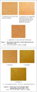
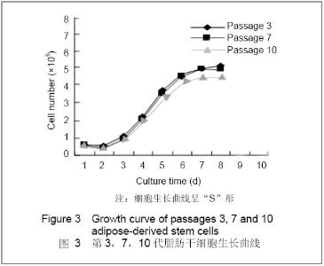
2.1 细胞形态学观察 原代细胞接种后两三个小时开始贴壁,初为圆形、椭圆形,细胞大小不等,12 h后可见部分细胞开始伸展为短梭形、三角形,24 h大部分细胞贴壁。细胞在最初三四天处于潜伏期,生长较缓慢,然后细胞进入增殖期,细胞开始形成团簇状增生灶,长梭形纤维样细胞数量明显增多,细胞逐渐彼此相连,铺满瓶底,长梭形、三角形细胞多见。7-9 d原代脂肪干细胞可达80%左右融合,见图1。传代细胞增殖速度较原代快,四五天可长至 80%融合。传代后细胞形态为长梭形,多次传代后,细胞形态基本保持不变,增殖旺盛,见图2。"

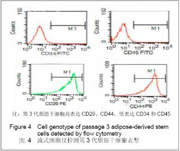
2.2 传代细胞增殖能力 用CCK-8法检测第3,7,10代细胞增殖能力。结果显示:传代培养的脂肪干细胞最初一两天细胞潜伏期逐渐向增殖期过渡,第4天进入对数生长期,随着时间增加而增殖,第7天进入平台期,细胞生长曲线呈“S”形,第3,7,10代细胞的生长曲线类似,见图3。"

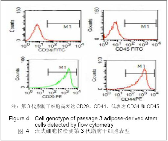
2.3 细胞周期 取3代细胞经过流式细胞仪检测结果显示:G1期细胞占绝大多数,达到87.16%,S期为 6.23%,这说明细胞正向增殖期过渡,处于活跃状态。 2.4 脂肪干细胞表型鉴定 流式细胞仪检测提示,第3,6代脂肪干细胞均高表达间充质干细胞的相关标志物CD29、CD44,阳性率超过90%;低表达造血干细胞相关标志物CD34和血细胞相关标志物CD45,阳性率超过5%,且CD29、CD44随着传代表达逐渐增高,CD34、CD45随细胞传代表达逐渐降低。见图4。"
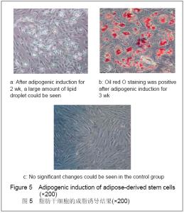
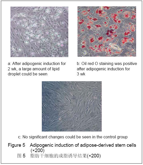
2.5 脂肪干细胞的成脂诱导结果 脂肪干细胞更换成脂诱导培养液后细胞增殖缓慢,诱导后5 d胞浆内出现少量脂滴并逐渐聚集,细胞形态亦明显变化,逐渐由长梭形变为多边形、椭圆形或不规则。2周时出现了大量的脂滴,见图5a,随着诱导时间的延长,脂滴逐渐增多增大,3周时油红O染色阳性,细胞内出现大量红染颗粒,见图5b,对照组脂肪干细胞无明显变化。证实脂肪干细胞可被诱导向脂肪分化,见图5c"
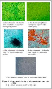
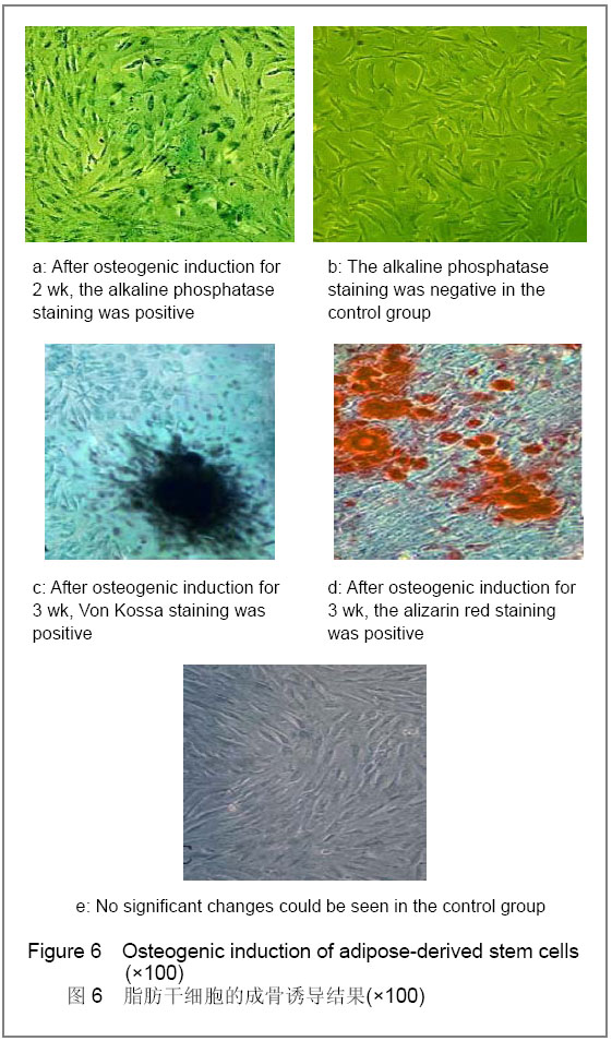
2.6 脂肪干细胞的成骨诱导结果 见图6。诱导脂肪干细胞更换成骨诱导培养液后细胞增殖稍慢,诱导后3- 5 d后,可见细胞形态逐渐改变,由长梭形变为类圆形、多边形或不规则形,并且体积有所增大;1周后细胞开始呈簇状生长,2周时细胞团逐渐增大。 2周后碱性磷酸酶染色阳性,可见胞浆内出现蓝色沉淀,见图6a,对照组细胞颜色及形态无明显改变仍呈成纤维细胞样外观,碱性磷酸酶染色为阴性,见图6b; Vonkossa染色见钙结节呈黑色,即Vonkossa染色阳性,见图6c。3周茜素红染色见钙结节呈桔红色,即茜素红染色阳性,见图6d;对照组均为阴性,见图6e。"
| [1] Zuk PA, Zhu M, Mizuno H, et al.Multilineage cellsfrom human adipose tissue:implications for cell based therapies.Tissue Eng,2001,7(2):211-228.[2] National Academy of Sciences,Institute of Laboratory Animal Resources,Commission on Life Sciences,National Research Council.Guide for the care and use of laboratory animals.[3] Zuk PA,Zhu M, Ashjian P, et al. Human adipose tissue is a source ofmultipotent stem cells. MolBiol Cell.2002;13(12): 4279-4295.[4] Williams KJ, Picou AA, Kish SL, et al. Isolation and characterization of porcine adipose tissue-derived adult stem cells. Cells Tissues Organs.2008;188(3): 251-258. [5] Miyazaki M, Zuk PA, Zou J, et al. Comparison of human mesenchymal stem cells derived from adipose tissue and bone marrow for ex vivo gene therapy in rat spinal fusion model. Spine. 2008;33(8): 863-869. [6] CherubinoM,Marra KG. Adipose-derived stem cells for soft tissue reconstruction. Regen Med.2009;4(1):109-117. [7] Anghileri E, Marconi S, Pignatelli A, et al. Neuronal differentiation potential of human adipose-derived mesenchymal stem cells. Stem Cells Dev. 2008;17(5): 909-916. [8] Zaminy A, RagerdiKashani I, Barbarestani M, et al. Osteogenic differentiation of rat mesenchymal stem cells from adipose tissue in comparison with bone marrow mesenchymal stem cells: melatonin as a differentiation factor. Iran Biomed J.2008;12(3): 133-141. [9] Gimble JM, Guilak F. Adipose-derived adult stemcells: isolation,characterization,anddifferentiation potential. Cytotherapy.2003,5(5):362-369.[10] Kim DH, Je CM, Sin JY, et al. Effect of partial hepatectomy on in vivo engraftment after intravenous administration of human adipose tissue stromal cells in mouse. Microsurgery.2003; 23(5):424-431.[11] Rangappa S, Fen C, Lee EH, et al. Transformation of adult mesenchymal stem cells isolated from the fatty tissue into cardiomyocytes. Ann Thorac Surg.2003;75(3):775-779.[12] Wang Y, Chen GH, Shao JH, et al. Shandong DaxueXuebao. 2005;43(7):578-581.[13] Sgodda M, Aurich H, Kleist S, et al. Hepatocyte differentiation of mesenchymal stem cells from rat peritoneal adipose tissue in vitro and in vivo. Exp Cell Res.2007;313(13):2875-2886. [14] Afizah H, Yang Z, Hui JH, et al. A comparison between the chondrogenic potential of human bone marrow stem cells (BMSCs) and adipose-derived stem cells (ADSCs) taken from the same donors. Tissue Eng.2007;13(4):659-666.[15] Cam pagnoli C, Roberts IA, Kumar S, et al. Identification ofmesenchymal stem/progenitor cells in human firsttrim ester fetal blood, liver and bone marrow.B lood, 2001,98(8):2396- 2402.[16] Safford KM, Hicok KC, Safford SD, et al. Neurogenic differentiation of murineand human adipose-derived stromal cells.BiochemBiophys Res Commun.2002;294(2):371-379[17] Gronthos S,Franklin DM, Leddy HA, et al. Surfaceproteincharacterization of human adipose tissue-derived stromal cells. Cell Physiol.2001;189(1):54-63.[18] Boquest AC, Shahdadfar A, Frønsdal K, et al.Isolation and transcription profiling of purified uncultured human stromal stem cells: alteration of gene expressionafter in vitrocellculture. MolBiolCell.2005;16(3):1131-1141. |
| [1] | Lyu Ruyue, Gu Lulu, Liu Qian, Zhou Siyi, Li Beibei, Xue Letian, Sun Peng. Regulatory mechanisms of exosome secretion and its application prospects in biomedicine [J]. Chinese Journal of Tissue Engineering Research, 2026, 30(1): 184-193. |
| [2] | Xu Canli, He Wenxing, Wang Yuping, Ba Yinying, Chi Li, Wang Wenjuan, Wang Jiajia. Research context and trend of TBK1 in autoimmunity, signaling pathways, gene expression, tumor prevention and treatment [J]. Chinese Journal of Tissue Engineering Research, 2026, 30(在线): 1-11. |
| [3] | Liu Xun, Ouyang Hougan, Pan Rongbin, Wang Zi, Yang Fen, Tian Jiaxuan . Optimal parameters for physical interventions in bone marrow mesenchymal stem cell differentiation [J]. Chinese Journal of Tissue Engineering Research, 2025, 29(31): 6727-6732. |
| [4] | Hu Enxi, He Wenying, Tao Xiang, Du Peijing, Wang Libin. Regulation of THZ1, an inhibitor of cyclin-dependent kinase 7, on stemness of glioma stem cells and its mechanism [J]. Chinese Journal of Tissue Engineering Research, 2025, 29(25): 5374-5381. |
| [5] | Lin Meiyu, Zhao Xilong, Gao Jing, Zhao Jing, Ruan Guangping. Action mechanism and progress of stem cells against ovarian granulosa cell senescence [J]. Chinese Journal of Tissue Engineering Research, 2025, 29(25): 5414-5421. |
| [6] | Tian Zhenli, Zhang Xiaoxu, Fang Xingyan, Xie Tingting. Effects of sodium arsenite on lipid metabolism in human hepatocytes and regulatory factors [J]. Chinese Journal of Tissue Engineering Research, 2025, 29(23): 4956-4964. |
| [7] | Han Fang, Shu Qing, Jia Shaohui, Tian Jun. Electrotactic migration and mechanisms of stem cells [J]. Chinese Journal of Tissue Engineering Research, 2025, 29(23): 4984-4992. |
| [8] | Hu Chen, Jiang Ying, Chen Jia, Qiao Guangwei, Dong Wen, Ma Jian. Preparation and characterization of alendronate/chitosan/polyvinyl alcohol composite hydrogel films [J]. Chinese Journal of Tissue Engineering Research, 2025, 29(22): 4720-4730. |
| [9] | Yang Chao, Luo Zongping. Small molecule drug TD-198946 enhances osteogenic differentiation of rat bone marrow mesenchymal stem cells [J]. Chinese Journal of Tissue Engineering Research, 2025, 29(13): 2648-2654. |
| [10] | Li Xiaofeng, Zhao Duo, Ouyang Qin, Pang Zixiang, Li Yuquan, Chen Qianfen. Protective effect of mangiferin on oxidative stress injury in rat bone marrow mesenchymal stem cells [J]. Chinese Journal of Tissue Engineering Research, 2025, 29(13): 2669-2674. |
| [11] | Hu Zezun, Yang Fanlei, Xu Hao, Luo Zongping. Effect of surface roughness of polydimethylsiloxane on osteogenic differentiation of bone marrow mesenchymal stem cells under stretching conditions [J]. Chinese Journal of Tissue Engineering Research, 2025, 29(10): 1981-1989. |
| [12] | Yang Zhihang, Sun Zuyan, Huang Wenliang, Wan Yu, Chen Shida, Deng Jiang. Nerve growth factor promotes chondrogenic differentiation and inhibits hypertrophic differentiation of rabbit bone marrow mesenchymal stem cells [J]. Chinese Journal of Tissue Engineering Research, 2025, 29(7): 1336-1342. |
| [13] | Huang Ting, Zheng Xiaohan, Zhong Yuanji, Wei Yanzhao, Wei Xufang, Cao Xudong, Feng Xiaoli, Zhao Zhenqiang. Effects of macrophage migration inhibitory factor on survival, proliferation, and differentiation of human embryonic stem cells [J]. Chinese Journal of Tissue Engineering Research, 2025, 29(7): 1380-1387. |
| [14] | Liu Haowen, Qiao Weiping, Meng Zhicheng, Li Kaijie, Han Xuan, Shi Pengbo. Regulation of osteogenic effects by bone morphogenetic protein/Wnt signaling pathway: revealing molecular mechanisms of bone formation and remodeling [J]. Chinese Journal of Tissue Engineering Research, 2025, 29(3): 563-571. |
| [15] | Zhou Shijie, Li Muzhe, Yun Li, Zhang Tianchi, Niu Yuanyuan, Zhu Yihua, Zhou Qinfeng, Guo Yang, Ma Yong, Wang Lining. Effect of Wenshen Tongluo Zhitong formula on mouse H-type bone microvascular endothelial cell/bone marrow mesenchymal stem cell co-culture system [J]. Chinese Journal of Tissue Engineering Research, 2025, 29(1): 8-15. |
| Viewed | ||||||
|
Full text |
|
|||||
|
Abstract |
|
|||||