Chinese Journal of Tissue Engineering Research ›› 2026, Vol. 30 ›› Issue (18): 4649-4662.doi: 10.12307/2026.750
Previous Articles Next Articles
Effects and molecular mechanisms of light exposure on neurotransmitter release in a rat model of heart failure
Ruan Lin1, Li Jia1, Shi Linan1, Zhu Hong1, Wu Dongning2, 3, Zheng Di1, Li Yupeng1, Zhao Yan1
- 1The Second Affiliated Hospital of Liaoning University of Traditional Chinese Medicine, Shenyang 110000, Liaoning Province, China; 2Xidan Outpatient, Guang’anmen Hospital, Chinese Academy of Chinese Medical Sciences, Beijing 100031, China; 3School of Chinese Medicine & State Key Laboratory of Mechanism and Quality of Chinese Medicine, Macau University of Science and Technology, Macao Special Administrative Region 999078, China
-
Received:2025-08-08Revised:2025-10-09Online:2026-06-28Published:2025-12-04 -
Contact:Li Yupeng, MS, Chief physician, The Second Affiliated Hospital of Liaoning University of Traditional Chinese Medicine, Shenyang 110000, Liaoning Province, China Co-corresponding author: Zhao Yan, PhD, Associate chief physician, The Second Affiliated Hospital of Liaoning University of Traditional Chinese Medicine, Shenyang 110000, Liaoning Province, China -
About author:Ruan Lin, MS, Chief physician, The Second Affiliated Hospital of Liaoning University of Traditional Chinese Medicine, Shenyang 110000, Liaoning Province, China -
Supported by:Xingliao Talent Program - Outstanding Young Talent Program, No. XLYC2007076 (to RL)
CLC Number:
Cite this article
Ruan Lin, Li Jia, Shi Linan, Zhu Hong, Wu Dongning, Zheng Di, Li Yupeng, Zhao Yan. Effects and molecular mechanisms of light exposure on neurotransmitter release in a rat model of heart failure[J]. Chinese Journal of Tissue Engineering Research, 2026, 30(18): 4649-4662.
share this article
Add to citation manager EndNote|Reference Manager|ProCite|BibTeX|RefWorks
2.1 实验动物数量分析 实验动物数量的确定参考了动物实验样本量估算的通用原则,确保实验设计的科学性和伦理合理性。根据实验设计和预实验结果,初始纳入数量为每组纳入8只SD大鼠,共5组,总计40只。在实验过程中,部分动物因手术并发症或自然死亡而脱失,具体脱失数量为4只,同时依据统计学的分组原则,最终进入结果分析的动物数量共计30只。 2.2 光暴露加剧心力衰竭进展的动物研究 2.2.1 光暴露对心力衰竭大鼠血清神经递质及激素的影响 各组大鼠血清神经递质的变化趋势见图1。相比于空白组,心力衰竭造模后各时间点大鼠去甲肾上腺素、5-羟色胺水平明显上升(P < 0.05),值得关注的是,光暴露可提升大鼠的血清去甲肾上腺素和5-羟色胺水平,两项指标均表现出类似的变化趋势:①2周时,光暴露模型组>模型组(P < 0.01),光暴露对照组>空白组(P < 0.05);②4周时,光暴露模型组>模型组(P < 0.05),而光暴露对照组>空白组(P > 0.05);③8周时,光暴露模型组>模型组(P < 0.05),而光暴露对照组>空白组(P > 0.05);④光暴露治疗组药物干预可有效降低光暴露鼠的血清去甲肾上腺素水平,在2,4,6,8周各时间点与空白组比较差异均无显著性意义。上述结果提示,光暴露会影响心力衰竭大鼠的神经递质浓度,加速心力衰竭大鼠去甲肾上腺素及5-羟色胺的释放。 为进一步研究光暴露对心力衰竭大鼠心脏相关激素指标的影响,研究考察了光暴露对于心力衰竭大鼠心房钠尿肽、脑钠肽、血管紧张素Ⅱ及内皮素1的变化水平,见图1K-AD。①2周时,心力衰竭造模后大鼠的血清脑钠肽、心钠素、血管紧张素Ⅱ及内皮素1的水平均显著升高(P < 0.05);但是,光暴露却会加剧心力衰竭模型大鼠血清激素水平的上升,光暴露模型组心钠素(P < 0.05)、脑钠肽、血管紧张素Ⅱ和内皮素1水平(P > 0.05)均高于模型组;②4,6周时,心力衰竭造模大鼠的血清脑钠肽、心钠素、血管紧张素Ⅱ及内皮素1的水平并未降低,仍然显著性高于空白组(P < 0.05),而此时光暴露对于心力衰竭模型大鼠血清激素水平的上升的刺激作"
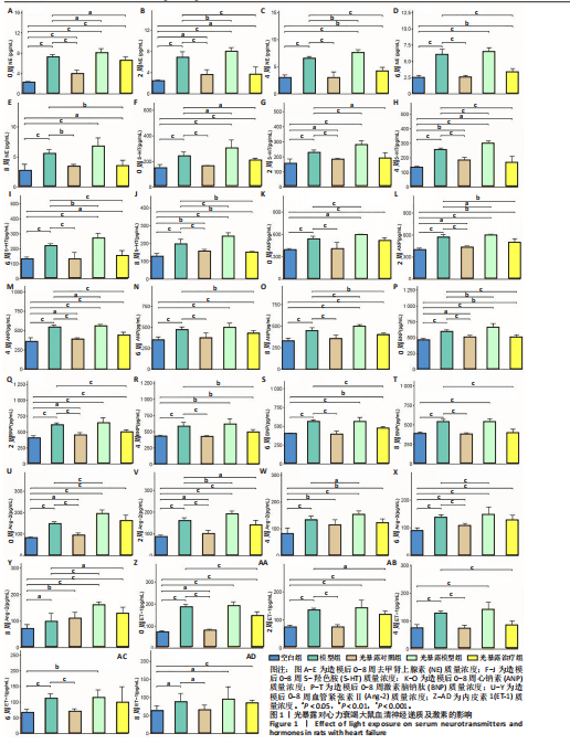
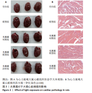
用被弱化,虽然光暴露模型组的心钠素、脑钠肽、血管紧张素Ⅱ及内皮素1仍然高于模型组,但组间对比差异已无显著性意义(P > 0.05);③8周时,模型组大鼠的血清脑钠肽和心钠素仍然高于空白组(P < 0.05),而血管紧张素Ⅱ及内皮素1同空白组相比差异无显著性意义(P > 0.05);④单纯光暴露对于大鼠血清激素的影响并不大,在整个实验期间,空白组同光暴露对照组上述4种激素的水平变化差异无显著性意义(P > 0.05);⑤同时,药物干预可迅速缓解光暴露所致的血管激素(血管紧张素Ⅱ及内皮素1)水平的上升:2周时,光暴露治疗组血管紧张素Ⅱ及内皮素1水平即已低于模型组(P < 0.05),并在4周时,同空白组比较差异无显著性意义(P > 0.05);另一方面,药物在缓解心力衰竭损伤的过程中,也会大幅降低血清脑钠肽和心钠素的水平,光暴露治疗组的脑钠肽水平在4周时、心钠素在6周时,开始显著性的低于模型组(P < 0.05);但在8周时,光暴露治疗组的血清脑钠肽和心钠素仍然显著性高于空白组。 基于上述结果可知,光暴露在心力衰竭的早期可以通过收缩血管调整血管内皮因子,加速心力衰竭的进展,随着时间延长其对血管功能的调节作用会逐渐减弱,但仍然会通过脑钠肽或者心钠素影响心力衰竭的进展。 2.2.2 光暴露对心力衰竭大鼠心脏病理形态的影响 大鼠心脏的组织形态如图2A所示。模型组和光暴露模型组的心脏组织都出现了明显的充血症状,可见心脏大体上出现了明显的毛细血管。从大体形态上看,光暴露治疗组、光暴露模型组同模型组相比并无显著性的差异。结果提示,在造模期间,光暴露虽然可以影响心脏的功能,然而单纯的光暴露,心脏的大体对比并不明显。为进一步展示大鼠的心脏内部形态变化,对各组大鼠的心脏进行了病理分析,药物干预对光暴露下心力衰竭大鼠心肌组织病理损伤的保护作用见图2B。无药物干预的条件下,光暴露模型组及模型组均呈现了明显的损伤,细胞出现了肿胀、核皱缩、空泡等现象;经过药物干预后,光暴露治疗组大鼠的心肌细胞损伤得到了明显的缓解;空白组内心肌空泡现象不多见。 2.2.3 光暴露对心力衰竭大鼠Nrf2/HO-1/NQO-1信号通路的调节作用 为考察在心力衰竭进展过程中光暴露的作用,理清其对Nrf2/HO-1/NQO-1信号通路的"
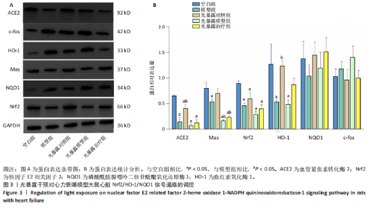
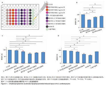
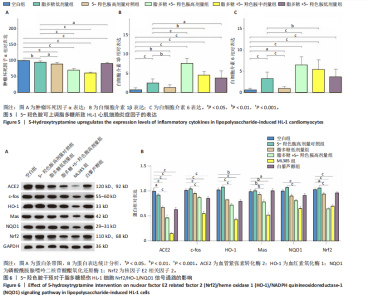
调节作用,研究考察了该信号通路上的6种蛋白的表达情况,见图3。心力衰竭造模会导致大鼠心脏c-fos上调,NQO1下调,与空白组相比差异无显著意义(P > 0.05);血管紧张素转换酶2,Mas、Nrf2及HO-1下调,与空白组相比差异有显著性意义(P < 0.05)。另一方面,光暴露也会影响上述蛋白的表达,如光暴露对照组的血管紧张素转换酶2低于空白组(P < 0.05)而高于模型组(P < 0.05)。相比于空白组,光暴露对照组的Mas、c-fos、HO-1均下调,但差异无显著性意义(P > 0.05);而在光暴露模型组内,血管紧张素转换酶2,Mas、Nrf2及HO-1相比于空白组均显著下调(P < 0.05)。相比于光暴露模型组,光暴露治疗组Nrf2/HO-1/NQO1信号通路上的3个靶点均产生了明显的上调作用(P < 0.05)。由上述结果提示,光暴露会抑制Nrf2/HO-1/NQO1信号通路,加速心力衰竭的进展。 基于上述结果可知,光暴露会提升心力衰竭模型大鼠的5-羟色胺水平,提升心钠素及脑钠肽的水平加剧心脏的损伤,而其潜在的机制为Nrf2/HO-1/NQO-1信号通路的抑制。可推测,血清5-羟色胺上升会通过抑制Nrf2/HO-1/NQO-1信号通路,加速心肌细胞的损伤。为了进一步验证5-羟色胺在心力衰竭进展中的作用,研究利用HL-1细胞模型考察了5-羟色胺对于脂多糖所致HL-1损伤模型的影响。 2.3 5-羟色胺通过Nrf2/HO-1/NQO-1加速HL-1心肌细胞损伤的机制研究 2.3.1 心力衰竭心肌细胞模型的构建 见图4A。研究考察不同浓度下脂多糖对于HL-1细胞增殖的影响,见图4B。由结果可以看出,脂多糖可损伤HL-1细胞,且呈现浓度相关(脂多糖高、中、低剂量组两两相比差异均有显著性意义),其中,脂多糖质量浓度在1 μg/mL时(脂多糖低剂量组)细胞的相对增殖率为88.25%。基于此次研究的结果、文献报道及预实验的结果[9],为降低脂多糖对于HL-1细胞损伤后相关蛋白表达的影响,此次研究以1 μg/mL脂多糖进行后续的实验研究。 2.3.2 5-羟色胺对于HL-1细胞损伤及相关炎症因子水平的影响 见图4C。在无脂多糖的条件下,即使较高的5-羟色胺浓度(500 mg/mL),也不会直接影响HL-1细胞的增殖率。而当以1 μg/mL脂多糖干预HL-1细胞后,5-羟色胺的添加会直接降低细胞的增殖率,且呈现浓度依赖关系,其中5-羟色胺高剂量条件下(500 mg/mL)影响最大,组间对比差异有显著性意义(P < 0.05);而5-羟色胺中剂量、低剂量及脂多糖低剂量组相比差异无显著性意义。上述结果提示,5-羟色胺可增加HL-1细胞的损伤。为进一步获得其损伤的机制,研究则分别考察了干预过程中各分组上清液内相关炎性细胞因子的水平变化,见图5。 基于获得的结果可以看出,伴随着脂多糖的添加,HL-1细胞内肿瘤坏死因子α、白细胞介素1β及白细胞介素6等炎性细胞因子的相对表达量显著上升(P < 0.001),而随着5-羟色胺的添加,同脂多糖组相比,5-羟色胺低剂量组细胞上清液内的肿瘤坏死因子α及白细胞介素1β的相对表达均显著增加 (P < 0.05),随着加入5-羟色胺浓度的上升,则白细胞介素6的表达开始逐渐上升(P < 0.05),提示5-羟色胺可增加脂多糖所致的HL-1细胞内白细胞介素6的浓度,且炎症细胞因子的表达呈剂量依赖关系,在肿瘤坏死因子α及白细胞介素1β也呈现了类似的趋势。另一方面,虽然5-羟色胺也可提升各组细胞内肿瘤坏死因子α、白细胞介素1β及白细胞介素6的相对表达量,但是它们同空白组相比,差异无显著性意义(P > 0.05)。 2.3.3 5-羟色胺通过抑制Nrf2/HO-1/NQO-1加速心力衰竭心肌细胞损伤的机制研究 基于获得的结果可知,5-羟色胺会增加脂多糖对于心肌细胞增殖的影响,同时进一步上调脂多糖所致相关炎性因子的表达,为进一步理清上述生物学现象的调控机制,利用挽救实验验证了5-羟色胺对于HL-1损伤模型的保护作用,见图4D。Nrf2抑制剂ML385会进一步降低5-羟色胺所致脂多糖损伤HL-1细胞的增殖,而白藜芦醇(Nrf2激动剂)则大幅提升上述细胞的增殖情况,抵消其所致的损伤状态。ML385降低了5-羟色胺所致脂多糖损伤HL-1细胞的核因子E2相关因子2、血红素氧化酶1、磷酸酰胺腺嘌呤二核苷酸醌氧化还原酶1蛋白的表达,而白藜芦醇则显著增加了上述蛋白的表达,见图6。上述结果提示,5-羟色胺是通过Nrf2极其下游的信号通过,加速HL-1细胞损伤的进展。"

"
"

| [1] 孙珂,王婷,李静颐,等. 心脏组织工程联合干细胞促进心肌修复的研究进展[J].中国循证心血管医学杂志,2024,16(2):237-240. [2] 董国菊,刘思雨. 射血分数保留心力衰竭中西医病证结合的分期诊断专家共识[J]. 中华中医药学刊,2023,41(5):254-258. [3] 张倩,卫晓红,陈洁,等. 慢性心力衰竭常用动物模型的研究进展及其在中医药研究中的应用[J]. 中国中药杂志,2023,48(3):614-624. [4] MENG D, LI HQ, DEISSEROTH K, et al. Neuronal activity regulates neurotransmitter switching in the adult brain following light-induced stress. Proc Natl Acad Sci U S A. 2018;115(20):5064-5071. [5] MADDALONI G, CHANG YJ, SENFT RA, et al. Adaptation to photoperiod via dynamic neurotransmitter segregation. Nature. 2024;632(8023): 147-156. [6] TEKIYEH MAROOF N, MEHRZADI S, NASEROLESLAMI M, et al. Apelin13 Loaded Nano-Niosomes Confer Cardioprotection in a Rat Model of Myocardial Ischemia Reperfusion by Targeting the Nrf2/HO-1 Pathway. J Cardiovasc Transl Res. 2025;18(3):529-543. [7] 张榕,刘春华,胡爽,等. 夜间光暴露小鼠肝脏非靶向代谢组学研究[J].预防医学,2021,33(2):130-134. [8] 李玮,郝翠芳.夜间光污染对雌性大鼠生殖功能的影响[J].中国妇幼保健,2021,36(6):1379-1381. [9] 刘鑫,朱浩彦,吴嘉贺,等. 基于生物信息学分析预测和验证实验性自身免疫性心肌炎的关键基因[J].武汉大学学报(医学版),2025, 46(1):27-35. [10] 徐建军.内质网应激介导自吞噬对脂多糖诱导HL-1心肌细胞损伤保护机制的研究[D].武汉:华中科技大学,2012. [11] 曹红,张小萌,许琮.镍纹蛋白样β对脂多糖诱导心肌细胞损伤的抑制作用及其机制[J].山东医药,2023,63(23):1-4. [12] 李凡凡,徐阳,王晓旭.紫草素调节Nrf2/HO-1信号通路对实验性大鼠肉芽肿性小叶性乳腺炎的治疗作用研究[J].中国临床解剖学杂志,2024,42(1):26-32. [13] 姚宁丰,佘仁夏,舒艺璇,等.白藜芦醇对脑出血后小胶质细胞功能的影响及其机制[J].解放军医学杂志,2023,48(4):420-430. [14] 曾佑成.基于铁死亡探讨白藜芦醇对脓毒症大鼠心肌损伤的作用机制的研究[D]. 石河子:石河子大学,2023. [15] 中华中医药学会慢性心力衰竭中医诊疗指南项目组.慢性心力衰竭中医诊疗指南(2022年)[J].中医杂志,2023,64(7):743-756. [16] CHICOS AB, KANNANKERIL PJ, KADISH AH, et al.Parasympathetic effects on cardiac electrophysiology during exercise and recovery in patients with left ventricular dysfunction. Am J Physiol Heart Circ Physiol. 2009;297(2):H743-H749. [17] HEIDENREICH PA, BOZKURT B, AGUILAR D, et al. 2022 AHA/ACC/HFSA Guideline for the Management of Heart Failure: A Report of the American College of Cardiology/American Heart Association Joint Committee on Clinical Practice Guidelines. Circulation. 2022;145(18): e895-e1032. [18] MARYAM, VARGHESE TP, B T.Unraveling the complex pathophysiology of heart failure: insights into the role of renin-angiotensin-aldosterone system (RAAS) and sympathetic nervous system (SNS). Curr Probl Cardiol. 2024;49(4):102411. [19] SAYER G, BHAT G.The Renin-Angiotensin-Aldosterone System and Heart Failure. Cardiol Clin. 2014;32(1):21-32. [20] MÖLLER PETRUN A, MARKOTA A.Angiotensin II-Real-Life Use and Literature Review. Medicina (Kaunas). 2024;60(9):1483. [21] YANG P, WU Y, LI F, et al. Activation of ETAR and ETBR in myocardial tissue characterizes heart failure induced by experimental autoimmune myocarditis. BMC Cardiovasc Disord. 2024;24(1):11. [22] 柯元南,Manthey J.慢性充血性心衰的血液动力学和儿茶酚胺,肾素活性及抗利尿激素的变化[J].中华内科杂志,1987,26(1):16-19. [23] 张浩华.精氨酸血管加压素受体拮抗剂在心力衰竭患者中应用效果的研究进展[J].实用心脑肺血管病杂志,2018,26(7): 9-13. [24] SHAO L, SHEN Y, REN C, et al. Inflammation in myocardial infarction: roles of mesenchymal stem cells and their secretome. Cell Death Discov. 2022;8(1):452. [25] ZHANG DY, ANDERSON AS.The Sympathetic Nervous System and Heart Failure. Cardiol Clin. 2014;32(1):33-45, [26] 郭艳琳.下丘脑室旁核CRH神经元激活在慢性充血性心力衰竭中的交感兴奋作用及机制研究[D].太原:山西医科大学,2011. [27] KANG YM, HE RL, YANG LM, et al. Brain tumour necrosis factor-α modulates neurotransmitters in hypothalamic paraventricular nucleus in heart failure. Cardiovasc Res. 2009;83(4):737-746. [28] BO W, CAI M, MA Y, et al. Manipulation of Glutamatergic Neuronal Activity in the Primary Motor Cortex Regulates Cardiac Function in Normal and Myocardial Infarction Mice. Adv Sci (Weinh). 2024; 11(20):e2305581. [29] TÄHKÄMÖ L, PARTONEN T, PESONEN AK.Systematic review of light exposure impact on human circadian rhythm.Chronobiol Int. 2019;36(2):151-170. [30] AN K, ZHAO H, MIAO Y, et al.A circadian rhythm-gated subcortical pathway for nighttime-light-induced depressive-like behaviors in mice. Nat Neurosci. 2020;23(7):869-880. [31] HAGE B, BRITTON B, DANIELS D, et al. Diminution of Heart Rate Variability in Bipolar Depression. Front Public Health. 2017;5:312. [32] MASON IC, GRIMALDI D, REID KJ,et al.Light exposure during sleep impairs cardiometabolic function. Proc Natl Acad Sci U S A. 2022; 119(12):e2113290119. [33] HUYNH P, HOFFMANN JD, GERHARDT T, et al. Myocardial infarction augments sleep to limit cardiac inflammation and damage. Nature. 2024;635(8037):168-177. [34] 闫赛强,王朝,张珣,等.疏肝健脾中药联合益生菌治疗失眠的临床疗效及对血清5-羟色胺,褪黑素,血管活性肠肽水平的影响[J].天津中医药,2024,41(3):287-293. [35] BRINDLEY RL, BAUER MB, BLAKELY RD, et al. Serotonin and Serotonin Transporters in the Adrenal Medulla: A Potential Hub for Modulation of the Sympathetic Stress Response. ACS Chem Neurosci. 2017;8(5): 943-954. [36] HWANG YK, OH JS. Interaction of the Vagus Nerve and Serotonin in the Gut–Brain Axis. Int J Mol Sci. 2025;26(3):1160. [37] 闻松,杨长坤.5-羟色胺及其受体拮抗剂在心血管疾病中的研究现状[J].河北医科大学学报,2021,42(12):1475-1481. [38] 杨荣军,史钰芳,王庆海.慢性心力衰竭与抑郁症共同发病机制及药物治疗的研究进展[J].中国全科医学,2022,25(5):625-630. [39] 刘倩,黄勇祥,郑昌博.5-羟色胺受体在心肌缺血再灌注损伤中作用的研究进展[J].中国药理学与毒理学杂志,2022,36(7):521-528. [40] ZHANG Q, WANG L, WANG S, et al. Signaling pathways and targeted therapy for myocardial infarction.Signal Transduct Target Ther. 2022; 7(1):78. [41] YU C, XIAO JH. The Keap1-Nrf2 System: A Mediator between Oxidative Stress and Aging. Oxid Med Cell Longev. 2021;2021:6635460. [42] 胡流芳,王迎,任汝静,等.Keap1-Nrf2/ARE信号通路的抗氧化应激作用及其调控机制[J].国际药学研究杂志,2016,43(1):146-152+166. [43] 陶卉,段晓宇,刘筱,等.Nrf2/HO-1通路通过调控内质网应激改善小鼠心肌细胞缺氧/复氧损伤的作用研究[J].蚌埠医学院学报, 2023,48(3):296-300. [44] 罗星,徐长庆.PTH,细胞内钙和CaSR在心肌损伤中的“三角关系”[J].中国病理生理杂志,2017,33(1):179-183. [45] KIM HJ, ZHENG M, KIM SK, et al. CO/HO-1 Induces NQO-1 Expression via Nrf2 Activation. Immune Netw. 2011;11(6):376-382. [46] 张玉琴,李鸷,李煌,等. 栝楼桂枝颗粒激活Nrf2信号通路减轻脑缺血再灌注损伤大鼠氧化应激损伤作用[J].中国实验方剂学杂志, 2017,23(21):112-116. [47] 卢志刚,黄家彬,徐忠诚,等.醒脑静注射液对急性脑梗死外周血单个核细胞Nrf2、HO-1、NQO1表达及临床疗效的影响[J].中药药理与临床,2016,32(5):98-101. [48] 纪新博,顾申红,麦华德,等. Prdx1过表达通过Nrf2/HO-1信号通路抑制氧化应激减轻自发性高血压大鼠心肌肥厚和纤维化[J].安徽医科大学学报,2023,58(2):196-201. [49] 张轶斐,白雪慧,曹梓静,等. 基于Nrf2/HO-1/NQO1信号通路探讨益肾通络方改善糖尿病肾脏病小鼠氧化应激损伤的机制[J].中国实验方剂学杂志,2025,31(5):41-51. [50] 唐玲,唐荣伟,赵臻怡,等.金丝桃苷调控Nrf2/HO-1/NQO1通路减轻大鼠肾缺血再灌注损伤[J].中国新药与临床杂志,2024,43(4): 285-290. [51] 郭君婷,赵婷婷,叶斯木·塔拉甫别克,等.基于Nrf2/HO-1信号通路调控的恰玛古多糖抗多柔比星心肌毒性的机制研究[J].药学学报,2024,59(4):930-938. [52] 刘金江,赵径,王娇,等.胺碘酮通过激活Keap1/Nrf2通路减轻大鼠心脏缺血再灌注损伤[J].岭南心血管病杂志,2024,30(3):316-322. [53] 陈伟,王海英,徐鹏,等.缺血后处理激活大鼠心肌缺血再灌注时Nrf2-ARE信号通路的机制:与ROS的关系[J].中华麻醉学杂志, 2015,35(8):998-1002. [54] 姚琪,唐关敏.虾青素调控Nrf2/HO-1/NQO1通路减轻心肌缺血再灌注大鼠氧化应激损伤[J]. 中国老年学杂志,2023,43(8):1953-1957. [55] 于馨雅,申元英,郭乐. Nrf2/HO-1通路在氧化应激和炎性反应中的作用[J].医学研究杂志,2023,52(7):19-22. [56] CHEN Y, ZHANG P, CHEN W, et al. Ferroptosis mediated DSS-induced ulcerative colitis associated with Nrf2/HO-1 signaling pathway. Immunol Lett. 2020;225:9-15. [57] LIU N, SUN S, WANG P, et al.The Mechanism of Secretion and Metabolism of Gut-Derived 5-Hydroxytryptamine. Int J Mol Sci. 2021; 22(15):7931. [58] 成龙,党正中,刘春亮,等. 5-羟基色氨酸生产、测定和代谢途径及其应用研究进展[J].经济动物学报,2024,28(1): 50-56. [59] PAQUELET GE, CARRION K, LACEFIELD CO, et al. Single-cell activity and network properties of dorsal raphe nucleus serotonin neurons during emotionally salient behaviors. Neuron. 2022;110(16):2664-2679.e8. [60] 田志锋,曾璇,严子涵,等. 5-羟色胺与缝隙连接的交互作用与抑郁症发病的关系及中药干预研究进展[J].中草药,2024,55(21): 7539-7546. [61] 李仲文,诸毅晖,宋孝军,等. 针刺通过调节睡眠结构改善失眠的神经递质机制研究进展[J]. 针刺研究,2023,48(6): 618-624. [62] 鲁楠,迟云鹏,陶淑慧,等. 5-羟色胺在心血管疾病中的研究进展[J].中华心血管病杂志,2023,51(2):208-214. [63] 闻松,杨长坤,江平. 5-羟色胺及其受体拮抗剂在心血管疾病中的研究现状[J].河北医科大学学报,2021,42(12):1475-1481. [64] BRATTELID T, QVIGSTAD E, BIRKELAND JA, et al.Serotonin responsiveness through 5-HT2A and 5-HT4 receptors is differentially regulated in hypertrophic and failing rat cardiac ventricle. J Mol Cell Cardiol. 2007;43(6):767-779. [65] BIRKELAND JA, SWIFT F, TOVSRUD N, et al. Serotonin increases L-type Ca2+ current and SR Ca2+ content through 5-HT4 receptors in failing rat ventricular cardiomyocytes. Am J Physiol Heart Circ Physiol. 2007; 293(4):H2367-H2376. [66] 刘敏科,金华.心血管疾病与肠道菌群和5-羟色胺信号系统关系的研究现状[J]. 中国临床药理学杂志,2020,36(23):3943-3946. [67] 彭仁聪,马培荣,伍崇信,等.参麦注射液联合新活素对缺血性心肌病患者神经内分泌激素与心肺运动功能的影响[J].吉林医学, 2024,45(9):2195-2199. [68] XU YX, ZHOU Y, HUANG Y, et al. Physical activity alleviates negative effects of bedroom light pollution on blood pressure and hypertension in Chinese young adults. Environ Pollut. 2022;313:120117. [69] XU YX, YU Y, HUANG Y,et al. Exposure to bedroom light pollution and cardiometabolic risk: A cohort study from Chinese young adults. Environ Pollut. 2022;294:118628. [70] 郭益雯,齐进,胡可嘉.室内外夜间灯光暴露的健康效应研究进展[J].环境与职业医学,2023,40(9):1102-1108. [71] OBAYASHI K, YAMAGAMI Y, TATSUMI S, et al. Indoor light pollution and progression of carotid atherosclerosis: A longitudinal study of the HEIJO-KYO cohort. Environ Int. 2019;133(Pt B):105184. [72] OBAYASHI K, SAEKI K, KURUMATANI N.et al.Light exposure at night is associated with subclinical carotid atherosclerosis in the general elderly population: The HEIJO-KYO cohort. Chronobiol Int. 2015;32(3):310-317. |
| [1] | Yu Huifen, Mo Licun, Cheng Leping. The position and role of 5-hydroxytryptamine in the repair of tissue injury [J]. Chinese Journal of Tissue Engineering Research, 2026, 30(5): 1196-1206. |
| [2] | Zou Yuxiong, Liu Xiaomeng, Liu Ying, Zhu Yue, Li Shuming, Guo Fangyang, Yu Xinyu, Nie Heyun, Liu Qian, Ao Meiying. Cerebral palsy decoction improves cerebral palsy in male and female young rats: mechanisms based on the “gut-brain-muscle” axis [J]. Chinese Journal of Tissue Engineering Research, 2026, 30(16): 4054-4066. |
| [3] | Wang Qifei, Du Xingbin, Kong Jianda. Neural physiological basis and exercise-induced mechanism of central fatigue [J]. Chinese Journal of Tissue Engineering Research, 2025, 29(32): 6979-6988. |
| [4] | Chen Jun, Jia Shaohui, Guo Chenggen, Xue Xinxuan, Dong Kunwei. Role and mechanism of resveratrol in delaying exercise-induced fatigue [J]. Chinese Journal of Tissue Engineering Research, 2025, 29(29): 6285-6294. |
| [5] | Song Jiamei, Liu Tingting, Yao Bin. The important role of the nervous system in regulating wound repair and scar healing [J]. Chinese Journal of Tissue Engineering Research, 2025, 29(18): 3877-3884. |
| [6] | Li Yanjie, Li Sijin, Hua Xiaoqiong, Qin Hewei, Jin Xiaoqin, Zhang Zhixin. Effects of Lipopharyngeal Qibi Formula on swallowing function and apoptosis in central cortical swallowing neurons in rats after stroke [J]. Chinese Journal of Tissue Engineering Research, 2024, 28(16): 2527-2533. |
| [7] | Cui Lei, Lei Xin, Niu Yu-hu, Chen Yan, Niu Bo, Liu Zhuo-la. Human umbilical cord-derived mesenchymal stem cell transplantation for the treatment of acute lung injury in rats [J]. Chinese Journal of Tissue Engineering Research, 2013, 17(14): 2563-2569. |
| Viewed | ||||||
|
Full text |
|
|||||
|
Abstract |
|
|||||