Chinese Journal of Tissue Engineering Research ›› 2026, Vol. 30 ›› Issue (33): 8751-8760.doi: 10.12307/2026.481
Previous Articles Next Articles
Targeted inhibition of poly(ADP-ribose) polymerase 1 attenuates neutrophil-mediated acute lung injury in mice
Su Tong, Wang Meng, Lyu Yanfang, Xu Xiaodi, Zhong Weixiang, Gao Wanjun, Gao Ge, Han Lulu
- Xuzhou Medical University, Xuzhou 221004, Jiangsu Province, China
-
Received:2025-11-13Revised:2026-03-20Online:2026-11-28Published:2026-06-16 -
Contact:Gao Ge, Associate professor, Xuzhou Medical University, Xuzhou 221004, Jiangsu Province, China Co-corresponding author: Han Lulu, Lecturer, Xuzhou Medical University, Xuzhou 221004, Jiangsu Province, China -
About author:Su Tong, MS, Assistant experimentalist, Xuzhou Medical University, Xuzhou 221004, Jiangsu Province, China -
Supported by:National Natural Science Foundation of China (Youth Science Fund), No. 82203172 (to HLL); 2024 Provincial Association for Science and Technology Young Science and Technology Talent Support Project, No. KY13062501 (to GG); 2024 University-level Young Science and Technology Innovation Team, No. KY14012403 (to GG); 2024 Xuzhou Medical University Outstanding Young Talent Peak Plan, No. RC5062503 (to GG)

CLC Number:
Cite this article
Su Tong, Wang Meng, Lyu Yanfang, Xu Xiaodi, Zhong Weixiang, Gao Wanjun, Gao Ge, Han Lulu. Targeted inhibition of poly(ADP-ribose) polymerase 1 attenuates neutrophil-mediated acute lung injury in mice[J]. Chinese Journal of Tissue Engineering Research, 2026, 30(33): 8751-8760.
share this article
Add to citation manager EndNote|Reference Manager|ProCite|BibTeX|RefWorks
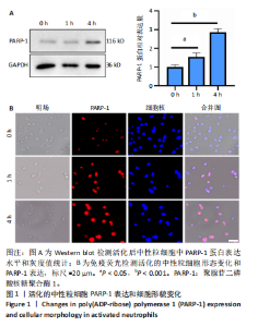
2.1 活化的中性粒细胞中PARP-1表达上调并伴随形貌改变 蛋白免疫印迹结果显示,与对照组(0 h)相比较,脂多糖处理以时间依赖性的方式诱导了PARP-1表达上调,中性粒细胞在活化4 h后PARP-1表达明显上调(P < 0.001),见图1A。 免疫荧光结果显示,与对照组近似圆形或稍显铺展的形态相比,经脂多糖处理后中性粒细胞发生了形态改变,呈纺锤形或不规则三角形的极化状态,同时这种形态的改变伴随着PARP-1蛋白水平的变化,PARP-1的荧光信号强度随着处理时间的延长明显增强,见图1B。"
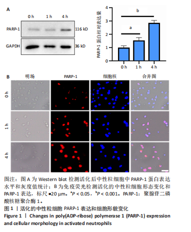
这些结果进一步证明了活化的中性粒细胞中PARP-1表达上调。 2.2 PARP-1低表达中性粒细胞系构建 Western blot结果显示敲低组HL-60 细胞中PARP-1蛋白表达水平显著低于对照组(P < 0.001),见图2A,B,表明PARP-1低表达中性粒细胞系构建成功。"
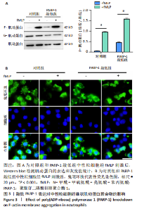
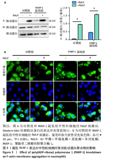
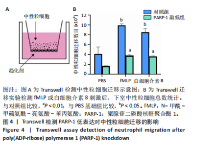
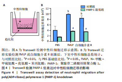
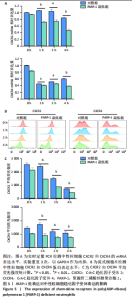
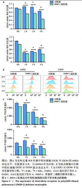
2.3 敲低PARP-1不影响中性粒细胞极化 细胞迁移的首要条件是细胞极化,当中性粒细胞极化时,首先将球状肌动蛋白(G-肌动蛋白)聚集到细胞膜形成纤维状肌动蛋白(F-肌动蛋白)聚合物,为细胞提供动力。为了确定PARP-1是否直接调节中性粒细胞的极化,对照组和敲低组的中性粒细胞经N-甲酰-甲硫氨酰-亮氨酸-苯丙氨酸刺激后,提取细胞膜蛋白,检测F-肌动蛋白的聚集情况。 Western blot结果显示,N-甲酰-甲硫氨酰-亮氨酸-苯丙氨酸刺激后,两组细胞间的F-肌动蛋白表达均明显上调(P < 0.001),见图3A。鬼笔环肽荧光染色显示,对照组和PARP-1敲低组中性粒细胞均表现出正常的极化形态,伪足中可见纤维状肌动蛋白(F-肌动蛋白)聚集,见图3B。 由此可见,敲低PARP-1表达并不影响N-甲酰-甲硫氨酰-亮氨酸-苯丙氨酸诱导的中性粒细胞的肌动蛋白聚合作用和极化过程。 2.4 敲低PARP-1抑制中性粒细胞迁移 Transwell迁移实验结果显示,与PBS刺激相比,趋化因子N-甲酰-甲硫氨酰-亮氨酸-苯丙氨酸或白细胞介素8刺激后,中性粒细胞迁移数量增加(P < 0.05)。与对照组相比,敲低PARP-1显著抑制中性粒细胞的迁移数量(P < 0.01),见图4A,B。上述结果表明敲低PARP-1能够抑制不同趋化因子诱导的中性粒细胞迁移,提示PARP-1通过非极化依赖途径调控中性粒细胞迁移。 2.5 敲低PARP-1抑制中性粒细胞趋化因子受体表达 中性粒细胞表面受体是感受外界刺激信号的关键,上述研究结果表明敲低PARP-1影响中性粒细胞的迁移,但并不影响其极化,因此PARP-1可能对中性粒细胞表面受体表达有影响,为了确定这些受体的表达,使用脂多糖分别刺激对照组和PARP-1敲低组dHL-60细胞,并检测C-X-C趋化因子受体2和C-X-C趋化因子受体4在基因及蛋白水平的表达。实时荧光定量PCR和流式细胞术检测结果显示,敲低PARP-1后,中性粒细胞C-X-C趋化因子受体2和C-X-C趋化因子受体4在mRNA及蛋白水平的表达均显著降低(P < 0.05或P < 0.01),见图5A-C。结果表明,PARP-1可能通过调控C-X-C趋化因子受体2和C-X-C趋化因子受体4的表达,影响活化后中性粒细胞的迁移能力。 2.6 敲低PARP-1抑制中性粒细胞的活化与炎症反应 为探究PARP-1对中性粒细胞活化及炎症因子释放的影响,采用脂多糖"
"
分别刺激对照组和PARP-1敲低组中性粒细胞,结果显示,脂多糖刺激能显著诱导中性粒细胞内活性氧的生成(P < 0.001),与对照组相比,敲低PARP-1有效抑制了脂多糖诱导的活性氧生成(P < 0.001),见图6A。 通过ELISA检测细胞上清液中白细胞介素1β水平,结果显示,在脂多糖刺激后PARP-1敲低组白细胞介素1β释放水平较对照组显著降低(P < 0.001),见图6B。"

中性粒细胞胞外陷阱在急性炎症中起关键作用,而瓜氨酸化组蛋白H3被广泛认为是中性粒细胞胞外陷阱形成的经典标志物[24]。为评估中性粒细胞胞外陷阱的形成,联合应用Sytox Green染色(胞外DNA染色)与瓜氨酸化组蛋白H3免疫荧光技术,分别从中性粒细胞胞外陷阱的最终产物及形成两个层面进行检测,结果显示,脂多糖刺激能诱导中性粒细胞产生弥散的DNA纤维网络并释放大量胞外DNA(P < 0.001);相比之下,敲低"
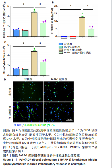
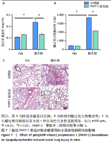
PARP-1显著抑制了胞外DNA的释放与中性粒细胞胞外陷阱的形成(P < 0.001),见图6C,D。 综上所述,敲低PARP-1能够抑制脂多糖诱导的活性氧生成与中性粒细胞胞外陷阱形成,并减少关键炎性因子白细胞介素1β的释放。这些结果共同表明,PARP-1对中性粒细胞的活化及炎症效应功能具有正向调控作用。 2.7 小鼠中性粒细胞过继转移模型证实阻断PARP-1减少中性粒细胞肺组织浸润,缓解脂多糖诱导的急性肺损伤 为了明确阻断中性粒细胞PARP-1对小鼠急性肺损伤的影响,通过尾静脉向野生型小鼠分别回输对照组与PARP-1敲低组中性粒细胞,随后腹腔注射脂多糖构建急性肺损伤模型。结果显示,与PBS溶剂处理组相比,脂多糖刺激后肺泡蛋白含量升高,提示脂多糖诱导小鼠发生肺水肿 (P < 0.05),相较于对照组,PARP-1敲低组显著降低了脂多糖刺激后的肺泡蛋白渗漏,减轻了肺水肿程度(P < 0.05),见图7A。 随后,通过检测髓过氧化物酶活性来评估肺部炎症,髓过氧化物酶活性是中性粒细胞浸润的一个指标[25],结果显示PARP-1敲低组基础髓过氧化物酶活性与对照组相当,但脂多糖刺激后PARP-1敲低组肺部髓过氧化物酶活性显著降低,肺部中性粒细"
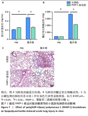
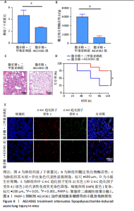
胞浸润减少(P < 0.05),见图7B。 肺组织苏木精-伊红染色显示,与对照组相比,在脂多糖刺激后PARP-1敲低组肺部结构损伤更轻,组织完整性更好,见图7C。 以上结果研究证实了PARP-1通过调控中性粒细胞的活化和迁移,促进中性粒细胞浸润加重急性肺损伤。 2.8 AG14361治疗减轻脂多糖诱导的小鼠急性肺损伤 为验证以PARP-1为靶点治疗急性肺损伤的可行性,利用PARP-1选择性抑制剂AG14361进行脂多糖诱导的急性肺损伤动物模型验证,结果显示,与二甲基亚砜溶剂对照处理相比,AG14361治疗降低了脂多糖刺激后的肺湿/干质量比,肺水肿程度减轻(P < 0.05),见图8A。同时,肺部髓过氧化物酶活性检测显示,AG14361处理后肺部中性粒细胞浸润被显著抑制(P < 0.001),见图8B。 苏木精-伊红染色显示,AG14361能有效维持肺组织结构的完整性,减轻肺部损伤,见图8C。生存分析表明,AG14361处理能显著延长小鼠生存期并提高生存率(P < 0.05),见图8D。 为进一步探究潜在机制,通过免疫荧光染色检测肺组织中与中性粒细胞募集相关的关键趋化因子受体,结果显示,抑制PARP-1可下调肺组织中C-X-C趋化因子受体2与C-X-C趋化因子受体4的表达,见图8E。 以上结果共同表明,选择性PARP-1抑制剂AG14361可能通过下调C-X-C趋化因子受体2与C-X-C趋化因子受体4的表达,减少中性粒细胞在肺组织的募集与浸润,从而有效缓解脂多糖诱导的急性肺损伤。"
| [1] WANG J, PENG X, YUAN N, et al. Interplay between pulmonary epithelial stem cells and innate immune cells contribute to the repair and regeneration of ALI/ARDS. Transl Res. 2024;272:111-125. [2] ZHENG J, LI Y, KONG X, et al. Exploring immune-related pathogenesis in lung injury: Providing new insights Into ALI/ARDS. Biomed Pharmacother. 2024;175:116773. [3] MOWERY NT, TERZIAN WTH, NELSON AC. Acute lung injury. Curr Probl Surg. 2020;57(5):100777. [4] MATTHAY MA, ARABI Y, ARROLIGA AC, et al. A New Global Definition of Acute Respiratory Distress Syndrome. Am J Respir Crit Care Med. 2024; 209(1):37-47. [5] SHANG L, ZHANG M, LI J, et al. Dachengqi decoction alleviates acute lung injury by suppressing HIF-1α-mediated glycolysis. J Ethnopharmacol. 2024;321:117410. [6] QI Y, YANG B, OUYANG H, et al. Advanced nanotherapies for precision treatment of inflammatory lung diseases. Bioact Mater. 2025;53:329-365. [7] PEI H, CHEN J, QU J, et al. S100A9 exacerbates sepsis-induced acute lung injury via the IL17-NFκB-caspase-3 signaling pathway. Biochem Biophys Res Commun. 2024;710:149832. [8] CAI Y, SHANG L, ZHOU F, et al. Macrophage pyroptosis and its crucial role in ALI/ARDS. Front Immunol. 2025;16:1530849. [9] ZHU J, ZHOU J, FENG B, et al. MSCs alleviate LPS-induced acute lung injury by inhibiting the proinflammatory function of macrophages in mouse lung organoid-macrophage model. Cell Mol Life Sci. 2024;81(1):124. [10] GROMMES J, SOEHNLEIN O. Contribution of neutrophils to acute lung injury. Mol Med. 2011;17(3-4):293-307. [11] ZHANG YL, ZHANG WX, YAN JQ, et al. Chalcone derivatives ameliorate lipopolysaccharide-induced acute lung injury and inflammation by targeting MD2. Acta Pharmacol Sin. 2022;43(1):76-85. [12] REUTERSHAN J, MORRIS MA, BURCIN TL, et al. Critical role of endothelial CXCR2 in LPS-induced neutrophil migration into the lung. J Clin Invest. 2006;116(3):695-702. [13] MOKRA D, DRGOVA A, PETRAS M, et al. N-acetylcysteine alleviates the meconium-induced acute lung injury. Adv Exp Med Biol. 2015;832:59-67. [14] PUENTES-PARDO JD, MORENO-SANJUAN S, CASADO J, et al. PARP-1 Expression Influences Cancer Stem Cell Phenotype in Colorectal Cancer Depending on p53. Int J Mol Sci. 2023;24(5):4787. [15] GHORAI A, SAHA S, RAO BJ. PARP-1 negatively regulates nucleolar protein pool and mitochondrial activity: a cell protective mechanism. Genes Environ. 2024;46(1):18. [16] KIM H, NAURA AS, ERRAMI Y, et al. Cordycepin blocks lung injury-associated inflammation and promotes BRCA1-deficient breast cancer cell killing by effectively inhibiting PARP. Mol Med. 2011;17(9-10):893-900. [17] HA HC, SNYDER SH. Poly(ADP-ribose) polymerase is a mediator of necrotic cell death by ATP depletion. Proc Natl Acad Sci U S A. 1999;96(24):13978-13982. [18] SETHI GS, DHARWAL V, NAURA AS. Poly(ADP-Ribose)Polymerase-1 in Lung Inflammatory Disorders: A Review. Front Immunol. 2017;8:1172. [19] ZHANG JN, MA Y, WEI XY, et al. Remifentanil Protects against Lipopolysaccharide-Induced Inflammation through PARP-1/NF-κB Signaling Pathway. Mediators Inflamm. 2019;2019:3013716. [20] HAN L, CHEN Y, HUANG N, et al. Cancer-educated neutrophils promote lung cancer progression via PARP-1-ALOX5-mediated MMP-9 expression. Cancer Biol Med. 2024;21(2):175-192. [21] KAPOOR K, SINGLA E, SAHU B, et al. PARP inhibitor, olaparib ameliorates acute lung and kidney injury upon intratracheal administration of LPS in mice. Mol Cell Biochem. 2015;400(1-2):153-162. [22] WANG G, CAO L, LIU X, et al. Oxidant Sensing by TRPM2 Inhibits Neutrophil Migration and Mitigates Inflammation. Dev Cell. 2016;38(5):453-462. [23] KABIR K, GELINAS JP, CHEN M, et al. Characterization of a murine model of endotoxin-induced acute lung injury. Shock. 2002;17(4):300-303. [24] ZHANG H, WU D, WANG Y, et al. Ferritin-mediated neutrophil extracellular traps formation and cytokine storm via macrophage scavenger receptor in sepsis-associated lung injury. Cell Commun Signal. 2024;22(1):97. [25] PENG X, HASSOUN PM, SAMMANI S, et al. Protective effects of sphingosine 1-phosphate in murine endotoxin-induced inflammatory lung injury. Am J Respir Crit Care Med. 2004;169(11):1245-1251. [26] LIANG L, PENG W, QIN A, et al. Intracellularly Synthesized Artificial Exosome Treats Acute Lung Injury. ACS Nano. 2024;18(32):21009-21023. [27] SHA HX, LIU YB, QIU YL, et al. Neutrophil extracellular traps trigger alveolar epithelial cell necroptosis through the cGAS-STING pathway during acute lung injury in mice. Int J Biol Sci. 2024;20(12):4713-4730. [28] BURKARD P, SCHONHART C, VÖGTLE T, et al. A key role for platelet GPVI in neutrophil recruitment, migration, and NETosis in the early stages of acute lung injury. Blood. 2023;142(17):1463-1477. [29] WANG G, HUANG X, LI Y, et al. PARP-1 inhibitor, DPQ, attenuates LPS-induced acute lung injury through inhibiting NF-κB-mediated inflammatory response. PLoS One. 2013;8(11):e79757. [30] HASSA PO, HOTTIGER MO. The functional role of poly(ADP-ribose)polymerase 1 as novel coactivator of NF-kappaB in inflammatory disorders. Cell Mol Life Sci. 2002;59(9):1534-1553. [31] SI MK, MITAKA C, TULAFU M, et al. Inhibition of poly (adenosine diphosphate-ribose) polymerase attenuates lung-kidney crosstalk induced by intratracheal lipopolysaccharide instillation in rats. Respir Res. 2013; 14(1):126. [32] ZEMANS RL, MATTHAY MA. What drives neutrophils to the alveoli in ARDS? Thorax. 2017;72(1):1-3. [33] ZOU S, JIE H, HAN X, et al. The role of neutrophil extracellular traps in sepsis and sepsis-related acute lung injury. Int Immunopharmacol. 2023;124(Pt A): 110436. [34] CAO T, LI AQ, ZHANG Y, et al. Norwogonin attenuates LPS-induced acute lung injury through inhibiting Src/AKT1/NF-κB signaling pathway. Phytomedicine. 2025;139:156432. [35] CALABRESE CR, ALMASSY R, BARTON S, et al. Anticancer chemosensitization and radiosensitization by the novel poly(ADP-ribose) polymerase-1 inhibitor AG14361. J Natl Cancer Inst. 2004;96(1):56-67. [36] LIAUDET L, PACHER P, MABLEY JG, et al. Activation of poly(ADP-Ribose) polymerase-1 is a central mechanism of lipopolysaccharide-induced acute lung inflammation. Am J Respir Crit Care Med. 2002;165(3):372-377. [37] MITTAL M, NEPAL S, TSUKASAKI Y, et al. Neutrophil Activation of Endothelial Cell-Expressed TRPM2 Mediates Transendothelial Neutrophil Migration and Vascular Injury. Circ Res. 2017;121(9):1081-1091. [38] BRUIN MAC, SONKE GS, BEIJNEN JH, et al. Pharmacokinetics and Pharmacodynamics of PARP Inhibitors in Oncology. Clin Pharmacokinet. 2022;61(12):1649-1675. [39] LI S, WANG L, WANG Y, et al. The synthetic lethality of targeting cell cycle checkpoints and PARPs in cancer treatment. J Hematol Oncol. 2022; 15(1):147. |
| [1] | Chen Yulin, He Yingying, Hu Kai, Chen Zhifan, Nie Sha Meng Yanhui, Li Runzhen, Zhang Xiaoduo , Li Yuxi, Tang Yaoping. Effect and mechanism of exosome-like vesicles derived from Trichosanthes kirilowii Maxim. in preventing and treating atherosclerosis [J]. Chinese Journal of Tissue Engineering Research, 2026, 30(7): 1768-1781. |
| [2] | Peng Zhiwei, Chen Lei, Tong Lei. Luteolin promotes wound healing in diabetic mice: roles and mechanisms [J]. Chinese Journal of Tissue Engineering Research, 2026, 30(6): 1398-1406. |
| [3] | Huang Weiqing, Liao Weimin, Du Tingting, Tian Xin. Atractylenolide III ameliorates myocardial injury in a mouse model of myocardial infarction [J]. Chinese Journal of Tissue Engineering Research, 2026, 30(36): 9560-9565. |
| [4] | Chen Weinan, Li Yusheng, Li Linzhen, Jiao Hongzhuo, Zhang Mingzhe, Wang Jianlong, Zhang Juntao. Effects of icariin-containing serum active ingredients on inflammatory chondrocytes: a metabolomics analysis [J]. Chinese Journal of Tissue Engineering Research, 2026, 30(36): 9429-9436. |
| [5] | Xiao Yang, Gao Zibo, Hu Yuxiang, Kang Zhixin, Zhang Chaoxuan, Huang Chengyu, Liu Honglin, Chen Kai, Wang Hongshen, Li Yongjin. Molecular mechanism by which the imbalance of the functional network of tissue inhibitors of metalloproteinases drives intervertebral disc degeneration [J]. Chinese Journal of Tissue Engineering Research, 2026, 30(35): 9258-9268. |
| [6] | Wang Yanfei, Jin Lianhai, Li Qingya, Fu Yuanfei, Tan Huangsheng, Deng Pengwei, Gao Kun. Synovial fluid exosome-mediated crosstalk between synoviocytes and chondrocytes in development and progression of knee osteoarthritis [J]. Chinese Journal of Tissue Engineering Research, 2026, 30(34): 9032-9040. |
| [7] | He Biyun, Wu Anqin, Yang Tao, Cao Ting, Xiao Yan, Guo Yi, Zeng Ling, Hua Zhaozhao. Probiotic supplementation regulates the gut microbiota to improve intestinal inflammation in gestational diabetes mellitus [J]. Chinese Journal of Tissue Engineering Research, 2026, 30(33): 8785-8793. |
| [8] | Hu Yingnan, Shi Wanwan, Wang Datao. Blood cells and the occurrence and progression of osteoporosis: biomarkers and emerging therapeutic strategies [J]. Chinese Journal of Tissue Engineering Research, 2026, 30(29): 7632-7638. |
| [9] | Zhao Yongjian, Ge Yunxiao, Yin Yunfei, Jiang Tingbo. Matrine promotes macrophage polarization to repair myocardial tissue injury in rats [J]. Chinese Journal of Tissue Engineering Research, 2026, 30(25): 6575-6583. |
| [10] | Liu Yufei, Chang Jinxia, Mi Baolai, Yuan Liang, Gu Hancheng, Cao Jianchun. Neutrophils and the repair of hard-to-heal wounds [J]. Chinese Journal of Tissue Engineering Research, 2026, 30(25): 6661-6668. |
| [11] | Zhang Xiaoxu, Tian Zhenli, Xie Tingting. Roles of pregnane X receptor in sodium arsenite-induced oxidative stress and inflammatory injury in human normal hepatocytes [J]. Chinese Journal of Tissue Engineering Research, 2026, 30(24): 6259-6266. |
| [12] | Jiang Chao, Che Yanjun. Biological mechanisms and future research trends of cartilaginous endplate degeneration [J]. Chinese Journal of Tissue Engineering Research, 2026, 30(23): 5915-5924. |
| [13] | Jiang Qiang, Ding Yu, Ding Zhili, Han Jiaheng. Mechanisms by which mitochondria-endoplasmic reticulum interaction stress mediates activation of inflammatory vesicles in nerve roots of lumbar intervertebral disc herniation rabbits modulated by acupotomy [J]. Chinese Journal of Tissue Engineering Research, 2026, 30(23): 6110-6121. |
| [14] | Li Jie, Liu Yang, Wang Dayu, Wan Qiang, Zhu Jiayi, Feng Wenjun, Chen Jinlun, Jie Ke, Huang Yiwei, Xin Pengfei, Zeng Jianchun, Zeng Yirong, Zhang Haitao. Rutin promotes osteogenic differentiation of MC3T3-E1 cells: regulating the formation of neutrophil extracellular traps [J]. Chinese Journal of Tissue Engineering Research, 2026, 30(19): 4972-4982. |
| [15] | Yang Ling, Dai Jiahui, Zhou Han, Yang Lin, Bian Bogao, Liu Gang. Moderate-intensity exercise improves renal injury and inflammatory response in mice with hyperuricemia [J]. Chinese Journal of Tissue Engineering Research, 2026, 30(18): 4638-4648. |
| Viewed | ||||||
|
Full text |
|
|||||
|
Abstract |
|
|||||