Chinese Journal of Tissue Engineering Research ›› 2026, Vol. 30 ›› Issue (23): 6110-6121.doi: 10.12307/2026.319
Previous Articles Next Articles
Mechanisms by which mitochondria-endoplasmic reticulum interaction stress mediates activation of inflammatory vesicles in nerve roots of lumbar intervertebral disc herniation rabbits modulated by acupotomy
Jiang Qiang1, Ding Yu2, Ding Zhili2, Han Jiaheng2
- 1Chinese PLA Medical School, Beijing 100853, China; 2Department of Orthopedics, TCM Senior Department, the Sixth Medical Center of Chinese PLA General Hospital, Beijing 100048, China
-
Received:2025-04-08Accepted:2025-06-17Online:2026-08-18Published:2026-01-05 -
Contact:Ding Yu, MD, Chief physician, Department of Orthopedics, TCM Senior Department, the Sixth Medical Center of Chinese PLA General Hospital, Beijing 100048, China -
About author:Jiang Qiang, MD candidate, Physician, Chinese PLA Medical School, Beijing 100853, China -
Supported by:National Natural Science Foundation of China, No. 82274637 (to DY)
CLC Number:
Cite this article
Jiang Qiang, Ding Yu, Ding Zhili, Han Jiaheng. Mechanisms by which mitochondria-endoplasmic reticulum interaction stress mediates activation of inflammatory vesicles in nerve roots of lumbar intervertebral disc herniation rabbits modulated by acupotomy[J]. Chinese Journal of Tissue Engineering Research, 2026, 30(23): 6110-6121.
share this article
Add to citation manager EndNote|Reference Manager|ProCite|BibTeX|RefWorks

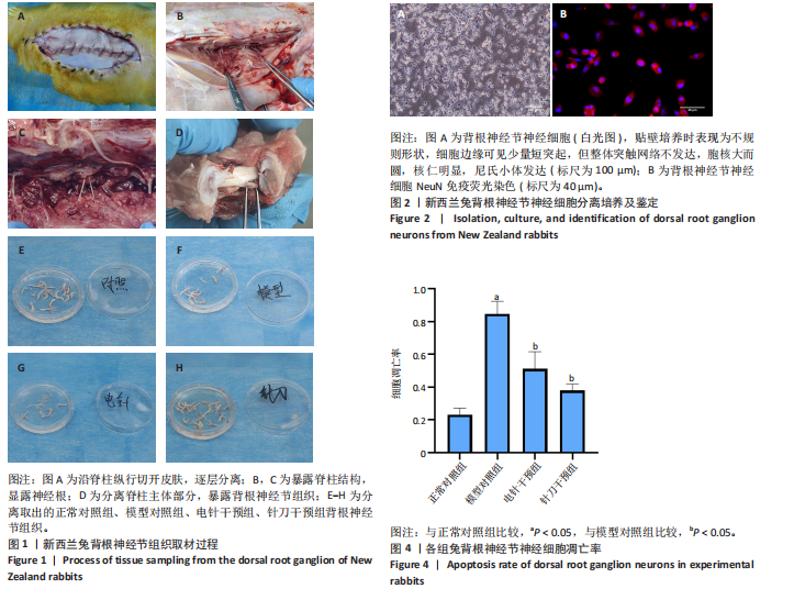
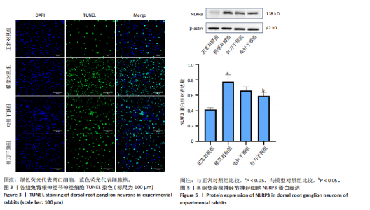
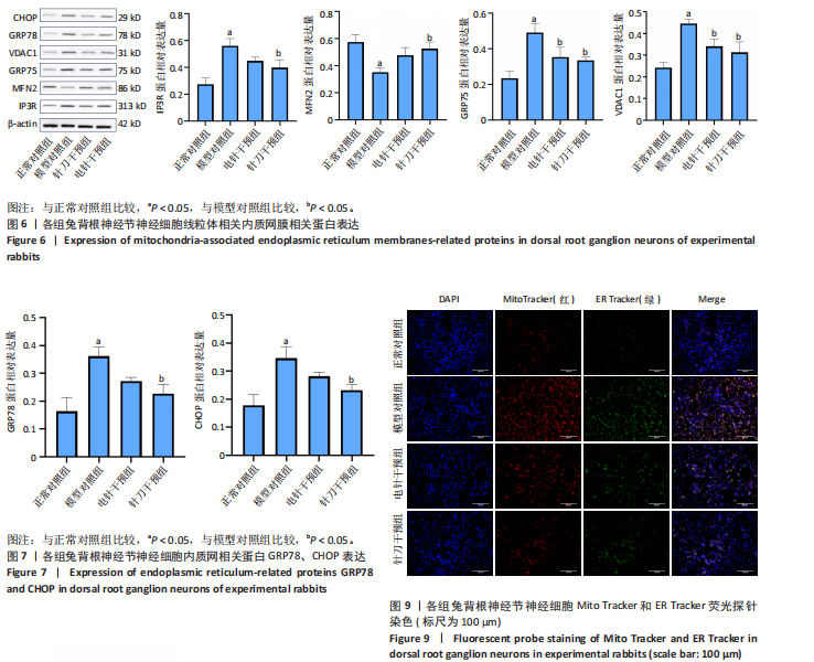
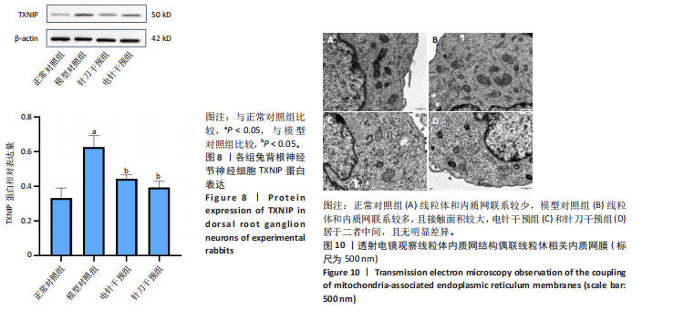
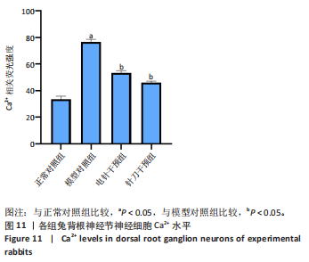
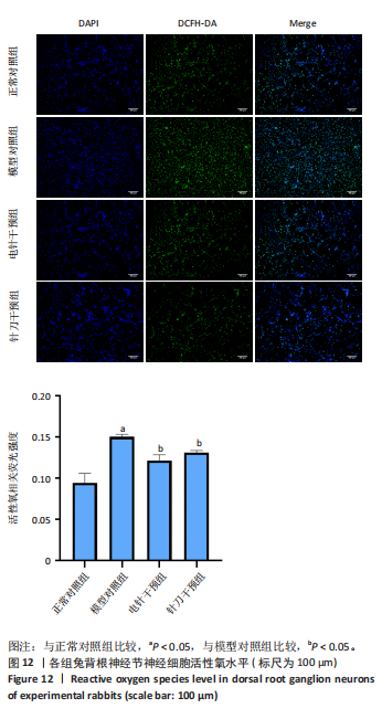
2.1 原代背根神经节细胞分离及鉴定结果 兔背根神经节神经细胞(白光图)贴壁培养时表现为不规则形状,细胞边缘可见少量短突起,但整体突触网络不发达,胞核大而圆,核仁明显,尼氏小体发达。NeuN免疫荧光结果显示:NeuN阳性率在90%以上,验证了分离培养的细胞是背根神经节细胞(图2)。 2.2 TUNEL检测结果 模型对照组细胞凋亡率明显高于正常对照组(P < 0.000 1);与模型对照组相比,针刀干预组(P=0.000 2)和电针干预组(P=0.001 9)的细胞凋亡率明显下降(图3,4)。 2.3 Western blot检测结果 2.3.1 各组细胞NLRP3蛋白表达 模型对照组细胞NLRP3 蛋白表达明显高于正常对照组(P=0.000 2);与模型对照组相比,针刀干预组细胞NLRP3蛋白表达明显下调(P=0.014 4);电针干预组细胞NLRP3蛋白表达相较于模型对照组无显著差异(P=0.124 1)(图5)。 2.3.2 各组细胞MAMs相关IP3R、MFN2、GRP75、VDAC1蛋白表达 模型对照组细胞IP3R(P=0.000 4)、GRP75(P=0.000 4)、VDAC1(P=0.000 3)蛋白表达明显高于正常对照组,MFN2(P=0.002 5)蛋白表达明显低于正常对照组;与模型对照组相比,针刀干预组细胞IP3R(P=0.013 2)、GRP75 (P=0.009 9)、VDAC1(P=0.000 3) 蛋白表达明显下调,MFN2(P=0.010 8)蛋白表达明显上调;与模型对照组相比,电针干预组细胞GRP75 (P=0.020 4)、VDAC1(P=0.019 4) 蛋白表达明显下调,IP3R(P=0.078 1)蛋白表达下调,但差异不明显,MFN2(P=0.057 2) 蛋白表达上调,但差异不明显(图6)。 2.3.3 各组细胞内质网相关GRP78和CHOP蛋白表达 模型对照组细胞GRP78(P=0.000 5)和CHOP(P=0.000 7)蛋白表达明显高于正常对照组;与模型对照组相比,针刀干预组细胞GRP78(P=0.006 5)和CHOP(P=0.008 5) 蛋白表达明显下调;与模型对照组相比,电针干预组细胞GRP78(P=0.054 1)和CHOP(P=0.127 6)蛋白表达下调,但差异不明显(图7)。 2.3.4 各组细胞TXNIP蛋白表达 模型对照组细胞TXNIP (P=0.000 3)蛋白表达明显高于正常对照组,与模型对照组相比,针刀干预组(P=0.001 5)和电针干细胞组(P=0.007 0)细胞TXNIP蛋白表达明显下调(图8)。 2.4 荧光探针染色(Mito Tracker和ER Tracker) 红色荧光代表线粒体,绿色荧光代表内质网,两者合图中提示线粒体-内质网共定位水平模型对照组最高,正常对照组最低;针刀干预组和电针干预组高于正常对照组,但低于模型对照组(图9)。 2.5 透射电镜观察线粒体内质网结构偶联MAMs 正常对照组线粒体和内质网联系/接触较少,模型对照组线粒体和内质网的联系/接触增强,针刀干预组和电针干预组线粒体和内质网的联系/接触较正常对照组增强,但低于模型对照组(图10)。 2.6 钙离子水平 模型对照组细胞钙离子水平明显高于正常对照组(P < 0.000 1),与模型对照组相比,针刀干预组(P < 0.000 1)和电针干预组(P < 0.000 1)细胞钙离子水平明显下降(图11)。 2.7 活性氧含量 模型对照组细胞活性氧水平明显高于正常对照组(P < 0.000 1),与模型对照组相比,针刀干预组(P=0.039 2)和电针干预组(P=0.004 4)细胞活性氧水平明显下降(图12)。"
"


"

"

"

"

"
"
| [1] FUKUHARA D, ONO K, KENJI T, et al. A Narrative Review of Full-Endoscopic Lumbar Discectomy Using Interlaminar Approach. World Neurosurg. 2022;168:324-332. [2] WONG T, PATEL A, GOLUB D, et al. Prevalence of Long-Term Low Back Pain After Symptomatic Lumbar Disc Herniation. World Neurosurg. 2023; 170:163-173. [3] ZHANG J, ZHAI X, WANG X, et al. The Effect of Thunder-Fire Moxibustion on Lumbar Disc Herniation: Study Protocol for a Randomized Controlled Trial. Front Public Health. 2022;10:930830. [4] LI W, DJURIC N, VLEGGEERT-LANKAMP CLA. A systematic review evaluating the association of atherosclerosis and lumbar degenerative disc disease. Brain Spine. 2024;4:103901. [5] WANG XZ, XUE KY, CHEN PN, et al. Ma’s bamboo-based medicinal moxibustion therapy of low back pain in lumbar disc herniation: study protocol for a randomized controlled trial. Trials. 2022;23(1):446. [6] KIM H, HONG JY, LEE J, et al. IL-1β promotes disc degeneration and inflammation through direct injection of intervertebral disc in a rat lumbar disc herniation model. Spine J. 2021;21(6):1031-1041. [7] 李楷,徐世红,赵军,等.小针刀配合手法治疗腰椎间盘突出症的临床疗效观察[J].中医临床研究,2023,15(9):105-108. [8] 朱文婷,郭长青,赵莉.超声可视化针刀联合火针治疗旁中央型腰椎间盘突出症的临床研究[J].中国中医急症,2023,32(12):2107-2111. [9] 程越生,黄问淼,曹晓琴,等.小针刀在改善腰椎间盘突出症患者疼痛程度及FRS、JOA评分中的应用[J].中国医学创新,2023,20(16):47-51. [10] 郑智文,朱俊琛,贺业霖,等.痛点与椎间孔点入路针刀松解术治疗腰椎间盘突出症的远期疗效:一项前瞻性研究[J].颈腰痛杂志,2023, 44(1):32-35. [11] 宋翔,张彩荣,左晓彤,等.不同针刀进针点治疗腰椎间盘突出症:随机对照研究[J].中国针灸,2022,42(1):35-40. [12] 黄祥宽.小针刀与推拿联合治疗腰椎间盘突出症伴有神经根疾病的临床效果及对患者炎性因子和生活质量的影响体会[J].中国实用医药, 2024,19(16):74-76. [13] 施颖初,吕伟剑,黄飞虎.针刺联合小针刀治疗腰椎间盘突出症临床研究[J].新中医,2022,54(9):176-179. [14] 胡江杉,李佳,黄重生,等.针刀治疗腰椎间盘突出症的机制研究进展[J].针灸临床杂志,2022,38(1):104-107. [15] LI Z, WANG B, TIAN L, et al. Methane-Rich Saline Suppresses ER-Mitochondria Contact and Activation of the NLRP3 Inflammasome by Regulating the PERK Signaling Pathway to Ameliorate Intestinal Ischemia-Reperfusio n Injury. Inflammation. 2024;47(1):376-389. [16] HUANG S, ZENG Z, CHU Y, et al. Mitigation of lipopolysaccharide-induced intestinal injury in rats by Chimonanthus nitens Oliv. essential oil via suppression of mitochondrial fusion protein mitofusin 2 (MFN2)-mediated mitochondrial-associated endoplasmic reticulum membranes (MAMs) formation. J Ethnopharmacol. 2025;337(2):118856. [17] WANG L, ZHANG D, JIANG B, et al. 4-Phenylbutyric Acid Attenuates Soybean Glycinin/β-Conglycinin-Induced IPEC-J2 Cells Apoptosis by Regulating the Mitochondria-Associated Endoplasmic Reticulum Membrane and NLRP-3. J Agric Food Chem. 2024;72(11):5926-5934. [18] 钟瑞丹,冉兵,魏俊.腰椎间盘退变与突出动物模型的研究进展[J].赣南医学院学报,2024,44(4):409-415. [19] HUANG SJ, YAN JQ, LUO H, et al. IL-33/ST2 signaling contributes to radicular pain by modulating MAPK and NF-κB activation and inflammatory mediator expression in the spinal cord in rat models of noncompressive lumber disk herniation. J Neuroinflammation. 2018;15(1):12. [20] KIM SJ, KIM WR, KIM HS, et al. Abnormal spontaneous activities on needle electromyography and their relation with pain behavior and nerve fiber pathology in a rat model of lumbar disc herniation. Spine (Phila Pa 1976). 2011;36(24):E1562-E1567. [21] ZHAO QX, WANG YH, WANG SC, et al. Protectin DX Attenuates Lumbar Radicular Pain of Non-compressive Disc Herniation by Autophagy Flux Stimulation via Adenosine Monophosphate-Activated Protein Kinase Signaling. Front Physiol. 2022;12:784653. [22] WANG YH, LI Y, WANG JN, et al. A Novel Mechanism of Specialized Proresolving Lipid Mediators Mitigating Radicular Pain: The Negative Interaction with NLRP3 Inflammasome. Neurochem Res. 2020; 45(8):1860-1869. [23] 何智菲,郭长青,张义,等.针刀干预对腰椎间盘突出疼痛模型大鼠背根神经节IL-1、IL-6和TNF-α的影响[J].针灸临床杂志,2016,32(3):73-76+91. [24] MELHAT AM, YOUSSEF ASA, ZEBDAWI MR, et al. Non-Surgical Approaches to the Management of Lumbar Disc Herniation Associated with Radiculopathy: A Narrative Review. J Clin Med. 2024;13(4):974. [25] LIN CH, HUANG YH, LIEN FC, et al. Percutaneous endoscopic lumbar discectomy versus open lumbar microdiscectomy for treating lumbar disc herniation: Using the survival analysis. Tzu Chi Med J. 2023;35(3):237-241. [26] CARVALHO MSR, PELLINO G, PEREIRA AMG, et al. Prevalence of urinary dysfunction after minimally invasive surgery for deep rectosigmoid endometriosis. Langenbecks Arch Surg. 2023;408(1):83. [27] CHAO-YANG G, PENG C, HAI-HONG Z. Roles of NLRP3 inflammasome in intervertebral disc degeneration. Osteoarthritis Cartilage. 2021;29(6):793-801. [28] BAI Z, LIU W, HE D, et al. Protective effects of autophagy and NFE2L2 on reactive oxygen species-induced pyroptosis of human nucleus pulposus cells. Aging (Albany NY). 2020;12(8):7534-7548. [29] ZHANG A, WANG K, DING L, et al. Bay11-7082 attenuates neuropathic pain via inhibition of nuclear factor-kappa B and nucleotide-binding domain-like receptor protein 3 inflammasome activation in dorsal root ganglions in a rat model of lumbar disc herniation. J Pain Res. 2017;10:375-382. [30] SUN Y, LENG P, SONG M, et al. Piezo1 activates the NLRP3 inflammasome in nucleus pulposus cell-mediated by Ca2+/NF-κB pathway. Int Immunopharmacol. 2020;85:106681. [31] ZHAO Y, QIU C, WANG W, et al. Cortistatin protects against intervertebral disc degeneration through targeting mitochondrial ROS-dependent NLRP3 inflammasome activation. Theranostics. 2020;10(15):7015-7033. [32] 吴子健,胡昭端,周晓红,等.通督活血汤含药血清可抑制椎间盘纤维环细胞的焦亡[J].中国组织工程研究,2021,25(14):2148-2154. [33] NASTO LA, ROBINSON AR, NGO K, et al. Mitochondrial-derived reactive oxygen species (ROS) play a causal role in aging-related intervertebral disc degeneration. J Orthop Res. 2013;31(7):1150-1157. [34] HUANG W, WANG TB, JIANG BG, et al. Mitochondrial biogenesis in ventral spinal cord and nerve following sciatic nerve axotomy. Int J Clin Exp Med. 2017;10(10):14386-14393. [35] ERUSTES AG, D’ELETTO M, GUARACHE GC, et al. Overexpression of α-synuclein inhibits mitochondrial Ca2+ trafficking between the endoplasmic reticulum and mitochondria through MAMs by altering the GRP75-IP3R interaction. J Neurosci Res. 2021;99(11):2932-2947. [36] SZYMAŃSKI J, JANIKIEWICZ J, MICHALSKA B, et al. Interaction of Mitochondria with the Endoplasmic Reticulum and Plasma Membrane in Calcium Homeostasis, Lipid Trafficking and Mitochondrial Structure. Int J Mol Sci. 2017;18(7):1576. [37] CHIPURUPALLI S, SAMAVEDAM U, ROBINSON N. Crosstalk Between ER Stress, Autophagy and Inflammation. Front Med (Lausanne). 2021;8:758311. [38] FAN Y, SIMMEN T. Mechanistic Connections between Endoplasmic Reticulum (ER) Redox Control and Mitochondrial Metabolism. Cells. 2019;8(9):1071. [39] HETZ C, ZHANG K, KAUFMAN RJ. Mechanisms, regulation and functions of the unfolded protein response. Nat Rev Mol Cell Biol. 2020;21(8):421-438. [40] ZHOU HY, SUN YY, CHANG P, et al. Curcumin Inhibits Cell Damage and Apoptosis Caused by Thapsigargin-Induced Endoplasmic Reticulum Stress Involving the Recovery of Mitochondrial Function Mediated by Mitofusin-2. Neurotox Res. 2022;40(2):449-460. |
| [1] | Zhao Jiaqing, Liu Dong, Lu Suni, Wang Dawei, Geng Xiaopeng, Ning Huaxiu. One-hole split endoscope surgery with the aid of digital 3D technology for highly migrated lumbar disc herniation [J]. Chinese Journal of Tissue Engineering Research, 2026, 30(9): 2278-2285. |
| [2] | Zeng Xuan, Weng Rui, Ye Shicheng, Tang Jiadong, Mo Ling, Li Wenchao. Two lumbar rotary manipulation techniques in treating lumbar disc herniation: a finite element analysis of biomechanical differences [J]. Chinese Journal of Tissue Engineering Research, 2026, 30(9): 2153-2161. |
| [3] | Chen Yulin, He Yingying, Hu Kai, Chen Zhifan, Nie Sha Meng Yanhui, Li Runzhen, Zhang Xiaoduo , Li Yuxi, Tang Yaoping. Effect and mechanism of exosome-like vesicles derived from Trichosanthes kirilowii Maxim. in preventing and treating atherosclerosis [J]. Chinese Journal of Tissue Engineering Research, 2026, 30(7): 1768-1781. |
| [4] | Wang Zhipeng, Zhang Xiaogang, Zhang Hongwei, Zhao Xiyun, Li Yuanzhen, Guo Chenglong, Qin Daping, Ren Zhen. A systematic review of application value of machine learning to prognostic prediction models for patients with lumbar disc herniation [J]. Chinese Journal of Tissue Engineering Research, 2026, 30(3): 740-748. |
| [5] | Yu Weijie, Cao Dongdong, Guo Tianci, Niu Puyu, Yang Jialin, Wang Simin, Liu Aifeng. Risk prediction models of recurrence after percutaneous endoscopic lumbar discectomy: a systematic review and meta-analysis [J]. Chinese Journal of Tissue Engineering Research, 2026, 30(3): 749-759. |
| [6] | Shi Gaolong, Ge Caijun, Chen Jianpeng, Wang Yuanbin, Fan Zelin, Yan Jun, Wang Qianliang. Mechanism by which the paraventricular nucleus of the hypothalamus is involved in chronic pain and anxiety in mice with lumbar disc herniation [J]. Chinese Journal of Tissue Engineering Research, 2026, 30(22): 5707-5715. |
| [7] | Yang Ling, Dai Jiahui, Zhou Han, Yang Lin, Bian Bogao, Liu Gang. Moderate-intensity exercise improves renal injury and inflammatory response in mice with hyperuricemia [J]. Chinese Journal of Tissue Engineering Research, 2026, 30(18): 4638-4648. |
| [8] | Gu Shan, Zhang Long, Li Zhigang. Relationship between inflammatory factors, white blood cells, and lumbar disc herniation [J]. Chinese Journal of Tissue Engineering Research, 2026, 30(18): 4782-4790. |
| [9] | Lyu Ruyue, Gu Lulu, Liu Qian, Zhou Siyi, Li Beibei, Xue Letian, Sun Peng. Regulatory mechanisms of exosome secretion and its application prospects in biomedicine [J]. Chinese Journal of Tissue Engineering Research, 2026, 30(1): 184-193. |
| [10] | Zhang Chunlin, Hou Zhaohua, Yan Xu, Jiang Yan, Fu Su, Ning Yongming, Li Dongzhe, Dong Chao, Liu Xiaokang, Wang Yongkui, Cao Zhengming, Yang Tengyue. Decompression mechanism of symmetrically adduction of lumbar decompression induced resorption of herniated nucleus pulpous [J]. Chinese Journal of Tissue Engineering Research, 2025, 29(9): 1810-1819. |
| [11] | Lu Qi, Sun Maji, Wang Xuezhi, Song Ting, Ma Yiming, Yuan Feng, Chen Hongliang. Two visual arthroplasty techniques for L5-S1 disc herniation: a half-year follow-up evaluation of clinical outcomes [J]. Chinese Journal of Tissue Engineering Research, 2025, 29(9): 1841-1847. |
| [12] | Feng Zhimeng, Sun Ning, Sun Zhaozhong, Li Yuefei, Liu Changzhen, Li Sa. Three-dimensional image reconstruction can safely assist one-hole split endoscope in treatment of #br# L5/S1 far lateral lumbar disc herniation [J]. Chinese Journal of Tissue Engineering Research, 2025, 29(9): 1876-1882. |
| [13] | Zhi Fang, Zhu Manhua, Xiong Wei, Lin Xingzhen. Analgesic effect of acupuncture in a rat model of lumbar disc herniation [J]. Chinese Journal of Tissue Engineering Research, 2025, 29(5): 936-941. |
| [14] | Ran Yaqin, Chen Xi, Xie Yanne, Yuan Jun. Mechanism and potential application strategies of pyroptosis in breast cancer treatment [J]. Chinese Journal of Tissue Engineering Research, 2025, 29(36): 7880-7888. |
| [15] | Yang Yu, Li Yinghao, Duo Zhuangzhi, Zhou Dingrong. Effect of overall functional physical exercise on lumbar biomechanics in patients with lumbar disc herniation after surgery [J]. Chinese Journal of Tissue Engineering Research, 2025, 29(33): 7096-7101. |
| Viewed | ||||||
|
Full text |
|
|||||
|
Abstract |
|
|||||