Chinese Journal of Tissue Engineering Research ›› 2026, Vol. 30 ›› Issue (22): 5739-5748.doi: 10.12307/2026.155
Previous Articles Next Articles
Effects of high-intensity interval training combined with estrogen therapy on skeletal muscle stem cells, myonuclear domain and ribosome function in ovariectomized rats
Sun Yuan1, Shu Jun1, Ren Shuang2, Wang Chenyu2
- 1Lianyungang Normal University, Lianyungang 222006, Jiangsu Province, China; 2Zhengzhou University of Aeronautics, Zhengzhou 450015, Henan Province, China
-
Received:2025-03-15Accepted:2025-08-14Online:2026-08-08Published:2025-12-27 -
Contact:Wang Chenyu, PhD, Professor, Zhengzhou University of Aeronautics, Zhengzhou 450015, Henan Province, China -
About author:Sun Yuan, MS, Associate professor, Lianyungang Normal University, Lianyungang 222006, Jiangsu Province, China -
Supported by:Henan Provincial Science and Technology Project, No. 232102321125 (to WCY); 2024 “Blue and Green Project” of Jiangsu Universities, No. [2024]14
CLC Number:
Cite this article
Sun Yuan, Shu Jun, Ren Shuang, Wang Chenyu. Effects of high-intensity interval training combined with estrogen therapy on skeletal muscle stem cells, myonuclear domain and ribosome function in ovariectomized rats[J]. Chinese Journal of Tissue Engineering Research, 2026, 30(22): 5739-5748.
share this article
Add to citation manager EndNote|Reference Manager|ProCite|BibTeX|RefWorks
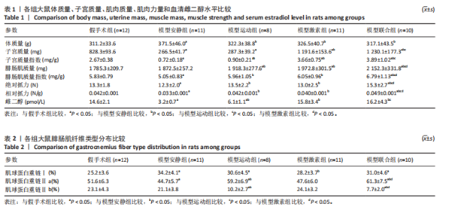
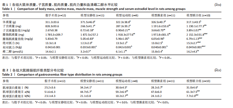
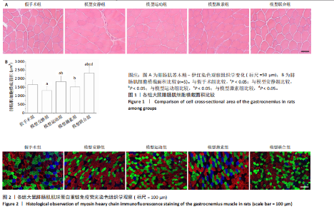
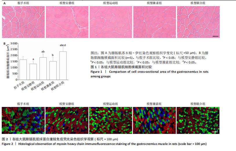
2.1 样本量分析 60只大鼠在实验过程中由于造模失败、运动损伤、过度疲劳、感染、意外死亡、拒绝训练等原因,共剔除8只,最终纳入统计的样本量为52只,即假手术组(n=12)、模型安静组(n=11)、模型运动组(n=8)、模型激素组(n=11)、模型联合组(n=10)。 2.2 各组大鼠体质量、子宫质量、肌肉质量、肌肉力量和血清雌二醇水平比较 与假手术组比较,模型安静组体质量增加(P < 0.05),子宫质量、子宫质量指数、腓肠肌质量指数、绝对抓力、相对抓力、血清雌二醇水平下降(P < 0.05)。与模型安静组比较,模型运动组和模型激素组体质量降低(P < 0.05),子宫质量指数、腓肠肌质量、腓肠肌质量指数、绝对抓力、相对抓力、血清雌二醇水平增加(P < 0.05)。与模型运动组和模型激素组比较,模型联合组腓肠肌质量、腓肠肌质量指数、绝对抓力和相对抓力升高 (P < 0.05)。见表1。 2.3 各组大鼠腓肠肌细胞横截面积比较 腓肠肌苏木精-伊红染色显示,肌纤维染成粉红色。假手术组细胞排列整齐有序,大小正常,模型安静组较假手术组细胞排列紊乱,细胞体积缩小,各干预组肌纤维排列均较模型安静组均有所改善,细胞体积增加,见图1A。 与假手术组比较,模型安静组腓肠肌细胞横截面积下降(P < 0.05)。与模型安静组比较,模型运动组和模型激素组细胞横截面积升高(P < 0.05)。与模型运动组和模型激素组比较,模型联合组腓肠肌细胞横截面积升高(P < 0.05),见图1B。 2.4 各组大鼠肌纤维类型分布比较 腓肠肌MHC免疫荧光染色显示,MHCⅠ、Ⅱa和Ⅱb型肌纤维分别呈现蓝色、绿色和红色。见图2。 与假手术组比较,模型安静组MHCⅠ比例增加 (P < 0.05),MHC Ⅱa比例下降(P < 0.05)。与模型安静组比较,模型运动组MHC Ⅱa比例增加,MHC Ⅱb比例下降(P < 0.05),模型激素组MHC Ⅰ比例降低(P < 0.05)。与模型运动组比较,模型联合组肌纤维分布无显著性变化(P > 0.05)。见表2。"
"

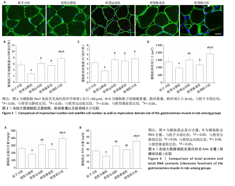
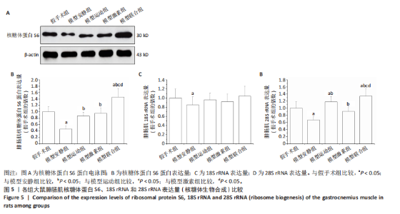
2.5 各组大鼠腓肠肌卫星细胞、肌核数量以及肌核域大小比较 腓肠肌Pax7免疫荧光染色显示,卫星细胞(Pax7+)呈粉色,肌核(DAPI+)呈蓝色,细胞外基质(laminin+)呈绿色。见图3A。 与假手术组比较,模型安静组卫星细胞和肌核数量以及肌核域大小下降(P < 0.05)。与模型安静组比较,模型运动组和模型激素组卫星细胞数量和肌核域大小升高(P < 0.05),肌核数量无显著性变化(P > 0.05)。与模型激素组比较,模型联合组卫星细胞数量和肌核域大小升高(P < 0.05),肌核数量无显著性变化(P > 0.05)。见图3B-D。 2.6 各组大鼠腓肠肌总蛋白和总RNA含量(核糖体功能)比较 与假手术组比较,模型安静组腓肠肌总蛋白和总RNA含量下降(P < 0.05)。与模型安静组比较,模型运动组和模型激素组腓肠肌总蛋白和总RNA含量增加(P < 0.05)。与模型运动组和模型激素组比较,模型联合组腓肠肌总蛋白和总RNA含量升高(P < 0.05)。见图4。 2.7 各组大鼠腓肠肌核糖体蛋白S6、18S rRNA和28S rRNA表达量(核糖体生物合成)比较 与假手术组比较,模型安静组腓肠肌核糖体蛋白S6、18S rRNA和28S rRNA表达量均下降(P < 0.05)。与模型安静组比较,模型运动组和模型激素组腓肠肌核糖体蛋白S6、28S rRNA表达量升高(P < 0.05)。与模型运动组和模型激素组比较,模型联合组腓肠肌核糖体蛋白S6、28S rRNA表达量升高(P < 0.05)。见图5。"
"
| [1] DAO T, GREEN AE, KIM YA, et al. Sarcopenia and muscle aging: A brief overview. Endocrinol Metab (Seoul). 2020;35(4):716-732. [2] LIU JH. Sarcopenia and menopause. Menopause. 2023;30(2):119-120. [3] SHEN Y, SHI Q, NONG K, et al. Exercise for sarcopenia in older people: A systematic review and network meta-analysis. J Cachexia Sarcopenia Muscle. 2023;14(3):1199-1211. [4] LU L, MAO L, FENG Y, et al. Effects of different exercise training modes on muscle strength and physical performance in older people with sarcopenia: a systematic review and meta-analysis. BMC Geriatr. 2021;21(1):e708. [5] DUPUIT M, MAILLARD F, PEREIRA B, et al. Effect of high intensity interval training on body composition in women before and after menopause: a meta-analysis. Exp Physiol. 2020;105(9):1470-1490. [6] MANDRUP CM, EGELUND J, NYBERG M, et al. Effects of high-intensity training on cardiovascular risk factors in premenopausal and postmenopausal women. Am J Obstet Gynecol. 2017;216(4):e11. [7] ATTWATERS M, HUGHES SM. Cellular and molecular pathways controlling muscle size in response to exercise. FEBS J. 2022;289(6): 1428-1456. [8] BACHMAN JF, CHAKKALAKAL JV. Satellite cells in the growth and maintenance of muscle. Curr Top Dev Biol. 2024;158:1-14. [9] SOUSA-VICTOR P, GARCÍA-PRAT L, MUÑOZ-CÁNOVES P. Control of satellite cell function in muscle regeneration and its disruption in ageing. Nat Rev Mol Cell Biol. 2022;23(3):204-226. [10] FANG J, SIA J, SOTO J, et al. Skeletal muscle regeneration via the chemical induction and expansion of myogenic stem cells in situ or in vitro. Nat Biomed Eng. 2021;5(8):864-879. [11] STEC MJ, KELLY NA, MANY GM, et al. Ribosome biogenesis may augment resistance training-induced myofiber hypertrophy and is required for myotube growth in vitro. Am J Physiol Endocrinol Metab. 2016;310(8):E652-661. [12] DAM TV, DALGAARD LB, RINGGAARD S, et al. Transdermal estrogen therapy improves gains in skeletal muscle mass after 12 weeks of resistance training in early postmenopausal women. Front Physiol. 2020;11:e596130. [13] PELLEGRINO A, TIIDUS PM, VANDENBOOM R. Mechanisms of estrogen influence on skeletal muscle: Mass, regeneration, and mitochondrial function. Sports Med. 2022;52(12):2853-2869. [14] DAM TV, DALGAARD LB, JOHANSEN FT, et al. Effects of transdermal estrogen therapy on satellite cell number and molecular markers for muscle hypertrophy in response to resistance training in early postmenopausal women. Eur J Appl Physiol. 2023;123(3):667-681. [15] NEDERVEEN JP, JOANISSE S, SÉGUIN CM, et al. The effect of exercise mode on the acute response of satellite cells in old men. Acta Physiol (Oxf). 2015;215(4):177-190. [16] 黄昱彬,凌丽,熊正爱.去卵巢大鼠肌肉衰减症模型构建及雌激素补充治疗的实验研究[J].陆军军医大学学报,2023,45(18): 1937-1946. [17] 袁国强,秦永生,彭朋.高强度间歇运动对自发性高血压模型大鼠病理性心脏肥大的影响及机制[J].中国组织工程研究,2020,24(23): 3708-3715. [18] HOSSEINI SA, SALEHI O, KEIKHOSRAVI F, et al. Mental health benefits of exercise and genistein in elderly rats. Exp Aging Res. 2022;48(1):42-57. [19] 杨瑞,曹凯,赵伟,等.高强度间歇训练影响绝经后骨质疏松模型大鼠骨健康的机制[J].中国组织工程研究,2024,28(32):5141-5147. [20] KITAJIMA Y, ONO Y. Estrogens maintain skeletal muscle and satellite cell functions. J Endocrinol. 2016;229(3):267-275. [21] TANG L, CAO W, ZHAO T, et al. Weight-bearing exercise prevents skeletal muscle atrophy in ovariectomized rats. J Physiol Biochem. 2021;77(2):273-281. [22] SHANG M, CAPPELLESSO F, AMORIM R, et al. Macrophage-derived glutamine boosts satellite cells and muscle regeneration. Nature. 2020;587(7835):626-631. [23] LIM C, NUNES EA, CURRIER BS, et al. An evidence-based narrative review of mechanisms of resistance exercise-induced human skeletal muscle hypertrophy. Med Sci Sports Exerc. 2022;54(9):1546-1559. [24] BAZGIR B, FATHI R, REZAZADEH VALOJERDI M, et al. Satellite cells contribution to exercise mediated muscle hypertrophy and repair. Cell J. 2017;18(4):473-484. [25] ENNS DL, TIIDUS PM. Estrogen influences satellite cell activation and proliferation following downhill running in rats. J Appl Physiol (1985). 2008;104(2):347-353. [26] DAMAS F, LIBARDI CA, UGRINOWITSCH C, et al. Early- and later-phases satellite cell responses and myonuclear content with resistance training in young men. PLoS One. 2018;13(1):e0191039. [27] GOH Q, SONG T, PETRANY MJ, et al. Myonuclear accretion is a determinant of exercise-induced remodeling in skeletal muscle. Elife. 2019;8:e44876. [28] AMAN F, EL KHATIB E, ALNEAIMI A, et al. Is the myonuclear domain ceiling hypothesis dead. Singapore Med J. 2023;64(7):415-422. [29] BAGLEY JR, DENES LT, MCCARTHY JJ, et al. The myonuclear domain in adult skeletal muscle fibres: Past, present and future. J Physiol. 2023;601(4):723-741. [30] SNIJDERS T, HOLWERDA AM, VAN LOON L, et al. Myonuclear content and domain size in small versus larger muscle fibres in response to 12 weeks of resistance exercise training in older adults. Acta Physiol (Oxf). 2021;231(4):e13599. [31] CHAN S, WłODARSKI T, STREIT JO, et al. The ribosome stabilizes partially folded intermediates of a nascent multi-domain protein. Nat Chem. 2022;14(10):1165-1173. [32] BARUTCU AR, WU M, BRAUNSCHWEIG U, et al. Systematic mapping of nuclear domain-associated transcripts reveals speckles and lamina as hubs of functionally distinct retained introns. Mol Cell. 2022; 82(5):1035-1052. [33] BAMMAN MM, ROBERTS BM, ADAMS GR. Molecular regulation of exercise-induced muscle fiber hypertrophy. Cold Spring Harb Perspect Med. 2018;8(6):e029751. [34] WEN Y, ALIMOV AP, MCCARTHY JJ. Ribosome biogenesis is necessary for skeletal muscle hypertrophy. Exerc Sport Sci Rev. 2016;44(3):110-115. [35] JAVED A, ORLOVA EV. Unravelling ribosome function through structural studies. Subcell Biochem. 2019;93:53-81. [36] LEE F, MUTHU V. From 18S to 28S rRNA gene: An improved targeted sarcocystidae PCR amplification, species identification with long DNA sequences. Am J Trop Med Hyg. 2021;104(4):1388-1393. [37] SHIRAKAWA Y, HIDE T, YAMAOKA M, et al. Ribosomal protein S6 promotes stem-like characters in glioma cells. Cancer Sci. 2020;111(6): 2041-2051. [38] NI C, BUSZCZAK M. The homeostatic regulation of ribosome biogenesis. Semin Cell Dev Biol. 2023;136:13-26. [39] HAMMARSTRÖM D, ØFSTENG S, KOLL L, et al. Benefits of higher resistance-training volume are related to ribosome biogenesis. J Physiol. 2020;598(3):543-565. [40] MUKUND K, SUBRAMANIAM S. Skeletal muscle: A review of molecular structure and function, in health and disease. Wiley Interdiscip Rev Syst Biol Med. 2020;12(1):e1462. |
| [1] | Cao Yong, Teng Hongliang, Tai Pengfei, Li Junda, Zhu Tengqi, Li Zhaojin. Interactions between cytokines and satellite cells in muscle regeneration [J]. Chinese Journal of Tissue Engineering Research, 2026, 30(7): 1808-1817. |
| [2] | Pan Dong, Yang Jialing, Tian Wei, Wang Dongji, Zhu Zheng, Ma Wenchao, Liu Na, Fu Changxi. Resistance exercise activates skeletal muscle satellite cells in aged rats: role of adiponectin receptor 1 pathway [J]. Chinese Journal of Tissue Engineering Research, 2026, 30(7): 1736-1746. |
| [3] | Hou Chaowen, Li Zhaojin, Kong Jianda, Zhang Shuli. Main physiological changes in skeletal muscle aging and the multimechanism regulatory role of exercise [J]. Chinese Journal of Tissue Engineering Research, 2026, 30(6): 1464-1475. |
| [4] | Yang Zhijie, Zhao Rui, Yang Haolin, Li Xiaoyun, Li Yangbo, Huang Jiachun, Lin Yanping, Wan Lei, HuangHongxing. Postmenopausal osteoporosis: predictive values of muscle mass, grip strength, and appendicular skeletal muscle index [J]. Chinese Journal of Tissue Engineering Research, 2026, 30(5): 1073-1080. |
| [5] | Huang Liuyan, Zhang Wenxi, Chen Shuwen, Yu Shimei, Dai Zhong, Zuo Changqing. Forskolin promotes C2C12 myoblast differentiation via regulating the ERK and Akt signaling pathways [J]. Chinese Journal of Tissue Engineering Research, 2026, 30(5): 1114-1121. |
| [6] | Li Qian, Li Zhenxing, Qiao Pengyan, Wang Pingzhi. Visual analysis of shear wave elastography in skeletal muscle research [J]. Chinese Journal of Tissue Engineering Research, 2026, 30(23): 6021-6029. |
| [7] | Yang Zijiang, Guo Chenggen, Deng Ziao, Xue Xinxuan. Postbiotic targeting muscle aging: mechanistic insights and application prospects of urolithin A [J]. Chinese Journal of Tissue Engineering Research, 2026, 30(22): 5804-5813. |
| [8] | Lu Anran, Wang Chenyu, Zhang Yan, Huang Huasheng. High-intensity interval training improves the function of exosomes derived from endothelial progenitor cells in spontaneously hypertensive rats [J]. Chinese Journal of Tissue Engineering Research, 2026, 30(18): 4627-4637. |
| [9] | Zan Bingxin, Zhao Yu, Dai Qinggang. Effect of raloxifene on alveolar bone resorption in ovariectomized rats [J]. Chinese Journal of Tissue Engineering Research, 2026, 30(18): 4594-4601. |
| [10] | Wang Shuo, Li Zhongshan, Che Tongtong, Xing Xinyang, Chen Zitong, Shi Yan. Effects of different sequential combinations of strength and endurance training on skeletal muscle function and aerobic metabolic capacity in young people [J]. Chinese Journal of Tissue Engineering Research, 2026, 30(18): 4602-4610. |
| [11] | Lu Biqiong, Wei Zhongjian. Skeletal muscle-derived exosome-mediated regulation of bone formation and role of exercise intervention [J]. Chinese Journal of Tissue Engineering Research, 2026, 30(13): 3379-3391. |
| [12] | Li Kaiying, Wei Xiaoge, Song Fei, Yang Nan, Zhao Zhenning, Wang Yan, Mu Jing, Ma Huisheng. Mechanism of Lijin manipulation regulating scar formation in skeletal muscle injury repair in rabbits [J]. Chinese Journal of Tissue Engineering Research, 2025, 29(8): 1600-1608. |
| [13] | Li Huayuan, Li Chun, Liu Junwei, Wang Ting, Li Long, Wu Yongli. Effect of warm acupuncture on PINK1/Parkin pathway in the skeletal muscle of rats with chronic fatigue syndrome [J]. Chinese Journal of Tissue Engineering Research, 2025, 29(8): 1618-1625. |
| [14] | Wang Xuanqiang, Zhang Wenyang, Li Yang, Kong Weiqian, Li Wei, Wang Le, Li Zhongshan, Bai Shi. Effects of chronic exposure to low-frequency pulsed magnetic fields on contractility and morphology of the quadriceps muscle in healthy adults [J]. Chinese Journal of Tissue Engineering Research, 2025, 29(8): 1634-1642. |
| [15] | Zhang Wenhua, Li Xun, Zhang Weichao, Li Xinying, Ma Guoao, Wang Xiaoqiang . Promoting myogenesis based on the SphK1/S1P/S1PR2 signaling pathway: a new perspective on improving skeletal muscle health through exercise [J]. Chinese Journal of Tissue Engineering Research, 2025, 29(6): 1265-1275. |
| Viewed | ||||||
|
Full text |
|
|||||
|
Abstract |
|
|||||