Chinese Journal of Tissue Engineering Research ›› 2024, Vol. 28 ›› Issue (30): 4915-4920.doi: 10.12307/2024.640
Research hotspots of artificial intelligence in the field of spinal deformity: visual analysis
Tao Guangyi1, Wang Linzi1, Yang Bin2, Huang Junqing2
- 1College of Bone Injury, Henan University of Chinese Medicine, Zhengzhou 450000, Henan Province, China; 2Department of Pain, Henan Provincial Hospital of TCM/Second Affiliated Hospital of Henan University of Chinese Medicine, Zhengzhou 450000, Henan Province, China
-
Received:2023-08-21Accepted:2023-09-20Online:2024-10-28Published:2023-12-28 -
Contact:Huang Junqing, Master’s supervisor, Chief physician, Department of Pain, Henan Provincial Hospital of TCM/Second Affiliated Hospital of Henan University of Chinese Medicine, Zhengzhou 450000, Henan Province, China -
About author:Tao Guangyi, Master candidate, College of Bone Injury, Henan University of Chinese Medicine, Zhengzhou 450000, Henan Province, China -
Supported by:National TCM Clinical Characteristics Technology Inheritance Talent Project (National TCM Education Letter (2019) No. 36) (to YB); Research Project of Henan National Clinical Research Base of Traditional Chinese Medicine, No. 2021JDZX2037 (to HJQ); Henan Province Physical Education Research Project, No. 202320 (to YB)
CLC Number:
Cite this article
Tao Guangyi, Wang Linzi, Yang Bin, Huang Junqing. Research hotspots of artificial intelligence in the field of spinal deformity: visual analysis[J]. Chinese Journal of Tissue Engineering Research, 2024, 28(30): 4915-4920.
share this article
Add to citation manager EndNote|Reference Manager|ProCite|BibTeX|RefWorks
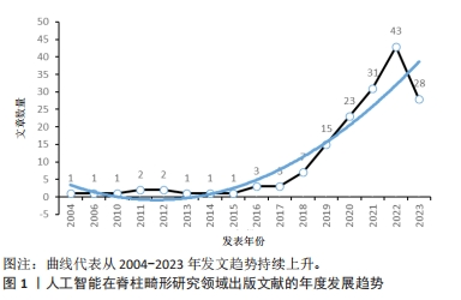
2.1 文献检索结果 总共检索到了268篇文章,按照纳排标准进行人工筛查,最终选出了183篇文章。经过阅读摘要和全文,剔除18篇,最终纳入165篇符合条件的文献,全部为研究原著类文章。 2.2 人工智能在治疗脊柱畸形领域文献发文趋势 在2018年此领域仅发表7篇论文,在几年内论文数量出现显著增加。从整体上看,论文发表数量呈上升趋势,表明学者们在人工智能治疗脊柱畸形领域所做的尝试与探索逐渐增多,其研究价值被学界众多研究者所重视,见图1。"

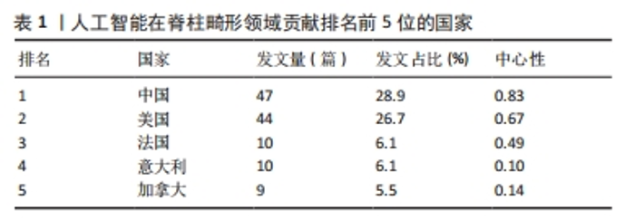
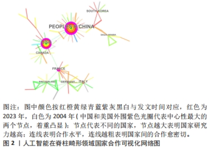
2.3 人工智能在脊柱畸形领域国家与作者之间的合著分析结果 从表1和图2可以看出,近20年来国际上对利用人工智能技术进行脊柱畸形领域的研究热情高涨,其中以亚洲和北美的论文发表数量最多。但从整体上看,各国联系强度比较零散,说明国际合作仍需加强。"
"

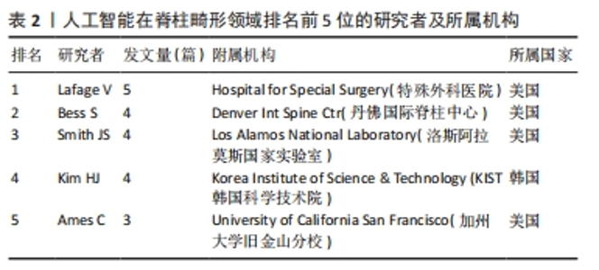
目前已有36个国家为人工智能技术在治疗脊柱畸形领域做出贡献。表1可见中国发表的文章数量最多,其次是美国、法国、意大利和加拿大。表2列出了5个最有成效的研究学者,其中发表文章数量最多的是Lafage V (5篇,3.03%),其次是Bess S,Smith JS,Kim HJ (4篇,2.42%),Ames C (3篇,1.82%),其中Kim HJ来自韩国,其他4个作者均来自美国。"
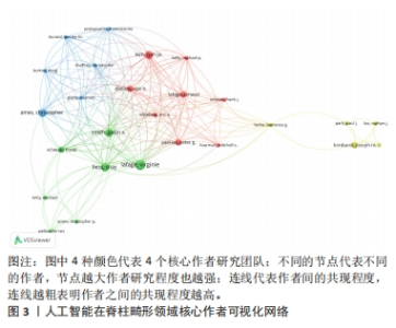
采用VOSviewer 1.6.19 软件通过对主要作者共现分析,得到了4个不同的颜色分组,代表着4个相关的研究团队,见图3。Lafage V (Hospital for Special Surgery,USA)、Bess S(Denver Int Spine Ctr,USA)、Smith JS(Los Alamos National Laboratory,USA)和Kim HJ(Korea Institute of Science & Technology,KR)等作者在这一领域的研究最为活跃。"

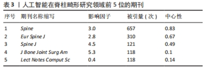
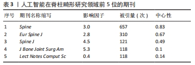
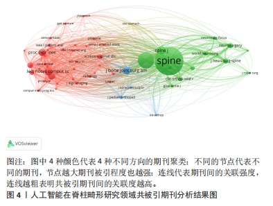
2.4 文献共被引与高被引文献深度分析结果 2.4.1 期刊共被引分析结果 所有论文共发表于98种期刊之上。表3显示了排名前5的期刊被引量。《Spine》(被引657次)在人工智能与脊柱畸形领域被引量最多。图4显示,聚类的期刊以临床脊柱与关节方面的期刊为主,探索脊柱畸形的致病机制、症状、治疗之间的关系,深入探索人工智能运用在脊柱畸形领域的算法优化和神经网络分析,为临床诊疗提供更有力的技术支撑。"

"
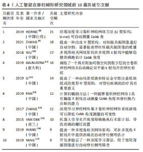
2.4.2 文献共被引分析结果 对文献共被引展开深入分析,利用VOSviewer 1.6.19软件,对人工智能在脊柱畸形领域从2004-2023年共被引次数前10名的文献进行分析[8-17],表4总结了人工智能在脊柱畸形领域前10位的共被引文献。这些文献都是这一领域的核心文献,它们的学术影响很大,某种程度上反映了这一领域的研究热点和前沿,是进行这一领域研究的重要参考。在被引数前10名的论文中,有2篇在《European Spine Journal》(期刊影响因子/JCR分区为2.8/Q1)杂志发表,而剩下的文章均发表在不同期刊。这些文章均发表于2015-2019年,排名前10位的共被引文献的发表年份,代表此领域处于快速发展阶段,研究热点新颖。"
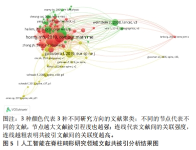
共被引频次最高的文献为HORNG等[8](2019,Comput Math Methods Med),共被引频次为21次,发表时间为2019年,该文章提出了一个自动系统测量脊柱弯曲使用前后(AP)视图脊柱X射线图像,采用多种U型架构(深度学习卷积神经网络的一种成熟模式)对椎骨进行分割;最后将椎骨的分割结果重构为完整的分割后的脊柱图像,并根据Cobb角(评估脊柱侧弯的角度)准则计算脊柱曲率。此文章共被引频次最高且发表年份新,可以看出该卷积神经网络的自动化方法受到此领域诸多学者的认可。 共被引频次第5名的文献提出了一种基于变形U型架构脊柱旁肌肉自动测量系统,它的残差模块可以在保留图像细节的同时直接返回梯度,使模型更容易训练,使磁共振图像中棘旁肌能够更加准确地分割[12]。 共被引频次第2,4,6,7名的文献也是提出一种训练策略和框架,利用U型架构(U-net,深度学习卷积神经网络的一种成熟模式)来自动测量影像学参数(Cobb角、棘旁肌准确分割等等),毫无疑问这是人工智能在脊柱畸形领域最大的研究热点。 共被引频次第3名的文献提出了多视图相关网络(Multi-View Correlation Network,MVC-Net)架构,该架构可以为多视图X射线片中的脊柱曲率估计提供全自动的端到端框架,使得网络能够通过利用两个视图的结构依赖性来缓解遮挡问题,为临床提供了有效的脊柱曲度评估框架[9, 11,13-14]。 共被引频次第8名的文献通过术前计算机断层扫描中的单个椎骨与术后电子计算机X射线片断层扫描技术融合来评估使用机器人引导的外科医生计划的可重复性,得出结论机器人引导可以高度准确地执行术前计划,从而实现准确的螺钉放置[15]。 共被引频次第9名的文献也关注于多视图相关网络(Multi-View Correlation Network,MVC-Net)架构,证明了MVC-Net在多视角X射线片中能提供准确的Cobb角度估计[16]。 共被引频次第10名的文献也开发了深度学习算法(但并非卷积神经网络),用于使用赤裸背部图像自动筛查脊柱侧凸和Cobb角等[17]。 文章采用VOSviewer 1.6.19软件生成参考文献共被引图谱,见图5。共被引文献被分为3种颜色(3大聚类),即人工智能在脊柱畸形领域的3大热点,包括利用U型架构(U-net,深度学习卷积神经网络的一种成熟模式)来自动测量影像学参数(Cobb角、棘旁肌准确分割等)、多视图相关网络(MVC-Net)架构(即脊柱曲度评估框架)、机器人引导脊柱手术。"

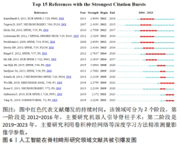
2.4.3 高被引文献深度分析结果 结合图6可知,文献共被引爆发研究共15篇文献,根据爆发时间分为2大的阶段:①第一阶段是2012-2016年,代表文献是KANTELHARDT等[18]于2011年发表,主要研究机器人引导和经皮椎弓根螺钉置入手术;②第二阶段是2019-2023年,代表文献是WU等[10]于2018年发表,主要研究多视图相关网络(Multi-View Extrapolation Net)架构在多视图X射线片中能够稳健准确地估计Cobb角度。对高共被引文献的年份进行分析,发现高共被引文献发表的时间位于2012-2021年。每个时间段都有相对应的研究热点,研究热点也随着时间发生改变。如在2012-2016时间段,机器人引导脊柱手术是其主要研究热点;而在2019-2023时间段,利用U型架构(U-net,深度学习卷积神经网络的一种成熟模式)来自动测量影像学参数、多视图相关网络(MVC-Net)架构(即脊柱曲度评估框架)是主要研究热点。"

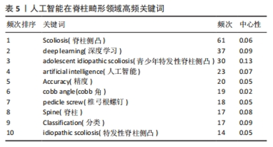
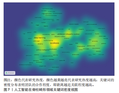
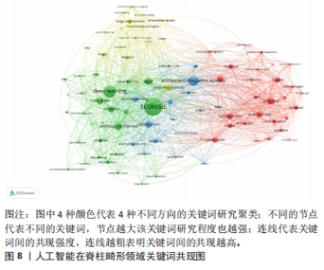
2.5 人工智能在脊柱畸形领域关键词分析结果 2.5.1 关键词共现分析 通过对关键词共现分析,能够全面展示该领域的热点和趋势[19-20]。表5总结了人工智能在脊柱畸形领域最常用的10个关键词,其中Scoliosis(脊柱侧凸)、adolescent idiopathic scoliosis(青少年特发性脊柱侧凸)分别是频次和中心性最高的关键词,说明人工智能可以高精度辅助脊柱畸形领域的手术,重点对脊柱畸形的分类与分型进行了讨论和研究,客观、真实地反映了目前人工智能在脊柱畸形领域的现状与发展趋势。图7,8显示,青少年早发性脊柱侧弯 (adolescent idiopathic scoliosis)、深度学习(deep learning)、分类(classification)、脊柱(spine)及精度(accuracy)等关键词的出现频次最高,其他的核心词都比较分散,链接的强度也较低。"

"
"

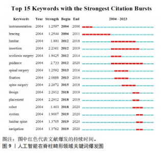
2.5.2 关键词爆发分析 为了更清楚地了解并掌握全世界对于人工智能在脊柱畸形领域的研究进展,对数据展开爆发关键词分析,见图9。根据爆发时间分为3个大的阶段,第一阶段是2004-2012年,主要爆发词为bracing(支撑);第二阶段是2012-2018年,主要爆发词有insertion(嵌入)、guidance(引导)等;第三阶段是2018-2023年,主要爆发词有robot(机器人)。"

| [1] DJURASOVIC M, GLASSMAN SD. Correlation of radiographic and clinical findings in spinal deformities. Neurosurg Clin N Am. 2007;18(2):223-227. [2] DIEBO BG, SHAH NV, BOACHIE-ADJEI O, et al. Adult spinal deformity. Lancet. 2019;394(10193):160-172. [3] MACKEL CE, JADA A, SAMDANI AF, et al. A comprehensive review of the diagnosis and management of congenital scoliosis. Childs Nerv Syst. 2018; 34(11):2155-2171. [4] DEVEZA LR, CHHABRA BN, HEYDEMANN J, et al. Comparison of baseline characteristics and postoperative complications in neuromuscular, syndromic and congenital scoliosis. J Pediatr Orthop B. 2023;32(4):350-356. [5] 陆洋阳,徐纯鑫,陈岑,等.基于三维运动捕捉系统建立青少年特发性脊柱侧弯胸廓容积与腹腔容积模型[J].中国组织工程研究,2022,36(26): 5807-5811. [6] RAMESH AN, KAMBHAMPATI C, MONSON JR, et al. Artificial intelligence in medicine. Ann R Coll Surg Engl. 2004;86(5):334-338. [7] BHATTAD PB, JAIN V. Artificial intelligence in modern medicine - the evolving necessity of the present and role in transforming the future of medical care. Cureus. 2020;12(5):e8041. [8] HORNG MH, KUOK CP, FU MJ, et al. Cobb angle measurement of spine from x-ray images using convolutional neural network. Comput Math Methods Med. 2019;2019:6357171. [9] ZHANG Q, DU Y, WEI Z, et al. Spine medical image segmentation based on deep learning. J Healthc Eng. 2021;2021:1917946. [10] WU H, BAILEY C, RASOULINEJAD P, et al. automated comprehensive adolescent idiopathic scoliosis assessment using mvc-net. med image anal. 2018;48:1-11. [11] GALBUSERA F, NIEMEYER F, WILKE HJ, et al. Fully automated radiological analysis of spinal disorders and deformities: a deep learning approach. Eur Spine J. 2019;28(5):951-960. [12] LI H, LUO H, LIU Y. Paraspinal muscle segmentation based on deep neural network. Sensors (Basel). 2019;19(12):2650. [13] PAN Y, CHEN Q, CHEN T, et al. Evaluation of a computer-aided method for measuring the Cobb angle on chest X-rays. Eur Spine J. 2019;28(12): 3035-3043. [14] ZHANG J, LI H, LV L, et al. Computer-aided cobb measurement based on automatic detection of vertebral slopes using deep neural network. Int J Biomed Imaging. 2017;2017:9083916. [15] VAN DIJK JD, VAN DEN ENDE RP, STRAMIGIOLI S, et al. Clinical pedicle screw accuracy and deviation from planning in robot-guided spine surgery: robot-guided pedicle screw accuracy. Spine (Phila Pa 1976). 2015; 40(17):E986-E991. [16] WANG L, XU Q, LEUNG S, et al. Accurate automated Cobb angles estimation using multi-view extrapolation net. Med Image Anal. 2019;58:101542. [17] YANG J, ZHANG K, FAN H, et al. Development and validation of deep learning algorithms for scoliosis screening using back images. Commun Biol. 2019;2:390. [18] KANTELHARDT SR, MARTINEZ R, BAERWINKEL S, et al. Perioperative course and accuracy of screw positioning in conventional, open robotic-guided and percutaneous robotic-guided, pedicle screw placement. Eur Spine J. 2011;20(6):860-868. [19] 魏锦强,黄登承,曹学伟,等.中医药治疗软骨疾病近20年文献知识图谱分析[J].中国组织工程研究,2021,25(20):3202-3209. [20] 叶慧,许悦,章雨桐,等.运动疗法在绝经后骨质疏松症中应用的可视化文献计量分析[J].中国骨质疏松杂志,2022,28(2):255-261, 268. [21] ANWAR SM, MAJID M, QAYYUM A, et al. Medical image analysis using convolutional neural networks: a review. J Med Syst. 2018;42(11):226. [22] FALK T, MAI D, BENSCH R, et al. U-Net: deep learning for cell counting, detection, and morphometry. Nat Methods. 2019;16(1):67-70. [23] ALOM MZ, YAKOPCIC C, HASAN M, et al. Recurrent residual U-Net for medical image segmentation. J Med Imaging (Bellingham). 2019;6(1): 014006. [24] ZHU G, LUO X, YANG T, et al. Deep learning-based recognition and segmentation of intracranial aneurysms under small sample size. Front Physiol. 2022;13:1084202. [25] HAN B, WEI Y, WANG Q, et al. Dual adaptive learning multi-task multi-view for graph network representation learning. Neural Netw. 2023;162:297-308. [26] WANG S, CHEN Z, DU S, et al. Learning deep sparse regularizers with applications to multi-view clustering and semi-supervised classification. IEEE Trans Pattern Anal Mach Intell. 2022;44(9):5042-5055. [27] ZIRAKI N, DORNAIKA F, BOSAGHZADEH A. Multiple-view flexible semi-supervised classification through consistent graph construction and label propagation. Neural Netw. 2022;146:174-180. [28] PERFETTI DC, KISINDE S, ROGERS-LAVANNE MP, et al. Robotic spine surgery: past, present, and future. Spine (Phila Pa 1976). 2022;47(13):909-921. [29] ZHANG Q, HAN XG, XU YF, et al. Robotic navigation during spine surgery. Expert Rev Med Devices. 2020;17(1):27-32. [30] 周立金,程云忠,海涌,等.全基因组关联分析在青少年特发性脊柱侧凸病因学研究中的进展[J].中国脊柱脊髓杂志,2022,32(3):269-273. [31] MAKKI N, ZHAO J, LIU Z, et al. Genomic characterization of the adolescent idiopathic scoliosis-associated transcriptome and regulome. Hum Mol Genet. 2021;29(22):3606 [32] BARLOW DP, BARTOLOMEI MS. Genomic imprinting in mammals. Cold Spring Harb Perspect Biol. 2014;6(2):a018382. [33] KHANSHOUR AM, KOU I, FAN Y, et al. Genome-wide meta-analysis and replication studies in multiple ethnicities identify novel adolescent idiopathic scoliosis susceptibility loci. Hum Mol Genet. 2018;27(22):3986-3998. [34] KIMES PK, LIU Y, NEIL HAYES D, et al. Statistical significance for hierarchical clustering. Biometrics. 2017;73(3):811-821. [35] PANG X, CAO J, CHEN S, et al. Unsupervised clustering reveals distinct subtypes of biliary atresia based on immune cell types and gene expression. Front Immunol. 2021;12:720841. [36] KARIM MR, BEYAN O, ZAPPA A, et al. Deep learning-based clustering approaches for bioinformatics. Brief Bioinform. 2021;22(1):393-415. |
| [1] | Ao Xiaojing, Li Kun, Liu Yuhang, Yang Xiaoxuan, Wang Xing, Li Zhijun, Ren Xiaoyan, Zhang Shaojie. Development and application of a three-dimensional digital visualization system for children’s neck acupoints [J]. Chinese Journal of Tissue Engineering Research, 2025, 29(9): 1834-1840. |
| [2] | Liang Haobo, Wang Zeyu, Ma Wenlong, Liu Hao, Liu Youwen. Hot issues in the field of joint revision: infection, rehabilitation nursing, bone defect, and prosthesis loosening [J]. Chinese Journal of Tissue Engineering Research, 2025, 29(9): 1963-1971. |
| [3] | Lyu Liting, Yu Xia, Zhang Jinmei, Gao Qiaojing, Liu Renfan, Li Meng, Wang Lu. Bibliometric analysis of research process and current situation of brain aging and exosomes [J]. Chinese Journal of Tissue Engineering Research, 2025, 29(7): 1457-1465. |
| [4] | Chang Jinxia, Liu Yufei, Niu Shaohui, Wang Chang, Cao Jianchun. Visualization analysis of macrophage polarization in tissue repair process [J]. Chinese Journal of Tissue Engineering Research, 2025, 29(7): 1486-1496. |
| [5] | Li Huijun, Li Huangyan, Zhang Yeting. Physical activity and cognition in older adults: research hotspot and topic evolution [J]. Chinese Journal of Tissue Engineering Research, 2025, 29(5): 1073-1080. |
| [6] | Wang Yuehui, Shang Jin, Yang Chen, Fu Dongge, Cao Can, Zhang Xiaodong, Wang Jingfu. Evaluation of FTA-LAMP direct extraction method for extracting DNA from Streptococcus mutans [J]. Chinese Journal of Tissue Engineering Research, 2025, 29(5): 1040-1049. |
| [7] | Dang Xiaowen, Huang Hailiang, Huang Lei, Wang Yajie . Research frontiers and hotspots of carbon nanomaterials in biomedical field over the past 10 years [J]. Chinese Journal of Tissue Engineering Research, 2025, 29(4): 752-760. |
| [8] | Yang Bin, Tao Guangyi, Yang Shun, Xu Junjie, Huang Junqing . Visualization analysis of research hotspots of artificial intelligence in field of spinal cord nerve injury and repair [J]. Chinese Journal of Tissue Engineering Research, 2025, 29(4): 761-770. |
| [9] | He Kai, Xing Wenhua, Liu Shengxiang, Bai Xianming, Zhou Chen, Gao Xu, Qiao Yu, He Qiang, Gao Zhiyu, Guo Zhen, Bao Aruhan, Li Chade. Constructing a model of degenerative scoliosis using finite element method: biomechanical analysis in etiology and treatment [J]. Chinese Journal of Tissue Engineering Research, 2025, 29(3): 572-578. |
| [10] | Song Haoran, Zhang Yuqiang, Gu Na, Zhi Xiaodong, Wang Wei. Visualization analysis of artificial intelligence in bone trauma research based on Citespace [J]. Chinese Journal of Tissue Engineering Research, 2025, 29(3): 493-502. |
| [11] | Xu Qiang, Qin Jialin, Lian Zeshuang, Wang Aoting, Li Ding, Wang Ye, Wang Junfang. Visual analysis of hot spots and trends in the study of ligamentum flavum ossification [J]. Chinese Journal of Tissue Engineering Research, 2025, 29(3): 628-636. |
| [12] | Zheng Xiaodong, Gao Shan, Han Wenjin, Liu Lijun, Jia Menglong, Yu Longtan. Visual analysis of treatment of adolescent idiopathic scoliosis [J]. Chinese Journal of Tissue Engineering Research, 2025, 29(3): 645-653. |
| [13] | Ma Xingyi, Li Huijing, Li Juan, Zhong Dongling, Zhang Yuan, Tang Hong, Li Yuxi, Jin Rongjiang. Visualization analysis of knowledge map and trends in glymphatic system research [J]. Chinese Journal of Tissue Engineering Research, 2025, 29(23): 5051-5060. |
| [14] | Wei Mengli, Zhong Yaping, Gui Huixian, Zhou Yiwen, Guan Yeming, Yu Shaohua. Sports injury prediction model based on machine learning [J]. Chinese Journal of Tissue Engineering Research, 2025, 29(2): 409-418. |
| [15] | Zhang Xiaoyun, Li Kunjian, Mo Jian, Chai Yuan, Huang Yourong . Current status and trends of research on animal models of postmenopausal osteoporosis: a bibliometric visualization analysis [J]. Chinese Journal of Tissue Engineering Research, 2025, 29(14): 3070-3080. |
| Viewed | ||||||
|
Full text |
|
|||||
|
Abstract |
|
|||||