Chinese Journal of Tissue Engineering Research ›› 2024, Vol. 28 ›› Issue (24): 3885-3889.doi: 10.12307/2024.617
Previous Articles Next Articles
Micro-CT analysis of distribution and structural characteristics of bone trabeculae in the calcaneus
Zhang Fengzhen1, Sun Ruifen2, Li Ziyu1, Wang Xing3, Li Kun1, Li Zhijun1, Zhang Shaojie1
- 1School of Basic Medicine, 3Digital Medicine Center, Inner Mongolia Medical University, Hohhot 010110, Inner Mongolia Autonomous Region, China; 2Second Affiliated Hospital of Inner Mongolia Medical University, Hohhot 010110, Inner Mongolia Autonomous Region, China
-
Received:2023-06-21Accepted:2023-08-07Online:2024-08-28Published:2023-11-21 -
Contact:Zhang Shaojie, MD, Professor, School of Basic Medicine, Inner Mongolia Medical University, Hohhot 010110, Inner Mongolia Autonomous Region, China -
About author:Zhang Fengzhen, Master candidate, Attending physician, Lecturer, School of Basic Medicine, Inner Mongolia Medical University, Hohhot 010110, Inner Mongolia Autonomous Region, China -
Supported by:Science and Technology Leading Talent and Innovation Team Construction Project of Inner Mongolia Autonomous Region Department of Education, No. NMGIRT2307 (to ZSJ); Key Campus Project of Inner Mongolia Medical University, No. YKD202ZD007 (to ZSJ); Research Project of Inner Mongolia Autonomous Region Mongolian Medicine Coordination and Innovation Center, No. MYYXTYB202101 (to ZSJ); Health Science and Technology Plan Project of Inner Mongolia Autonomous Region, No. 202201219 (to ZSJ); Youth Innovation Team Project of Inner Mongolia Medical University, No. QNLC-2020025 (to ZSJ); National Natural Science Foundation of China, No. 81660358 (to ZSJ); Natural Science Foundation of Inner Mongolia Autonomous Region, No. 2019MS08017 (to ZSJ); National Natural Science Foundation of China, No. 81860382 (to WX); National Natural Science Foundation of China, No. 81860383 (to LZJ); Natural Science Foundation of Inner Mongolia Autonomous Region, No. 2020MS03061 (to WX); Natural Science Foundation of Inner Mongolia Autonomous Region, No. 2020LH08021 (to LZJ)
CLC Number:
Cite this article
Zhang Fengzhen, Sun Ruifen, Li Ziyu, Wang Xing, Li Kun, Li Zhijun, Zhang Shaojie. Micro-CT analysis of distribution and structural characteristics of bone trabeculae in the calcaneus[J]. Chinese Journal of Tissue Engineering Research, 2024, 28(24): 3885-3889.
share this article
Add to citation manager EndNote|Reference Manager|ProCite|BibTeX|RefWorks
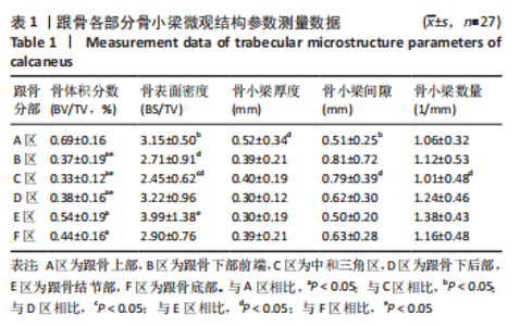
2.1 跟骨骨小梁形态及分布特征 在跟骨Micro-CT扫描影像图像上可以观察到,跟骨表面皮质层很薄,内部充满大量骨松质,在Gissane角的骨皮质明显增厚。Gissane角位于距骨外侧突的下方,代表跟骨前后关节面之间的夹角[16]。骨外表面有滋养孔,滋养孔从皮质骨表面向骨松质内部深入。除中和三角区以外,在骨松质其他部分偶可见因骨小梁减少或消失形成的空洞,在1例标本扫描矢状图跟骨上部观测到1个椭圆形、约17.44 mm×2.86 mm的空洞,其周围的微结构与正常骨小梁结构对比未见明显不同。 跟骨上部(A区)主要走行平行跟骨上面的骨小梁;跟骨下部前端(B区),以Gissane角为顶点、骰关节面为底的锥体,其内主要走行致密的层状骨小梁;中和三角区(C区)主要走行稀疏的杆状骨小梁;跟骨下后部(D区)主要走行网状的骨小梁;跟骨结节部(E区)主要走行平行跟骨结节的骨小梁;跟骨底部(F区)主要走行平行跟骨底面的骨小梁,与B、D区骨小梁交织成网状,见图1。 依据骨小梁的走行特点,将跟骨矢状面分为6个部分:A区,从距下关节后面至跟骨结节上;B区,从距下关节向前至骰关节面;C区,以Gissane角为顶点与B、D、F区交界形成的三角区域;D区,从距下关节后面至跟骨结节;E区,从跟腱附着处到跟骨结节后下方;F区,从跟骨结节到足底。 2.2 感兴趣区域骨小梁结构参数 在跟骨矢状面,以7 mm为标准的相同厚度下,各部分选择1个49 mm2等面积的感兴趣区,并测量其BV/TV、BS/TV、骨小梁厚度、骨小梁间隙和骨小梁数量,测量结果见表1。"

2.2.1 骨小梁BV/TV测量结果分析 A区骨小梁的BV/TV值大于B、C、D、E、F区,E区大于B、C、D区。A、E区和B、C、D区比较,A区和E、F区比较,差异均有显著性意义(P < 0.05),其余部位骨小梁的BV/TV相互比较差异均无显著性意义(P > 0.05),见图4。"
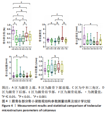

2.2.2 骨小梁BS/TV测量结果分析 E区骨小梁的BS/TV值大于B、C、F区,A、D区骨小梁的BS/TV值大于C区。C区和A、D、E区比较,B、F区和E区比较,差异均有显著性意义(P < 0.05),其余部位骨小梁的BS/TV相互比较差异均无显著性意义(P > 0.05),见图4。 2.2.3 骨小梁厚度测量结果分析 A区骨小梁厚度值大于E区,差异有显著性意义(P < 0.05),其余部位骨小梁厚度相互比较差异均无显著性意义(P > 0.05),见图4。 2.2.4 骨小梁间隙测量结果分析 C区骨小梁间隙值大于A、E区,差异均有显著性意义(P < 0.05),其余部位骨小梁间隙相互比较差异无显著性意义(P < 0.05),见图4。 2.2.5 骨小梁数量测量结果分析 E区骨小梁数量值大于C区,差异有显著性意义(P < 0.05),其余部位的骨小梁数量相互比较差异无显著性意义(P < 0.05),见图4。"

| [1] HALLINAN JTPD, WANG W, PATHRIA MN, et al. The peroneus longus muscle and tendon: a review of its anatomy and pathology. Skeletal Radiol. 2019;48(9):1329-1344. [2] ALSAYEDNOOR J, METCALF L, ROCHESTER J, et al. Comparison of HR-pQCT- and microCT-based finite element models for the estimation of the mechanical properties of the calcaneus trabecular bone. Biomech Model Mechanobiol. 2018;17(6):1715-1730. [3] CALLENS SJP, TOUROLLE NÉ BETTS DC, MÜLLER R, et al. The local and global geometry of trabecular bone. Acta Biomater. 2021;130:343-361. [4] 徐光华,刘鸿宇,张立夫,等.跆拳道运动对跟骨骨皮质厚度与骨小梁应力分布的影响[J].中国组织工程研究,2021,25(35):5582-5587. [5] SAMELSON EJ, BROE KE, XU H, et al. Cortical and trabecular bone microarchitecture as an independent predictor of incident fracture risk in older women and men in the Bone Microarchitecture International Consortium (BoMIC): a prospective study [published correction appears in Lancet Diabetes Endocrinol. 2019;7(1):e1] [published correction appears in Lancet Diabetes Endocrinol. 2019 Jun;7(6):e18]. Lancet Diabetes Endocrinol. 2019;7(1):34-43. [6] BERNHARDT R, KUHLISCH E, SCHULZ MC, et al. Comparison of bone-implant contact and bone-implant volume between 2D-histological sections and 3D-SRµCT slices. Eur Cell Mater. 2012;23:237-248. [7] 马剑雄,赵杰,何伟伟,等.高分辨率外周定量计算机断层扫描评估骨小梁微结构和骨强度的研究进展[J].生物医学工程学杂志,2018,35(3): 468-474. [8] SOLDATI E, ROSEREN F, GUENOUN D, et al. Multiscale Femoral Neck Imaging and Multimodal Trabeculae Quality Characterization in an Osteoporotic Bone Sample. Materials (Basel). 2022;15(22):8048. [9] IBRAHIM N, PARSA A, HASSAN B, et al. Comparison of anterior and posterior trabecular bone microstructure of human mandible using cone-beam CT and micro CT. BMC Oral Health. 2021;21(1):249. [10] CLARK DP, BADEA CT. Advances in micro-CT imaging of small animals. Phys Med. 2021;88:175-192. [11] 朱芳芳. CBCT测量骨小梁孔隙率评价骨小梁结构的应用研究[D].合肥:安徽医科大学,2021. [12] SHEVROJA E, CAFARELLI FP, GUGLIELMI G, et al. DXA parameters, Trabecular Bone Score (TBS) and Bone Mineral Density (BMD), in fracture risk prediction in endocrine-mediated secondary osteoporosis. Endocrine. 2021;74(1):20-28. [13] CHU L, HE Z, QU X, et al. Different subchondral trabecular bone microstructure and biomechanical properties between developmental dysplasia of the hip and primary osteoarthritis. J Orthop Translat. 2019; 22:50-57. [14] ALMHDIE-IMJABBAR A, PODSIADLO P, LJUHAR R, et al. Trabecular bone texture analysis of conventional radiographs in the assessment of knee osteoarthritis: review and viewpoint. Arthritis Res Ther. 2021;23(1):208. [15] YU YE, HU YJ, ZHOU B, et al. Microstructure Determines Apparent-Level Mechanics Despite Tissue-Level Anisotropy and Heterogeneity of Individual Plates and Rods in Normal Human Trabecular Bone. J Bone Miner Res. 2021; 36(9):1796-1807. [16] 张喻磊,胡家军,王臻.小切口解剖钢板联合加压螺栓微创术对跟骨关节内骨折患者足功能恢复的促进作用[J].临床研究,2020,28(12):46-47. [17] 田国富,李君基,李文杰.人体跟骨模型的建立及其骨折力学研究[J].机械设计与制造,2021(11):52-55. [18] 林娟颖,刘晓颖,邢立杰,等.基于有限元法的跟骨生物力学分析[J].医用生物力学,2018,33(1):37-41. [19] GALLUZZO M, GRECO F, PIETRAGALLA M, et al. Calcaneal fractures: radiological and CT evaluation and classification systems. Acta Biomed. 2018;89(1-S):138-150. [20] 魏霞,刘海玉.2200例健康体检者超声桡骨骨密度检测结果分析[J].影像研究与医学应用,2020,4(2):144-145. [21] NICOLIELO LFP, VAN DESSEL J, JACOBS R, et al.Relationship between trabecular bone architecture and early dental implant failure in the posterior region of the mandible. Clin Oral Implants Res. 2020;31(2):153-161. [22] SAERS JPP, RYAN TM, STOCK JT. Baby steps towards linking calcaneal trabecular bone ontogeny and the development of bipedal human gait. J Anat. 2020;236(3):474-492. [23] 徐光华,刘鸿宇,张立夫,等.跆拳道运动对跟骨骨皮质厚度与骨小梁应力分布的影响[J].中国组织工程研究,2021,25(35):5582-5587. [24] BURT LA, SCHIPILOW JD, BOYD SK. Competitive trampolining influences trabecular bone structure, bone size, and bone strength. J Sport Health Sci. 2016;5(4):469-475. [25] VAN AS C, KOEDAM M, MCLUSKEY A, et al. Loss of Anti-Müllerian Hormone Signaling in Mice Affects Trabecular Bone Mass in a Sex- and Age-Dependent Manner. Endocrinology. 2022;163(11):bqac157. [26] OKADA R, YAMATO K, KAWAKAMI M, et al. Low magnetic field promotes recombinant human BMP-2-induced bone formation and influences orientation of trabeculae and bone marrow-derived stromal cells. Bone Rep. 2021;14:100757. [27] ATHAVALE SA, JOSHI SD, JOSHI SS. Internal architecture of calcaneus: correlations with mechanics and pathoanatomy of calcaneal fractures. Surg Radiol Anat. 2010;32(2):115-122. [28] GONG H, WANG L, FAN Y, et al. Apparent- and Tissue-Level Yield Behaviors of L4 Vertebral Trabecular Bone and Their Associations with Microarchitectures. Ann Biomed Eng. 2016;44(4):1204-1223. [29] 吴宇航,郑利钦,张彪,等.去势大鼠骨质疏松性骨小梁的压缩断裂仿真[J].中国组织工程研究,2020,24(15):2387-2392. [30] SANDINO C, MCERLAIN DD, SCHIPILOW J, et al. Mechanical stimuli of trabecular bone in osteoporosis: A numerical simulation by finite element analysis of microarchitecture. J Mech Behav Biomed Mater. 2017;66:19-27. [31] RIEGER R, AUREGAN JC, HOC T. Micro-finite-element method to assess elastic properties of trabecular bone at micro- and macroscopic level. Morphologie. 2018;102(336):12-20. [32] MAQUER G, MUSY SN, WANDEL J, et al. Bone volume fraction and fabric anisotropy are better determinants of trabecular bone stiffness than other morphological variables. J Bone Miner Res. 2015;30(6):1000-1008. [33] 杨锐敏,吴文正,郑永泽,等.不同松质骨体积分数影响股骨近端表观力学响应的有限元分析[J].中国组织工程研究,2021,25(36):5765-5770. [34] 沛泽,罗守华,陈功,等.基于MicroCT的骨小梁参数测量系统的应用效果分析[J].中国医疗设备,2016,31(4):45-48+39. [35] YU Q, LI Z, LI J, et al. Calcaneal fracture maps and their determinants. J Orthop Surg Res. 2022;17(1):39. |
| [1] | Niu Hegang, Yang Kun, Zhang Jingjing, Yan Yizhu, Zhang Yinshun. Design of a new posterior atlas fracture reduction and internal fixation system [J]. Chinese Journal of Tissue Engineering Research, 2024, 28(9): 1399-1402. |
| [2] | Wang Menghan, Qi Han, Zhang Yuan, Chen Yanzhi. Three kinds of 3D printed models assisted in treatment of Robinson type II B2 clavicle fracture [J]. Chinese Journal of Tissue Engineering Research, 2024, 28(9): 1403-1408. |
| [3] | Yang Cekai, Cai Zhuoyan, Chen Ming, Liu Hao, Weng Rui, Cui Jianchao, Zhang Shuncong, Yao Zhensong. Relationship between degeneration of paraspinal muscle and refractures in postmenopausal women treated by percutaneous vertebroplasty [J]. Chinese Journal of Tissue Engineering Research, 2024, 28(9): 1414-1419. |
| [4] | Dai Yuexing, Zheng Liqin, Wu Minhui, Li Zhihong, Li Shaobin, Zheng Desheng, Lin Ziling. Effect of vessel number on computational fluid dynamics in vascular networks [J]. Chinese Journal of Tissue Engineering Research, 2024, 28(8): 1206-1210. |
| [5] | Tong Yibo, Li Minghui. Influencing factors of adjacent vertebral re-fracture in patients with osteoporotic vertebral fractures after percutaneous vertebroplasty [J]. Chinese Journal of Tissue Engineering Research, 2024, 28(8): 1241-1246. |
| [6] | Xue Xiaofeng, Wei Yongkang, Qiao Xiaohong, Du Yuyong, Niu Jianjun, Ren Lixin, Yang Huifeng, Zhang Zhimin, Guo Yuan, Chen Weiyi. Finite element analysis of osteoporosis in proximal femur after cannulated screw fixation for femoral neck fracture [J]. Chinese Journal of Tissue Engineering Research, 2024, 28(6): 862-867. |
| [7] | Huang Peizhen, Dong Hang, Cai Qunbin, Lin Ziling, Huang Feng. Finite element analysis of anterograde and retrograde intramedullary nail for different areas of femoral shaft fractures [J]. Chinese Journal of Tissue Engineering Research, 2024, 28(6): 868-872. |
| [8] | Tan Nengxian, Wu Wenzheng, Zheng Churong, Luo Lieliang, Gu Peng, Ouyang Chongzhi, Zheng Xiaohui. Finite element analysis of different fixation methods of partially threaded cannulated screws for treating vertical femoral neck fractures [J]. Chinese Journal of Tissue Engineering Research, 2024, 28(6): 873-878. |
| [9] | Wang Mingming, Zhang Zhong, Sun Jianhua, Zhao Gang, Song Hua, Yan Huadong, Lyu Bin. Finite element analysis of three different minimally invasive fixation methods for distal tibial fractures with soft tissue injury [J]. Chinese Journal of Tissue Engineering Research, 2024, 28(6): 879-885. |
| [10] | Wu Zhonghan, Wang Jingkun, Li Tao, Xu Xinzhong, Yu Shuisheng, Cheng Li, Tian Dasheng, Tang Jian, Jing Juehua. Proximal femoral nail antirotation for femoral intertrochanteric fractures with lateral wall integrity and lateral wall risk [J]. Chinese Journal of Tissue Engineering Research, 2024, 28(6): 911-916. |
| [11] | Zheng Jiafa, Song Xiufeng, Li Hongzhi, Zhou Jinming, Guan Shengyi, Yu He. Open reduction and internal fixation via the para-Achilles tendon approach for the treatment of posterior malleolus sandwich fractures [J]. Chinese Journal of Tissue Engineering Research, 2024, 28(6): 934-938. |
| [12] | Abuduwupuer·Haibier, Alimujiang·Yusufu, Maihemuti·Yakufu, Maimaitimin·Abulimiti, Tuerhongjiang·Abudurexiti. Meta-analysis of efficacy and safety of terlipatide and bisphosphate in the treatment of postmenopausal osteoporosis fractures [J]. Chinese Journal of Tissue Engineering Research, 2024, 28(4): 639-645. |
| [13] | Zhang Qianlong, Maihemuti•Yakufu, Song Chenhui, Liu Xiuxin, Ren Zheng, Liu Yuzhe, Muyashaer•Abudushalamu, Sajidan•Aikebaier, Ran Jian. Finite element analysis of the effect of the distribution position and content of bone cement on the stress and displacement of reverse femoral intertrochanteric fracture [J]. Chinese Journal of Tissue Engineering Research, 2024, 28(3): 336-340. |
| [14] | Wang Chunhong, Lu Ming. Universal stepwise rehabilitation training promotes the functional recovery of quadriceps femoris after intertrochanteric femoral fracture surgery [J]. Chinese Journal of Tissue Engineering Research, 2024, 28(26): 4216-4220. |
| [15] | Wang Jiaqi, Luo Xiaozhong, Tong Yi, Lu Xiaobo, Shi Weixiang, Zhou Xin, Wu Gang, Ding Yong, Zhang Caidong. Biomechanical analysis on Vancouver BI periprosthetic femoral fractures fixed by a customized anatomical plate system [J]. Chinese Journal of Tissue Engineering Research, 2024, 28(24): 3807-3813. |
| Viewed | ||||||
|
Full text |
|
|||||
|
Abstract |
|
|||||