Chinese Journal of Tissue Engineering Research ›› 2023, Vol. 27 ›› Issue (16): 2480-2487.doi: 10.12307/2023.176
Previous Articles Next Articles
Characterization and osteogenic ability of Mg-F membrane/icariin membrane/beta-tricalcium phosphate scaffolds fabricated by coating process combined with 3D printing
Xue Peng1, Du Bin1, Liu Xin1, Sun Guangquan1, Cheng Tongfei2, Chen Hao1, He Shuai1
- 1Affiliated Hospital of Nanjing University of Chinese Medicine, Nanjing 210029, Jiangsu Province, China; 2Institute of Chemical Industry, Nanjing University of Science and Technology, Nanjing 210094, Jiangsu Province, China
-
Received:2022-04-07Accepted:2022-05-24Online:2023-06-08Published:2022-11-10 -
Contact:Du Bin, MD, Professor, Chief TCM physician, Doctoral supervisor, Affiliated Hospital of Nanjing University of Chinese Medicine, Nanjing 210029, Jiangsu Province, China -
About author:Xue Peng, Doctoral candidate, Affiliated Hospital of Nanjing University of Chinese Medicine, Nanjing 210029, Jiangsu Province, China -
Supported by:National Natural Science Foundation of China, No. 82074471 (to DB); Jiangsu Provincial Health and Health Commission Scientific Research Project, No. NK2019027 (to DB); Jiangsu Province Postgraduate Practice Innovation Program, No. SJCX22_0769 (to XP)
CLC Number:
Cite this article
Xue Peng, Du Bin, Liu Xin, Sun Guangquan, Cheng Tongfei, Chen Hao, He Shuai. Characterization and osteogenic ability of Mg-F membrane/icariin membrane/beta-tricalcium phosphate scaffolds fabricated by coating process combined with 3D printing[J]. Chinese Journal of Tissue Engineering Research, 2023, 27(16): 2480-2487.
share this article
Add to citation manager EndNote|Reference Manager|ProCite|BibTeX|RefWorks

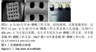
2.1 各组支架的形貌结构 3D打印β-磷酸三钙支架结构较为规则,孔隙连通率好,孔隙大小为300-400 μm,见图1A,B;淫羊藿素膜为淡黄色薄膜,Mg-F膜为黄色膜,见图1C。"
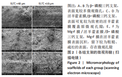
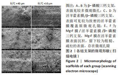
β-磷酸三钙支架表面见较多微观微孔,见图2A;淫羊藿素膜/β-磷酸三钙支架表面可见较为致密的淫羊藿素膜覆盖原微观孔隙,见图2B;Mg-F膜在淫羊藿素膜表面沉积,留下较为粗糙、疏松的表面,存在微观孔隙,见图2C。"

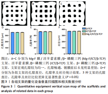
2.2 各组支架的孔隙率、孔隙体积、丝径 量化扫描图显示3种支架宏观孔隙较为均一,孔隙畅通,镀膜前后未见明显差别,见图3A-C。相较于淫羊藿素膜/β-磷酸三钙支架、β-磷酸三钙支架而言,Mg-F膜/淫羊藿素膜/β-磷酸三钙支架孔隙直径和孔隙率稍降低、丝径稍增加,但差异均无显著性意义(P > 0.05),见图3D-F。表明镀膜后镀层厚度会影响支架相应结构参数,但不会导致统计学意义的差异。"

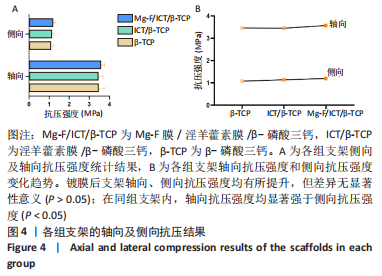
2.3 各组支架的抗压强度 3种支架的轴向抗压强度均超过了松质骨的抗压强度,整体抗压性能优异,任意支架的轴向抗压强度均高于侧向抗压强度(P < 0.05),且镀膜后支架的轴向/侧向抗压强度均提高,尤其是Mg-F膜修饰支架后支架抗压强度有明显提升,但组间差异无显著性意义(P > 0.05),见图4。"
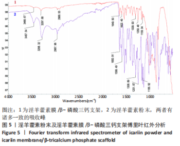
2.4 淫羊藿素膜/β-磷酸三钙支架的傅里叶红外分析结果 在淫羊藿素粉末的傅里叶红外光谱中发现,酮羟基的特殊吸收峰在1 650.91 cm-1,1 599.49 cm-1区域是芳香环的吸收峰,芳香醚的吸收峰在1 308.89 cm-1,在1 300 cm-1处观察到了C-O拉伸振动的耦合,酚羟基的弯曲振动为1 251.87 cm-1,叔醇中羟基的特征吸收峰为1 156.92 cm-1,在3 467.54 cm-1和3 298.32 cm-1处的峰值为O-H拉伸振动,在2 967.96 cm-1处观察到C-H的拉伸振动,见图5。一般羟基吸收峰出现的地方频率高(大于3 000 cm-1),所以大于3 000 cm-1的吸收峰通常表明分子中存在羟基。"

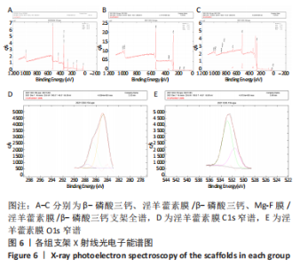
在淫羊藿素膜的傅里叶红外光谱中发现,酮羟基的吸收峰为1 649.50 cm-1,芳香环的区域吸收峰1 602.49 cm-1,酚羟基的弯曲振动为1 258.48 cm-1,叔醇羟基的特征吸收峰在1 159.04 cm-1处,O-H在3 466.67 cm-1和3 297.68 cm-1处有明显的拉伸振动;在2966.55 cm-1处观察到C-H的拉伸振动,见图5。结果表明淫羊藿素膜和淫羊藿素粉末的傅里叶红外光谱是相同的,在傅里叶红外光谱中可以找到淫羊藿素的特殊官能团。傅里叶红外验证了低能电子束沉积法在β-磷酸三钙支架上制备的淫羊藿素膜与淫羊藿素粉末具备一样的化学结构,化学组成未发生改变,不影响生物活性。 2.5 各组支架的表面元素成分X射线光电子能谱分析 见图6。"
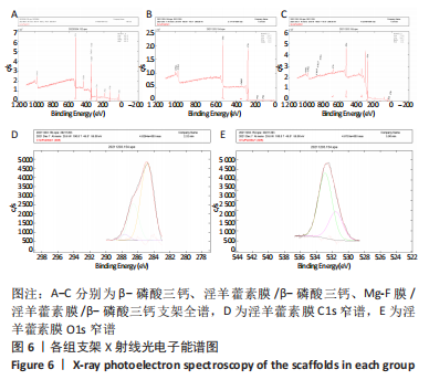

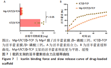
β-磷酸三钙以O、C、Ca、P元素为主,符合β-磷酸三钙支架打印材料的元素构成。通过低能电子束沉积法在普通支架表面沉积淫羊藿素膜后,支架上淫羊藿素膜的表面元素仍与原成分一样。淫羊藿素膜/β-磷酸三钙支架由于淫羊藿素膜的加入,元素构成比例发生变化,C、O元素显著增加,占绝大部分比例。这是由于此时支架表面绝大部分都被淫羊藿素膜覆盖所致,检测到部分Ca、P元素说明部分β-磷酸三钙支架表面局部未能沉积淫羊藿素所致,这可能与支架表面不光滑导致局部沉积淫羊藿素膜不均有关。同时表明淫羊藿素膜较为致密平滑,覆盖全面,这与扫描电镜下观察到淫羊藿素膜覆盖原支架微观孔隙的结果相符。通过淫羊藿素膜/β-磷酸三钙支架的X射线光电子能谱窄谱可知,镀膜后该支架表面淫羊藿素膜C1s的复合峰可以拟合到3个特征峰,这3特征峰对应于脂肪族在284.87 eV处的C-C键,286.56 eV应该对应于C-O,287.64 eV对应于C=O。淫羊藿素膜O1s的复合峰元素分别在531.58 eV对应于C=O,532.90 eV处对应-OH。通过拟合得到的特征峰与文献报道的淫羊藿素本身结构相对应[19]。这进一步表明,通过低能电子束沉积镀膜技术在支架上制备的淫羊藿素膜保留了淫羊藿素化学结构。 由于Mg-F为无机物,需X射线光电子能谱分析镀膜后Mg-F膜/淫羊藿素膜/β-磷酸三钙支架表面元素。Mg-F膜/淫羊藿素膜/β-磷酸三钙支架的X射线光电子能谱全谱图显示,该支架含有O、Mg、Ca、F、P等元素,Mg与F元素的出现表明脉冲激光沉积镀膜后支架上Mg-F缓释膜的存在。而镀膜后仍存在其他元素也表明Mg-F膜是一层结构疏松的结构,未完全覆盖原支架,在支架表面留下了孔隙利于淫羊藿素释放,这与扫描电镜下观察到粗糙、疏松、有孔隙的Mg-F膜结果相符。 2.6 载药支架的淫羊藿素结合力及体外释放曲线 Mg-F膜/淫羊藿素膜/β-磷酸三钙支架的淫羊藿素结合力显著优于淫羊藿素膜/β-磷酸三钙支架(P < 0.05),见图7A。支架的淫羊藿素释放曲线显示:淫羊藿素膜/β-磷酸三钙支架在21 d时淫羊藿素累积释放量约到总量的50.77%,在经历过一个明显的突释阶段后,释放曲线逐渐趋缓,30 d时累积释放量达到约60.92%,且趋于平稳;相较于淫羊藿素膜/β-磷酸三钙支架,Mg-F膜/淫羊藿素膜/β-磷酸三钙支架在各个时间点的淫羊藿素释放较为缓慢,释放曲线平滑近似控释效果,直至60 d时释放量约达到57.12%,预计至90 d时仍有约30%的药物尚未释放,显著延长了淫羊藿素的中晚期释放,见图7B。"

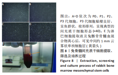
2.7 兔骨髓间充质干细胞的提取及培养 P0代细胞夹杂着大量形状不一的杂细胞,见图8A;通过贴壁筛选法,P1代细胞以梭形贴壁细胞为主,但可见较多杂细胞,见图8B。P2代细胞内杂细胞明显减少,细胞较规则,见图8C。P3代细胞贴壁良好,呈鱼群状、梭形排列,呈现典型的间充质干细胞形态,见图8D。图8E为分离得到的单核细胞层。"

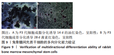
14 d的油红染色与28 d的茜素红染色结果显示,制备的细胞具有成脂分化与成骨分化的能力,见图9。"

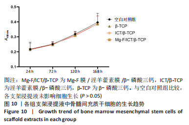
2.8 各组支架的细胞相容性 4组细胞的A450 nm值均随时间推移而增长,与空白对照组相比,3个支架组A450 nm值无明显变化,见图10,提示3个支架浸提液中的细胞生长情况良好,支架无细胞毒性。"
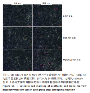
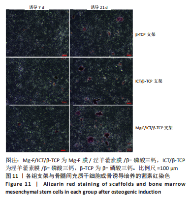
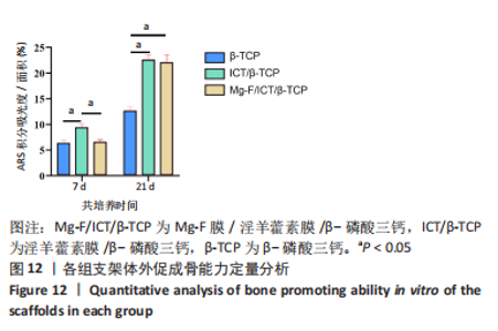
2.9 各组的支架体外促成骨分化能力 成骨诱导7 d时,3组支架的茜素红染色显示均有钙结节形成,但淫羊藿素膜/β-磷酸三钙支架组更多,Mg-F膜/淫羊藿素膜/β-磷酸三钙支架组次之,依据淫羊藿素缓释曲线可知,这与淫羊藿素膜/β-磷酸三钙支架组淫羊藿素膜的早期释放较多有关;当诱导进行到21 d时,茜素红染色显示,Mg-F膜/淫羊藿素膜/β-磷酸三钙组和淫羊藿素膜/β-磷酸三钙支架组钙结节数及钙结节成熟度明显优于β-磷酸三钙支架组,见图11。定量分析结果示,成骨诱导第7天时,淫羊藿素膜/β-磷酸三钙支架体外促成骨能力最优;成骨诱导第21天时,淫羊藿素膜/β-磷酸三钙支架及Mg-F膜/淫羊藿素膜/β-磷酸三钙支架体外促成骨能力已无差异,见图12。"
"

| [1] HINES JT, JO WL, CUI Q, et al. Osteonecrosis of the Femoral Head: an Updated Review of ARCO on Pathogenesis, Staging and Treatment.J Korean Med Sci. 2021;36(24):e177. [2] GUGGENBUHL P, ROBIN F, CADIOU S, et al. Etiology of avascular osteonecrosis of the femoral head. Morphologie. 2021;105(349): 80-84. [3] REZUS E, TAMBA BI, BADESCU MC, et al. Osteonecrosis of the Femoral Head in Patients with Hypercoagulability-From Pathophysiology to Therapeutic Implications. Int J Mol Sci. 2021;22(13):6801. [4] CUI Q, JO WL, KOO KH, et al. ARCO Consensus on the Pathogenesis of Non-traumatic Osteonecrosis of the Femoral Head. J Korean Med Sci. 2021;36(10):e65. [5] HOOGERVORST P, CAMPBELL JC, SCHOLZ N, et al. Core Decompression and Bone Marrow Aspiration Concentrate Grafting for Osteonecrosis of the Femoral Head. J Bone Joint Surg Am. 2022;104(Suppl 2):54-60. [6] ANDRONIC O, WEISS O, SHOMAN H, et al. What are the outcomes of core decompression without augmentation in patients with nontraumatic osteonecrosis of the femoral head? Int Orthop. 2021; 45(3):605-613. [7] LIGH CA, NELSON JA, FISCHER JP, et al. The Effectiveness of Free Vascularized Fibular Flaps in Osteonecrosis of the Femoral Head and Neck: A Systematic Review. J Reconstr Microsurg. 2017;33(3):163-172. [8] RICHARD MJ, DIPRINZIO EV, LORENZANA DJ, et al. Outcomes of free vascularized fibular graft for post-traumatic osteonecrosis of the femoral head. Injury. 2021;52(12):3653-3659. [9] MURAB S, HAWK T, SNYDER A, et al. Tissue Engineering Strategies for Treating Avascular Necrosis of the Femoral Head. Bioengineering (Basel). 2021;8(12):200. [10] 刘锌,杜斌,孙光权,等.多孔β磷酸三钙-聚吡咯-生物素-淫羊藿素微球复合支架促进骨髓间充质干细胞的募集[J].中国组织工程研究,2020,24(34):5532-5537. [11] 张军,任跃明,张双庆.淫羊藿素抗骨质疏松作用及其给药系统研究进展[J].中国药学杂志,2021,56(10):781-784. [12] 李时斌,夏天,章晓云,等.淫羊藿活性单体成分调控骨质疏松症相关信号通路影响骨吸收与骨形成的稳态[J].中国组织工程研究, 2022,26(11):1772-1779. [13] RONDANELLI M, FALIVA MA, TARTARA A, et al. An update on magnesium and bone health. Biometals. 2021;34(4):715-736. [14] 张竞心,刘林枫,张士文,等.镁离子促进骨再生的分子机制[J].中国组织工程研究,2022,26(33):5384-5392. [15] CIOSEK Z, KOT K, KOSIK-BOGACKA D, et al. The Effects of Calcium, Magnesium, Phosphorus, Fluoride, and Lead on Bone Tissue. Biomolecules. 2021;11(4):506. [16] LIU S, ZHOU H, LIU H, et al. Fluorine-contained hydroxyapatite suppresses bone resorption through inhibiting osteoclasts differentiation and function in vitro and in vivo. Cell Prolif. 2019;52(3): e12613. [17] AL-EESA NA, FERNANDES SD, HILL RG, et al. Remineralising fluorine containing bioactive glass composites. Dent Mater. 2021;37(4):672-681. [18] 李贝贝.PLA基复合薄膜的几种制备工艺及其抗菌性能研究[D].南京:南京理工大学,2020. [19] CHENG T, CAO J, JIANG X, et al. Study of Icaritin Films by Low-Energy Electron Beam Deposition. Eurasian Chem-Techno. 2021;23(2):77-87. [20] CAO J, LIU X, JIANG X, et al. Studies of magnesium - hydroxyapatite micro/nano film for drug sustained release. Appl Surf Sci. 2021;565: 150598. [21] 薛鹏,杜斌,王礼宁,等.可控释淫羊藿苷-β-磷酸三钙复合支架的制备[J].中国组织工程研究,2018,22(6):865-870. [22] NUGRAHA AP, RANTAM FA, NARMADA IB, et al. Gingival-Derived Mesenchymal Stem Cell from Rabbit (Oryctolagus cuniculus): Isolation, Culture, and Characterization. Eur J Dent. 2021;15(2):332-339. [23] 薛鹏.3D打印淫羊藿苷-β-磷酸三钙复合材料对兔股骨头坏死骨修复的实验研究[D].南京:南京中医药大学,2018. [24] JANA A, DAS M, BALLA VK. In vitro and in vivo degradation assessment and preventive measures of biodegradable Mg alloys for biomedical applications. J Biomed Mater Res A. 2022;110(2):462-487. [25] BALLOUZE R, MARAHAT MH, MOHAMAD S, et al. Biocompatible magnesium-doped biphasic calcium phosphate for bone regeneration. J Biomed Mater Res B Appl Biomater. 2021;109(10):1426-1435. [26] SALAMANCA E, PAN YH, SUN YS, et al. Magnesium Modified beta-Tricalcium Phosphate Induces Cell Osteogenic Differentiation In Vitro and Bone Regeneration In Vivo. Int J Mol Sci. 2022;23(3):1717. [27] DENG C, XU L, ZHANG Y, et al. The value of the hedgehog signal in osteoblasts in fluoride-induced bone-tissue injury. J Orthop Surg Res. 2021;16(1):160. [28] CAO J, LIAN R, JIANG X. Magnesium and fluoride doped hydroxyapatite coatings grown by pulsed laser deposition for promoting titanium implant cytocompatibility. Appl Surf Sci. 2020;515:146069. [29] LAI Y, CAO H, WANG X, et al. Porous composite scaffold incorporating osteogenic phytomolecule icariin for promoting skeletal regeneration in challenging osteonecrotic bone in rabbits. Biomaterials. 2018;153: 1-13. [30] QIN Y, LIU A, GUO H, et al. Additive manufacturing of Zn-Mg alloy porous scaffolds with enhanced osseointegration: In vitro and in vivo studies. Acta Biomater. 2022;145:403-415. [31] QU M, WANG C, ZHOU X, et al. Multi-Dimensional Printing for Bone Tissue Engineering. Adv Healthc Mater. 2021;10(11): e2001986. [32] PARMAKSIZ M, ELCIN AE, ELCIN YM. Biomimetic 3D-Bone Tissue Model. Methods Mol Biol. 2021;2273:239-250. [33] MAIA FR, BASTOS AR, OLIVEIRA JM, et al. Recent approaches towards bone tissue engineering. Bone. 2022;154:116256. [34] PEDRERO SG, LLAMAS-SILLERO P, SERRANO-LOPEZ J. A Multidisciplinary Journey towards Bone Tissue Engineering. Materials (Basel). 2021; 14(17):4896. [35] QI J, YU T, HU B, et al. Current Biomaterial-Based Bone Tissue Engineering and Translational Medicine. Int J Mol Sci. 2021;22(19): 10223. |
| [1] | Huang Yanni, Yang Hua, Yang Dongmei, Hu Xulin, Gao Hong, Huang Yina. Rat bone defect repaired with polytrimethylene carbonate/beta-tricalcium phosphate microsphere scaffold [J]. Chinese Journal of Tissue Engineering Research, 2023, 27(12): 1856-1862. |
| [2] | Shen Zhen, Guo Ying, Jiang Ziwei, Zhang Yan, Li Zige, Chen Zehua, Ye Xiangling, Chen Guoqian. Comparison of the effects between two routes of total flavones of Rhizoma Drynariae administration on large segmental bone defects in rats based on bone tissue engineering technique [J]. Chinese Journal of Tissue Engineering Research, 2022, 26(27): 4346-4352. |
| [3] | Liu Yin, Liu Qin, Chen Jiao, Gu Xianyang, Chen Jiawen, Ma Minxian. Properties of injectable gluconolactone-sodium alginate/beta-tricalcium phosphate/polyethylene glycol composite hydrogel scaffold [J]. Chinese Journal of Tissue Engineering Research, 2022, 26(27): 4308-4313. |
| [4] | Wang Jinsi, Wang Shengfa, Wu Zhuguo, He Xiaoling, Wang Xinyu, Luo Xiaoyu, Zhao Yi, Zhang Jingying. Design and biological activity of beta-tricalcium phosphate biomimetic bone scaffold based on triply periodic minimal surfaces [J]. Chinese Journal of Tissue Engineering Research, 2022, 26(21): 3291-3297. |
| [5] | Li Yaoming, Jiang Hong, Shi Yongfang. Preparation and characterization of chitosan/polylactic acid/hydroxyapatite/polyvinyl alcohol composite bone scaffold [J]. Chinese Journal of Tissue Engineering Research, 2022, 26(18): 2888-2893. |
| [6] | Chen Feng, Zhang Xiaoyun, Chen Yueping, Liao Jianzhao, Li Jiajun, Song Shilei, Lai Yu. Molecular mechanism of anhydroicaritin in the treatment of osteoarthritis: an analysis based on network pharmacology and bioinformatics [J]. Chinese Journal of Tissue Engineering Research, 2021, 25(23): 3704-3710. |
| [7] | Li Xinping, Cui Qiuju, Zeng Shuguang, Ran Gaoying, Zhang Zhaoqiang, Liu Xianwen, Fang Wei, Xu Shuaimei. Effect of modification of β-tricalcium phosphate/chitosan hydrogel on growth and mineralization of dental pulp stem cells [J]. Chinese Journal of Tissue Engineering Research, 2021, 25(22): 3493-3499. |
| [8] | Liao Jian, Huang Xiaolin, Zhou Qian, Cheng Yuting, Huo Hua, Li Fang, Wu Chao, Shi Qianhui, Liao Yunmao, Liang Xing. Preparation and characterization of calcined bone/chitosan composite material [J]. Chinese Journal of Tissue Engineering Research, 2020, 24(22): 3452-3459. |
| [9] | Li Zhi-ge, Wang Yi, Qi Yuan-yuan, Che Xiao-qiang, Liu Bin. Polyvinyl alcohol and its composite materials for tissue engineering scaffolds [J]. Chinese Journal of Tissue Engineering Research, 2013, 17(34): 6193-6199. |
| Viewed | ||||||
|
Full text |
|
|||||
|
Abstract |
|
|||||