[1] 姚依村,梁伟国 ,叶冬平.细胞骨架与力学信号传导[J].中国组织工程研究,2014,18(7):1109-1114.
[2] CROSS SE, JIN YS, RAO J, et al. Nanomechanical analysis of cells from cancer patients. Nat Nanotechnol. 2007;2(12):780-783.
[3] DULIŃSKA I, TARGOSZ M, STROJNY W, et al. Stiffness of normal and pathological erythrocytes studied by means of atomic force microscopy. J Biochem Biophys Methods. 2006;66(1-3):1-11.
[4] JIN H, XING X, ZHAO H, et al. Detection of erythrocytes influenced by aging and type 2 diabetes using atomic force microscope. Biochem Biophys Res Commun. 2010;391(4):1698-1702.
[5] URAY K, MAJOR E, LONTAY B. MicroRNA Regulatory Pathways in the Control of the Actin-Myosin Cytoskeleton. Cells. 2020;9(7):1649.
[6] YAM PT, WILSON CA, JI L, et al. Actin-myosin network reorganization breaks symmetry at the cell rear to spontaneously initiate polarized cell motility. J Cell Biol. 2007;178(7):1207-1221.
[7] VAN HAASTERT PJM. Unified control of amoeboid pseudopod extension in multiple organisms by branched F-actin in the front and parallel F-actin/myosin in the cortex. PLoS One. 2020;15(12):e0243442.
[8] MESSI Z, BORNERT A, RAYNAUD F, et al. Traction Forces Control Cell-Edge Dynamics and Mediate Distance Sensitivity during Cell Polarization. Curr Biol. 2020;30(9):1762-1769.e5.
[9] BRÜCKNER BR, NÖDING H, SKAMRAHL M, et al. Mechanical and morphological response of confluent epithelial cell layers to reinforcement and dissolution of the F-actin cytoskeleton. Prog Biophys Mol Biol. 2019;144:77-90.
[10] CASCIONE M, DE MATTEIS V, TOMA CC, et al. Morphomechanical and structural changes induced by ROCK inhibitor in breast cancer cells. Exp Cell Res. 2017;360(2):303-309.
[11] SCHILLERS H, WÄLTE M, URBANOVA K, et al. Real-time monitoring of cell elasticity reveals oscillating myosin activity. Biophys J. 2010;99(11): 3639-3646.
[12] VARGA B, FAZAKAS C, WILHELM I, et al. Elasto-mechanical properties of living cells. Biochem Biophys Rep. 2016;7:303-308.
[13] 徐丽萌,罗文宇,张书伟,等.ANLN缺失后Hela细胞的力学特性与骨架蛋白变化分析[J].中国组织工程研究,2020,24(1):87-92.
[14] PERTZ O, HODGSON L, KLEMKE RL, et al. Spatiotemporal dynamics of RhoA activity in migrating cells. Nature. 2006;440(7087):1069-1072.
[15] DULYANINOVA NG, HOUSE RP, BETAPUDI V, et al. Myosin-IIA heavy-chain phosphorylation regulates the motility of MDA-MB-231 carcinoma cells. Mol Biol Cell. 2007;18(8):3144-3155.
[16] 王润生,刘义灏,毛克亚 .磁性纳米颗粒细胞内吞评价方法研究及进展[J].中国组织工程研究,2018,22(18):2921-2926.
[17] ISHIHARA H, MARTIN BL, BRAUTIGAN DL, et al. Calyculin A and okadaic acid: inhibitors of protein phosphatase activity. Biochem Biophys Res Commun. 1989;159(3):871-877.
[18] LIU YJ, LE BERRE M, LAUTENSCHLAEGER F, et al. Confinement and low adhesion induce fast amoeboid migration of slow mesenchymal cells. Cell. 2015;160(4):659-672.
[19] BURRIDGE K, WENNERBERG K. Rho and Rac take center stage. Cell. 2004;116(2):167-179.
[20] 刘晓晨,王改凤.运动预适应与力竭运动大鼠心肌损伤:Rho/ROCK信号通路的作用[J].中国组织工程研究,2020,24(11):1714-1719.
[21] EVEN C, ABRAMOVICI G, DELORT F, et al. Mutation in the Core Structure of Desmin Intermediate Filaments Affects Myoblast Elasticity. Biophys J. 2017;113(3):627-636.
[22] KOPFER KH, JÄGER W, MATTHÄUS F. A mechanochemical model for rho GTPase mediated cell polarization. J Theor Biol. 2020;504:110386.
[23] FODOR É, MEHANDIA V, COMELLES J, et al. Spatial Fluctuations at Vertices of Epithelial Layers: Quantification of Regulation by Rho Pathway. Biophys J. 2018;114(4):939-946.
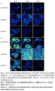
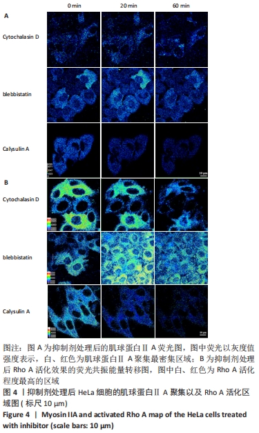
|