[1] PUPPI D, CHIELLINI F, PIRAS A M, et al. Polymeric materials for bone and cartilage repair. Prog Polym Sci. 2010;35(4): 403-440.
[2] REZWAN K,CHEN QZ,BLAKER JJ,et al.Biodegradable and bioactive porous polymer/inorganic composite scaffolds for bone tissue engineering.Biomaterials.2006;27(18): 3413-3431.
[3] CARLETTI E, MOTTA A, MIGLIARESI C.Scaffolds for tissue engineering and 3D cell culture. Methods Mol Biol. 2011;695: 17.
[4] CUI L, XU W, GUO X, et al.Synthesis of strontium hydroxyapatite embedding ferroferric oxide nano-composite and its application in Pb2+ adsorption.J Mol Liq.2014;197:40-47.
[5] LIU X, ZHU C, LI Y, et al.The Preparation and In Vitro Evaluations of a Nanoscaled Injectable Bone Repair Material. J Nanomater.2015;2015(4):1-8.
[6] RENGIER F, MEHNDIRATTA A, TENGGKOBLIGK HV, et al. 3D printing based on imaging data: review of medical applications. Int J Comput Assist Radiol Surg.2010;5(4):335.
[7] PEREIRA TF, SILVA MAC, OLIVEIRA MF, et al.Effect of process parameters on the properties of selective laser sintered Poly(3-hydroxybutyrate) scaffolds for bone tissue engineering. Vir Phys Prototyp.2012;7(4):275-285.
[8] BOSE S, VAHABZADEH S, BANDYOPADHYAY A.Bone tissue engineering using 3D printing.Mater Today. 2013; 16(12): 496-504.
[9] AMINI AR, LAURENCIN CT, NUKAVARAPU SP.Bone Tissue Engineering: Recent Advances and Challenges.Crit Rev Biomed Eng.2012;40(5):363-408.
[10] MARÍA GF, BERTA RF, JAIME RC, et al.Effects of intravenous administration of allogenic bone marrow- and adipose tissue-derived mesenchymal stem cells on functional recovery and brain repair markers in experimental ischemic stroke.Stem Cell Res Ther.2013;4(1):1-12.
[11] DOMINICI M, LE BK, MUELLER I, et al.Minimal criteria for defining multipotent mesenchymal stromal cells.The International Society for Cellular Therapy position statement. Cytotherapy. 2006;8(4):315.
[12] YIN Z, CHEN X, SONG HX, et al.Electrospun scaffolds for multiple tissues regeneration in vivo through topography dependent induction of lineage specific differentiation. Biomaterials. 2015;44:173.
[13] TODA M, OHNO J, SHINOZAKI Y, et al.Osteogenic potential for replacing cells in rat cranial defects implanted with a DNA/protamine complex paste.Bone.2014;67(5):237-245.
[14] WU S, LIU X, YEUNG KWK, et al.Biomimetic porous scaffolds for bone tissue engineering. Mater Sci Eng R.2014;80:1-36.
[15] POLO-CORRALES L, LATORRE-ESTEVES M, RAMIREZ-VICK JE.Scaffold Design for Bone Regeneration.J Nanosci Nanotechnol.2014;14(1):15-56.
[16] MANDRYCKY C, WANG Z, KIM K, et al.3D Bioprinting for Engineering Complex Tissues.Biotechnol Adv. 2016;34(4): 422-434.
[17] MIRONOV V, VISCONTI RP, KASYANOV V, et al.Organ printing: tissue spheroids as building blocks.Biomaterials. 2009;30(12):2164.
[18] WOO KM, SEO J, ZHANG R, et al.Suppression of apoptosis by enhanced protein adsorption on polymer/hydroxyapatite composite scaffolds.Biomaterials.2007;28(16):2622-2630.
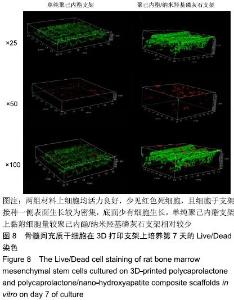
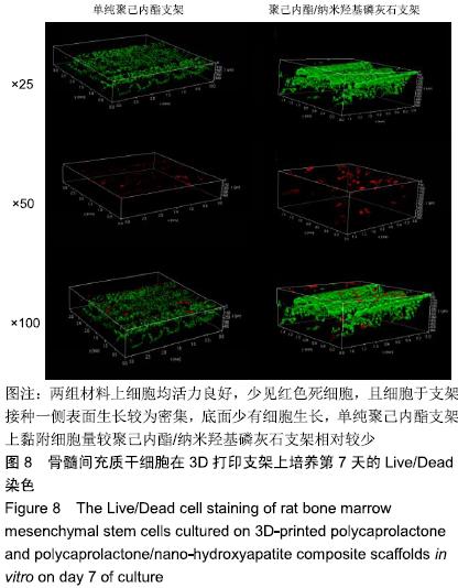
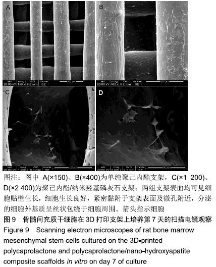
|