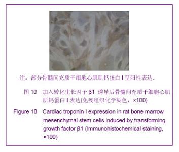
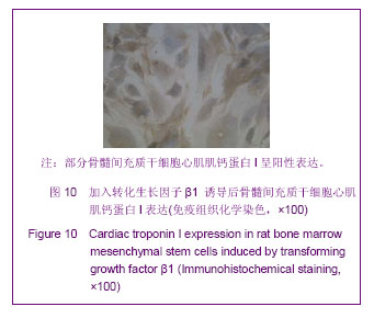
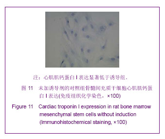
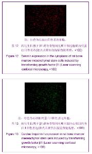
| [1] 王彤,符岳,方向龆,等.体外诱导骨髓问充质干细胞向心肌细胞的分化和鉴定[J].中山大学学报:医学科学版, 2007,28(z1): 9-11.[2] 吕洋,吴志刚,王海萍,等.转化生长因子β诱导骨髓干细胞分化为心肌样细胞的研究[J].中华老年心脑血管病杂志,2012,14(1): 62-65[3] Huang YL,Kuang J, Hu YZ, et al. Bone marrow stromal cell transplantation combined with angiotensin-converting enzyme inhibitor treatment in rat with acute myocardial infarction and the role of insulin-like growth factor-1.Cytotherapy.2012;14(5): 563-569.[4] Krausgrill B, Vantler M, Burst V, et al. Influence of cell treatment with PDGF-BB and reperfusion on cardiac persistence of mononuclear and mesenchymal bone marrow cells after transplantation into acute myocardial infarction in rats.Cell Transplant.2009;18(8):847-853.[5] Alshammary S, Fukushima S, Miyagawa S, et al. Impact of cardiac stem cell sheet transplantation on myocardial infarction.Surg Today.2013, Epub ahead of print [6] Mezey E.The therapeutic potential of bone marrow-derived stromal cells.J Cell Biochem. 2011;112(10):2683-2687.[7] Ling SK,Wang R, Dai ZQ, et al. Pretreatment of rat bone marrow mesenchymal stem cells with a combination of hypergravity and 5-azacytidine enhances therapeutic efficacy for myocardial infarction.Biotechnol Prog.2011;27(2):473-482.[8] Haghani K, Bakhtiyari S, Nouri AM.In vitro study of the differentiation of bone marrow stromal cells into cardiomyocyte-like cells.Mol Cell Biochem. 2012;361(1-2): 315-320.[9] 王海萍,张雷,李秀华.大鼠骨髓间充质干细胞体外诱导分化为心肌样细胞[J].解剖学报, 2007,38(1):70-74.[10] Bierie B,Moses HL.TGF-beta and caneer.Cytokine Growth Factor Rev.2006;17(l/2):29-40.[11] Antoon R,Yeger H,Loai Y,et al.Impact of bladder-derived acellular matrix, growth factors,and extracellular matrix constituents on the survival and multipotency of marrow-derived mesenchymal stem cells.J Biomed Mater Res A.2012; 100(1):72-83. [12] Sun Z, Wang Y, Gong X, et al. Secretion of rat tracheal epithelial cells induces mesenchymal stem cells to differentiate into epithelial cells.Cell Biol Int. 2012;36(2): 169-175.[13] Li TS,Hayashi M,Ito H,et al.Regeneration of infarcted myocardium by intramyocardial implantation of ex vivo transforming growth factor- beta- preprogrammed bone marrow stem cells.Circulation.2005;111(19): 2438-2445. [14] Lawson-Smith MJ, McGeachie JK.The identification of myogenic cells in skeletal muscle, with emphasison the use of tritiated thymidine autoradiography and desmin antibodies. Anat.1998;192(1): 161-171.[15] 史绍蓉,王娟.α-心肌肌球蛋白重链与原肌球蛋白-1在不同负荷运动中的差异表达研究[J].北京体育大学学报, 2009, 32(10): 51-54. |