Chinese Journal of Tissue Engineering Research ›› 2026, Vol. 30 ›› Issue (27): 6995-7001.doi: 10.12307/2026.368
Previous Articles Next Articles
Finite element analysis of four Kirschner wire fixation methods for treating patellar transverse fractures
Li Yongwei, Ye Hong
- The First Affiliated Hospital of Nanping, Fujian Medical University, Nanping 353000, Fujian Province, China
-
Received:2025-06-16Accepted:2025-09-03Online:2026-09-28Published:2026-04-16 -
Contact:Ye Hong, Chief physician, The First Affiliated Hospital of Nanping, Fujian Medical University, Nanping 353000, Fujian Province, China -
About author:Li Yongwei, Attending physician, The First Affiliated Hospital of Nanping, Fujian Medical University, Nanping 353000, Fujian Province, China -
Supported by:Fujian Natural Science Foundation, No. 2024J011602 (to LYW)
CLC Number:
Cite this article
Li Yongwei, Ye Hong . Finite element analysis of four Kirschner wire fixation methods for treating patellar transverse fractures[J]. Chinese Journal of Tissue Engineering Research, 2026, 30(27): 6995-7001.
share this article
Add to citation manager EndNote|Reference Manager|ProCite|BibTeX|RefWorks
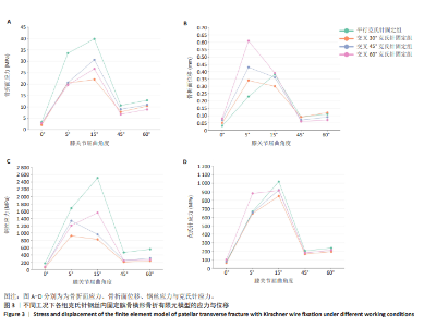
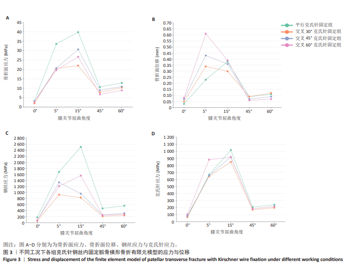
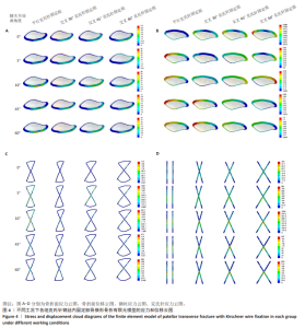
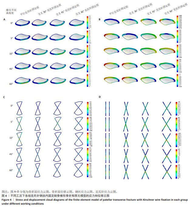
克氏针钢丝内固定髌骨横形骨折有限元模型分析结果,见图3,4所示。在整个步态周期中,4组模型分析结果在膝关节屈曲5°和15°时表现出了明显差异,而其他3种工况下的结果差异较小,因此,该文重点讨论膝关节屈曲5°和15°两种工况。 4组模型的骨折面应力在2.06-40.00 MPa之间,平行克氏针固定组在5种工况中均表现出最大的骨折面应力,而且这种差异在膝关节屈曲5°和15°时更加明显;交叉30°克氏针固定组膝关节屈曲15°时的骨折面应力明显小于交叉45°克氏针固定组、交叉60°克氏针固定组。 4组模型的骨折面位移在0.03-0.61 mm之间,交叉60°克氏针固定组膝关节屈曲5°和15°时均表现出最大的骨折面位移,平行克氏针固定组膝关节屈曲5°时的骨折面位移最小,交叉30°克氏针固定组膝关节屈曲15°时的骨折面位移最小。 4组内固定模型的钢丝应力在56.80-2 511.00 MPa之间,平行克氏针固定组膝关节屈曲5°,15°时的钢丝应力最大,交叉30°克氏针固定组膝关节屈曲5°,15°时的钢丝应力最小。 4组内固定模型的克氏针应力在65.67-1 018.00 MPa之间,交叉60°克氏针固定组膝关节屈曲5°时的克氏针应力最大,平行克氏针固定组膝关节屈曲15°的克氏针应力最大,交叉30°克氏针固定组膝关节屈曲5°和15°的克氏针应力最小。"
"
| [1] LARSEN P, COURT-BROWN CM, VEDEL JO, et al. Incidence and Epidemiology of Patellar Fractures. Orthopedics. 2016;39(6):e1154-e1158. [2] HENRICHSEN JL, WILHEM SK, SILJANDER MP, et al. Treatment of Patella Fractures. Orthopedics. 2018;41(6):e747-e755. [3] HARGETT DI, SANDERSON BR, LITTLE MTM. Patella Fractures: Approach to Treatment. J Am Acad Orthop Surg. 2021;29(6):244-253. [4] ZHANG JZ, LIU Z. Standardized evaluation and treatment of patellar fractures. Zhongguo Gu Shang. 2013;26(6):445-448. [5] STEINMETZ S, BRÜGGER A, CHAUVEAU J, et al. Practical guidelines for the treatment of patellar fractures in adults. Swiss Med Wkly. 2020;150:w20165. [6] MA XY, LIU B, ZHOU DP, et al. Treatment for transverse patella fractures with minimally invasive techniques (Review). Exp Ther Med. 2022;23(3):192. [7] MENG D, XU P, SHEN D, et al. A clinical comparison study of three different methods for treatment of transverse patellar fractures. J Orthop Sci. 2019;24(1):142-146. [8] SCHUETT DJ, HAKE ME, MAUFFREY C, et al. Current Treatment Strategies for Patella Fractures. Orthopedics. 2015;38(6):377-384. [9] MICHELITSCH C, SOMMER C. Reduction techniques for osteosynthesis of intra-articular fractures. Unfallchirurg. 2019;122(2):110-119. [10] DELLA ROCCA GJ. Displaced patella fractures. J Knee Surg. 2013;26(5): 293-299. [11] LEBRUN CT, LANGFORD JR, SAGI HC. Functional outcomes after operatively treated patella fractures. J Orthop Trauma. 2012;26(7):422-426. [12] DIETZ SO, HESSMANN MH, GERCEK E, et al. [Patella fracture]. Oper Orthop Traumatol. 2009;21(2):206-220. [13] KAKAZU R, ARCHDEACON MT. Surgical Management of Patellar Fractures. Orthop Clin North Am. 2016;47(1):77-83. [14] SCHNABEL B, SCHARF M, SCHWIEGER K, et al. Biomechanical comparison of a new staple technique with tension band wiring for transverse patella fractures. Clin Biomech (Bristol, Avon). 2009;24(10):855-859. [15] DARGEL J, GICK S, MADER K, et al. Biomechanical comparison of tension band- and interfragmentary screw fixation with a new implant in transverse patella fractures. Injury. 2010;41(2):156-160. [16] BERNINGER MT, FROSCH KH. Change in the treatment of patellar fractures. Unfallchirurgie (Heidelb). 2022;125(7):518-526. [17] LEE SY, CHOI JY, LEE HI, et al. The Comparison of Postoperative Outcomes Open and Closed Reduction for Patellar Fractures. J Knee Surg. 2020; 33(1):73-77. [18] TAYLOR BC, MEHTA S, CASTANEDA J, et al. Plating of patella fractures: techniques and outcomes. J Orthop Trauma. 2014;28(9):e231-235. [19] MÜLLER EC, FROSCH KH. Functional Outcomes of Revision Osteosynthesis after Failure of Surgical Treatment of Patellar Fractures. J Knee Surg. 2021;34(1):80-86. [20] SAYUM FILHO J, LENZA M, TAMAOKI MJ, et al. Interventions for treating fractures of the patella in adults. Cochrane Database Syst Rev. 2021;2(2): Cd009651. [21] SMITH ST, CRAMER KE, KARGES DE, et al. Early complications in the operative treatment of patella fractures. J Orthop Trauma. 1997;11(3): 183-187. [22] SHYMON SJ, JANSSON H, SCHNEIDERMAN BA, et al. Functional Outcomes of Patella Fractures Treated With Anterior Plate Osteosynthesis at One Year. J Orthop Trauma. 2021;35(1): e1-e6. [23] XUE Z, QIN H, DING H, et al. Two-Tension-Band Technique in Revision Surgery for Fixation Failure of Patellar Fractures. Med Sci Monit. 2016; 22:2736-2741. [24] SCOLARO J, BERNSTEIN J, AHN J. Patellar fractures. Clin Orthop Relat Res. 2011;469(4): 1213-1215. [25] WILD M, WINDOLF J, FLOHÉ S. Fractures of the patella. Unfallchirurg. 2010;113(5):401-11; quiz 12. [26] CAMARDA L, MORELLO S, BALISTRERI F, et al. Non-metallic implant for patellar fracture fixation: A systematic review. Injury. 2016;47(8): 1613-1617. [27] HSU KL, CHANG WL, YANG CY, et al. Factors affecting the outcomes of modified tension band wiring techniques in transverse patellar fractures. Injury. 2017;48(12):2800-2806. [28] BURKHART TA, ANDREWS DM, DUNNING CE. Finite element modeling mesh quality, energy balance and validation methods: a review with recommendations associated with the modeling of bone tissue. J Biomech. 2013;46(9):1477-1488. [29] OEFNER C, HERRMANN S, KEBBACH M, et al. Reporting checklist for verification and validation of finite element analysis in orthopedic and trauma biomechanics. Med Eng Phys. 2021;92:25-32. [30] YE Y, YOU W, ZHU W, et al. The Applications of Finite Element Analysis in Proximal Humeral Fractures. Comput Math Methods Med. 2017; 2017:4879836. [31] MENGONI M. Biomechanical modelling of the facet joints: a review of methods and validation processes in finite element analysis. Biomech Model Mechanobiol. 2021;20(2):389-401. [32] ZHANG ZH, QI YS, WEI BG, et al. Application strategy of finite element analysis in artificial knee arthroplasty. Front Bioeng Biotechnol. 2023;11: 1127289. [33] ZHU JJ, DU ZP, CAO CP, et al. Effects of different reduction patterns on stress distribution in patients with intertrochanteric fractures with intramedullary nail fixation: a finite element analysis. Front Bioeng Biotechnol. 2025;13: 1507774. [34] GOFFIN JM, PANKAJ P, SIMPSON AH. The importance of lag screw position for the stabilization of trochanteric fractures with a sliding hip screw: A subject-specific finite element study. J Orthop Res. 2013;31(4):596-600. [35] MU JX, XIANG SY, MA QY, et al. Selection of internal fixation method for femoral intertrochanteric fractures using a finite element method. World J Clin Cases. 2021;9(22):6343-6356. [36] LING M, ZHAN S, JIANG D, et al. Where should Kirschner wires be placed when fixing patella fracture with modified tension-band wiring? A finite element analysis. J Orthop Surg Res. 2019;14(1):14. [37] BARCIK J, ERNST M, BUCHHOLZ T, et al. The absence of immediate stimulation delays bone healing. Bone. 2023;175:116834. [38] DUAN ZW, LU H. Effect of Mechanical Strain on Cells Involved in Fracture Healing. Orthop Surg. 2021;13(2):369-375. [39] KENWRIGHT J, GARDNER T. Mechanical influences on tibial fracture healing. Clin Orthop Relat Res. 1998;(355 Suppl):S179-190. [40] LENIHAN J, RAMOS-PASCUAL S, SILVESTROS P, et al. Novel techniques demonstrate superior fixation of simple transverse patella fractures - A biomechanical study. Injury. 2020;51(6):1288-1293. [41] ZHANG Z, SUN F, ZHANG T, et al. An innovative anti-rotation tension band wiring for treating transverse patellar fractures: finite element analysis and mechanical testing. J Orthop Surg Res. 2024;19(1):416. [42] VAJGEL A, CAMARGO IB, WILLMERSDORF RB, et al. Comparative finite element analysis of the biomechanical stability of 2.0 fixation plates in atrophic mandibular fractures. J Oral Maxillofac Surg. 2013;71(2):335-342. [43] 黄晓燕,薛锋,殷诺,等.双螺栓与克氏针张力带固定治疗髌骨横形骨折模型的有限元分析[J].生物骨科材料与临床研究,2018,15(4):6-8,82. [44] MADEN M, MURAT DULGEROGLU A, BACAKSIZ T, et al. Does pin configuration matter in modified tension band wiring for transverse patellar fracture? A biomechanical study. Knee. 2022;39:300-307. [45] KIM Y, KWON M, RYU JY, et al. Biomechanical Analysis of the Kirschner-Wire Depth of the Modified Tension Band Wiring Technique in Transverse Patellar Fractures: An Experimental Study Using the Finite-Element Method. Clin Orthop Surg. 2021;13(3):315-319. [46] 刘峰,冯毅.步态周期下不同克氏针张力带治疗髌骨横行骨折的有限元分析[J].中国组织工程研究,2022,26(9):1367-1371. [47] 蔡昊,周海波,虞嘉欢,等.空心螺钉联合“8”、“0”张力带钢丝内固定治疗髌骨横行骨折模型的有限元分析与比较[J].中国临床解剖学杂志,2022,40(4):454-459. |
| [1] | Zhou Daobin, Wang Kehao, Xie Yang, Ning Rende. Biomechanical characteristics of volar locking plate only versus combined dorsal mini-plate fixation of distal radius fractures with dorsal ulnar fragment [J]. Chinese Journal of Tissue Engineering Research, 2026, 30(9): 2255-2261. |
| [2] | Chen Huiting, Zeng Weiquan, Zhou Jianhong, Wang Jie, Zhuang Congying, Chen Peiyou, Liang Zeqian, Deng Weiming. Tail anchoring technique of vertebroplasty in treatment of osteoporotic vertebral compression fractures with intravertebral cleft: a finite element analysis [J]. Chinese Journal of Tissue Engineering Research, 2026, 30(9): 2145-2152. |
| [3] | Zeng Xuan, Weng Rui, Ye Shicheng, Tang Jiadong, Mo Ling, Li Wenchao. Two lumbar rotary manipulation techniques in treating lumbar disc herniation: a finite element analysis of biomechanical differences [J]. Chinese Journal of Tissue Engineering Research, 2026, 30(9): 2153-2161. |
| [4] | Cheng Qisheng, Julaiti·Maitirouzi, Xiao Yang, Zhang Chenwei, Paerhati·Rexiti. Finite element analysis of novel variable-diameter screws in modified cortical bone trajectory of lumbar vertebrae [J]. Chinese Journal of Tissue Engineering Research, 2026, 30(9): 2162-2171. |
| [5] | Wu Hongxu, Liu Xuanyu, Wang Taoyu, Wang Shiyao, Cheng Jingyi, Zhang Mingwen, Zhang Yinxia, Liu Zhihua, Wang Xiaojie. Finite element simulation of scoliosis with muscle unit introduction: verification of correction effect under bidirectional load [J]. Chinese Journal of Tissue Engineering Research, 2026, 30(9): 2172-2181. |
| [6] | Liu Jiafu, Ren Ruxia, Liao Zhouwei, Zhou Xiali, Wu Yihong, Zhang Shaoqun. Three-dimensional finite element analysis of cervical spine biomechanical characteristics in a rat model of cervical vertigo [J]. Chinese Journal of Tissue Engineering Research, 2026, 30(9): 2182-2190. |
| [7] | Liu Wenlong, Dong Lei, Xiao Zhengzheng, Nie Yu. Finite element analysis of tibial prosthesis loosening after fixed-bearing unicompartmental knee arthroplasty for osteoporosis [J]. Chinese Journal of Tissue Engineering Research, 2026, 30(9): 2191-2198. |
| [8] | Zheng Wangyang, Fei Ji, Yang Di, Zhao Lang, Wang Lingli, Liu Peng, Li Haiyang. Finite element analysis of the force changes of the supraspinatus tendon and glenohumeral joint during the abduction and flexion of the humerus [J]. Chinese Journal of Tissue Engineering Research, 2026, 30(9): 2199-2207. |
| [9] | Cai Qirui, Dai Xiaowei, Zheng Xiaobin, Jian Sili, Lu Shaoping, Liu Texi, Liu Guoke, Lin Yuanfang. Mechanical effects of Long’s traction orthopedic method on cervical functional units: quantitative analysis of biomechanical model of head and neck [J]. Chinese Journal of Tissue Engineering Research, 2026, 30(9): 2208-2216. |
| [10] | Rao Jingcheng, Li Yuwan, Zheng Hongbing, Xu Zhi, Zhu Aixiang, Shi Ce, Wang Bing, Yang Chun, Kong Xiangru, Zhu Dawei. Biomechanical differences between the new proximal femoral stable intramedullary nail and traditional intramedullary nail#br# [J]. Chinese Journal of Tissue Engineering Research, 2026, 30(9): 2217-2225. |
| [11] | Chen Long, Wang Xiaozhen, Xi Jintao, Lu Qilin. Biomechanical performance of short-segment screw fixation combined with expandable polyetheretherketone vertebral body replacement in osteoporotic vertebrae [J]. Chinese Journal of Tissue Engineering Research, 2026, 30(9): 2226-2235. |
| [12] | Yan Xiangning, Chen Lei, Chen Yonghuan, Wang Chao, Li Xiaosheng. Influence of different depths and loads on knee joint mechanics and peripheral muscle force characteristics during squatting [J]. Chinese Journal of Tissue Engineering Research, 2026, 30(9): 2236-2247. |
| [13] | Zhang Zizheng, Luo Wang, Liu Changlu. Application value of finite element analysis on unicompartmental knee arthroplasty for medial knee compartmental osteoarthritis [J]. Chinese Journal of Tissue Engineering Research, 2026, 30(9): 2313-2322. |
| [14] | Zhao Feifan, Cao Yujing. Risk factors and coping strategies of internal fixation failure in treatment of intertrochanteric fracture with proximal femoral nail antirotation [J]. Chinese Journal of Tissue Engineering Research, 2026, 30(9): 2323-2333. |
| [15] | Zhang Xianxu, Ma Zhong, Liu Xin, Huang Lei, Shen Wenxiang, Luo Zhiqiang . Lumbar fusion combined with unilateral fixation for lumbar degenerative diseases: biomechanics, technical evolution, and clinical applications [J]. Chinese Journal of Tissue Engineering Research, 2026, 30(9): 2334-2342. |
| Viewed | ||||||
|
Full text |
|
|||||
|
Abstract |
|
|||||