Chinese Journal of Tissue Engineering Research ›› 2026, Vol. 30 ›› Issue (16): 4030-4037.doi: 10.12307/2026.700
Previous Articles Next Articles
Cone beam computed tomography of the distance and position of the root apex of the mandibular second molar relative to the mandibular canal
Weng Zhirong1, 2, Gegentana1, 2
- 1 Affiliated Hospital of Inner Mongolia Medical University, Hohhot 010050, Inner Mongolia Autonomous Region, China; 2 School of Stomatology, Inner Mongolia Medical University, Hohhot 010059, Inner Mongolia Autonomous Region, China
-
Received:2025-06-16Accepted:2025-08-20Online:2026-06-08Published:2025-11-25 -
Contact:Gegentana, MD, Professor, Chief physician, Affiliated Hospital of Inner Mongolia Medical University, Hohhot 010050, Inner Mongolia Autonomous Region, China; School of Stomatology, Inner Mongolia Medical University, Hohhot 010059, Inner Mongolia Autonomous Region, China -
About author:Weng Zhirong, MS candidate, Physician, Affiliated Hospital of Inner Mongolia Medical University, Hohhot 010050, Inner Mongolia Autonomous Region, China; School of Stomatology, Inner Mongolia Medical University, Hohhot 010059, Inner Mongolia Autonomous Region, China
CLC Number:
Cite this article
Weng Zhirong, Gegentana. Cone beam computed tomography of the distance and position of the root apex of the mandibular second molar relative to the mandibular canal[J]. Chinese Journal of Tissue Engineering Research, 2026, 30(16): 4030-4037.
share this article
Add to citation manager EndNote|Reference Manager|ProCite|BibTeX|RefWorks
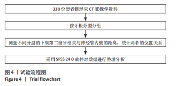
2.1 参与者数量分析 按纳排标准纳入330例患者的影像学资料,其中男性164例,女性166例;青年组200例,中年组103例,老年组27例。左右侧共660颗下颌第二磨牙,其中三根型共6颗,由于数目较少,故该根型只统计其数量;其他型属于下颌第二磨牙牙根形态的变异,此次资料中未检出,故不做研究。 2.2 试验流程图 见图4。"
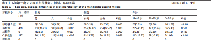
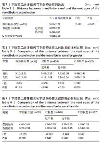
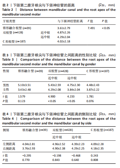
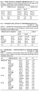
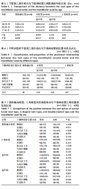
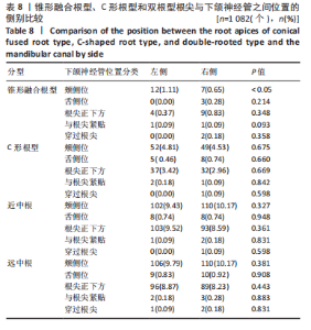
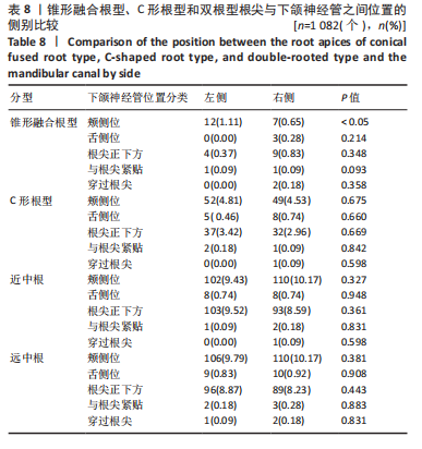
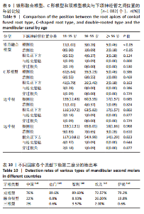
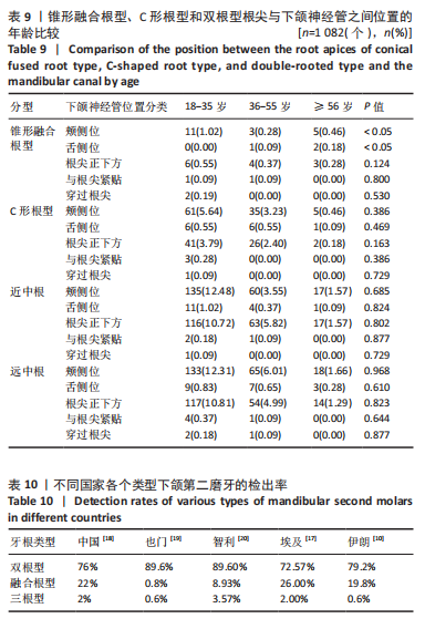
2.3 下颌第二磨牙牙根形态 下颌第二磨牙常见双根型,其次是C形根型,次之是锥形融合根形,最后是三根型,检出率依次为64.85%,28.33%,5.91%,0.9%。锥形融合根型、双根型及C形根型的分布在性别、年龄的比较中差异有统计学意义。在不同性别中,锥形融合根型和C形根型女性占比较大(P < 0.05),男性下颌第二磨牙以双根型为主(P < 0.05)。在不同年龄中,锥形融合根型和C形根型在青年组检出率较高(P < 0.05),左右侧分布差异无统计学意义(P > 0.05),见表1。 2.4 下颌第二磨牙各牙根根尖与下颌神经管的距离 不同类型下颌第二磨牙根尖距神经管的距离差异有显著性意义(P < 0.05)。锥形融合根型的根尖距下颌神经管的距离最近,其次是C形根型,次之是双根型远中根,最后是双根型近中根,见表2。 在性别中,男性双根型下颌第二磨牙近中根和远中根根尖到神经管的距离均大于女性(P < 0.05),不同侧别和年龄间差异无显著性意义(P > 0.05),但该距离有随着年龄增大而增加的趋势,见表3-5。 2.5 下颌第二磨牙各牙根根尖与下颌神经管的位置 下颌神经管位于下颌第二磨牙牙根颊侧、根尖正下方、舌侧、与根尖紧贴及穿过根尖的占比依次为50.64%,42.78%,4.72%,1.20%,0.64%。根尖紧贴和位于神经管内的情况占锥形融合根与神经管位置总分布的10.24%;占C形根2.14%;占双根型远中根1.87%;占近中根0.93%,见表6。 在锥形融合根中,女性神经管位于颊侧位及根尖正下方的占比高于男性(P < 0.05);左侧神经管位于颊侧位的占比高于右侧(P < 0.05);青年组神经管位于颊侧位的占比高于中年组和老年组(P < 0.05);老年组神经管位于舌侧位占比高于青年组和中年组(P < 0.05)。在C形根中,女性神经管位于根尖正下方的占比高于男性(P < 0.05),见表7-9。"
"
"
"
"
| [1] 蓝炎涛, 张丽霞.下颌第三磨牙阻生类型与第二磨牙远中邻面龋坏的相关性研究[J]. 现代医院,2021,21(1):159-161+164. [2] DEEPA K, UJJWAL K, ARCHANA M, et al. Association of Prevalence of Dental Caries in Mandibular Second Molar with Impacted Third Molar.J Nepal Health Res Counc. 2021;19(2):259-263. [3] 冯璐, 张岚, 黄定明. 中国西部地区人群下颌后牙与下牙槽神经管距离关系的三维研究[C].中华口腔医学会牙体牙髓病学专业委员会第十四次全国牙体牙髓病学学术大会论文汇编,2022. [4] Kovisto T, Ahmad M, Bowles WR. Proximity of the Mandibular Canal to the Tooth Apex. J Endod. 2011;37(3):311-315. [5] CHONG BS, QUINN A, PAWAR RR, et al. The anatomical relationship between the roots of mandibular second molars and the inferior alveolar nerve. Int Endod J. 2015;48(6):549-555. [6] PATEL R, CLARKSON E. Implant surgery update for the general practitioner: dealing with common postimplant surgery complications. Dent Clin North Am. 2021;65(1):125-134. [7] CEBRIÁN F, PEÑA-CARDELLES JF, AKHONDI S, et al. Inferior alveolar nerve damage related to dental implant placement. A systematic review and meta-analysis. Med Oral Patol Oral Cir Bucal. 2025;7:27125. [8] 庞显卓, 薛明. 下颌后牙区显微根尖手术中颏神经的管理[J].中国实用口腔科杂志,2023,16(5):526-529. [9] BADR O, TALAL Z. Mental Nerve Anterior Loop Detection in Panoramic and Cone Beam Computed Tomography Radiograph for Safe Dental Implant Placement.Cureus.2022;14(10): e30687-e30687. [10] MARYAM J, SAEED R, FARNAZ J, et al. Anatomic Features of C-shaped Mandibular Second Molars in a Selected Iranian Population Using CBCT.Iran Endod J. 2018;13(1):120-125. [11] ADIBI S, PAKNAHAD M. Comparison of cone-beam computed tomography and osteometric examination in preoperativeassessment of the proximity of the mandibular canal to the apices of the teeth. Br J Oral Maxillofac Surg. 2017;55(3):246-250. [12] ALMANSOUR MI, AL-ZUBAIDI SM, ENIZY AS, et al. Comprehensive evaluation of root and root canal morphology of mandibular second molars in a Saudi subpopulation evaluated by cone-beam computed tomography. BMC Oral Health. 2022;22(1):267. [13] 吴康汝, 吴新智,张振标.人体测量方法[M].北京:科学出版社, 1984:12-13. [14] MICHAŁ P, ALEKSANDRA P, KRZYSZTOF S, et al. The influence of age, sex, and tooth type on the anatomical relationship between tooth roots and the mandibular canal. Imaging Sci Dent. 2021;51(4):373-382. [15] 谢霓, 王九龙, 朱友家. 下颌第二磨牙牙根及根管形态学研究[J].临床口腔医学杂志,2015,31(7):415-417. [16] 邵俊杰, 倪洁丽, 李琥, 等. 下颌神经管结构、位置与分支的CBCT影像研究[J]. 口腔生物医学,2018,9(3):148-151. [17] SABER SM, SEOUD MAE, SADAT SMAE, et al. Root and canal morphology of mandibular second molars in an Egyptian subpopulation: a cone-beam computed tomography study. BMC Oral Health. 2023;23(1):217. [18] ZHANG R, WANG H, TIAN YY, et al. Use of cone-beam computed tomography to evaluate root and canal morphology of mandibular molars in Chinese individuals. Int Endod J. 2011;44(11):990-999. [19] SENAN EM, MADFA AA, ALHADAINY HA. Root and Canal Configuration of Mandibular First Molars in a Yemeni Population: A Cone-beam Computed Tomography. Eur Endod J. 2020;5(1):10-17. [20] ANDRES T, REINHILDE J, PAUL L, et al.Characterization of mandibular molar root and canal morphology using cone beam computed tomography and its variability in Belgian and Chilean population samples. Imaging Sci Dent. 2015;45(2):95-101. [21] YANG L, HAN J, WANG Q, et al. Variations of root and canal morphology of mandibular second molars in Chinese individuals:a cone-beam computed tomography study. BMC Oral Health. 2022;22(1): 274. [22] 袁曦玉, 孟凡琦, 马丽, 等. 维吾尔族成人下颌第二磨牙牙根及根管形态的CBCT研究[J]. 口腔颌面修复学杂志,2023,24(3):199-205. [23] GUO Q, WANG Q, YANG Y, et al. Root and root canal morphology of mandibular second permanent molars in the Gansu province population: A CBCT study. Aust Endod J. 2023:49 Suppl 1:27-32. [24] 黎祺, 黄少宏. 岭南地区广府民系人群下颌第二恒磨牙牙根和根管形态的锥形束CT研究[J]. 国际口腔医学杂志,2019,46(6):640-649. [25] SRIVASTAVA S, ALHARBI HM, ALHARBI AS, et al. Assessment of the Proximity of the Inferior Alveolar Canal with the Mandibular Root Apices and Cortical Plates—A Retrospective Cone Beam Computed Tomographic Analysis.J Pers Med. 2022;12(11):1784. [26] SHOKRY SM, ALSHAIB SA, AL MOHAIMEED ZZ, et al. Assessment of the Inferior Alveolar Nerve Canal Course Among Saudis by Cone Beam Computed Tomography (Pilot Study).J Maxillofac Oral Surg. 2019;18(3):452-458. [27] 杨偲偲, 程波, 尹苗, 等. 下颌第一、第二磨牙与神经管相对位置的CBCT影像分析[J]. 口腔医学研究,2024,40(9):810-815. [28] BÜRKLEIN S, GRUND C, SCHÄFER E. Relationship between Root Apices and the Mandibular Canal: A Cone-beam Computed Tomographic Analysis in a German Population. J Endod. 2015;41(10):1696-1700. [29] KOIVISTO T, CHIONA D, MILROY LL, et al. Mandibular Canal Location: Cone-beam Computed Tomography Examination. J Endod. 2016;42(7):1018-1021. [30] DAGER MM, MCNAMARA JA, BACCETTI T, et al. Aging in the craniofacial complex. Angle Orthod. 2008;78(3):440-444. [31] 高璇. 下颌第二磨牙根尖与下颌管间距离及相关因素的研究[D].太原:山西医科大学,2022. [32] CASTRO R, GUIVARC’H M, FOLETTI JM, et al. Endodontic-related inferior alveolar nerve injuries: A review and a therapeutic flow chart.J Stomatol Oral Maxillofac Surg. 2018;119(5):412-418. [33] 高璇, 李霞. 牙髓相关性下牙槽神经损伤的原因及诊治研究进展[J]. 山东医药,2022,62(24):98-101. [34] MATTHIAS P, ANSELM P. Permanent mimic musculature and nerve damage caused by sodium hypochlorite: a case report. Oral Surg Oral Med Oral Pathol Oral Radiol Endod. 2008;106(3):e80-83. [35] BERK KM, ECEM DG. Inferior alveolar nerve injury due to the extrusion of calcium hydroxide during endodontic treatment: A case report.Aust Endod J. 2022;48(2):342-346. [36] LIU H, LI Y, SHEN Y. Persistent paresthesia of inferior alveolar nerve after accidental extrusion of calcium hydroxide paste containing iodoform into the mandibular canal.J Dent Sci. 2024;19(1):720-721. [37] ALMASLAMANI M, ALHASSAWI S, PRASAD P, et al. Diagnosis and Management of Inferior Alveolar Nerve Injury Caused by Calcium Hydroxide Extrusion during Root Canal Treatment: Case Report. J Pharm Bioallied Sci. 2024;16(Suppl 5):S4900-S4904. [38] 唐蓓, 赵文俊, 王虎, 等. 根管超填导致下牙槽神经损伤2例[J].国际口腔医学杂志,2020,47(3):293-296. [39] 廖洪林, 方仲瀚, 张艳艳, 等.牙种植术后三叉神经创伤性神经病理性疼痛的诊断与防治[J]. 国际口腔医学杂志,2023,50(6):729-738. [40] TURKYILMAZ I. Persistent numbness of the lower lip and chin due to inferior alveolar nerve injury after implant placement: A clinical report. Prim Dent J. 2024;13(4):66-68. [41] 蔡锋, 高文鼎, 程志刚. 种植体损伤下牙槽神经1例及文献回顾[J].临床口腔医学杂志,2024,40(2):117-118. [42] NAVOT G, EYAL R, LARS B, et al. Medico-legal aspects of altered sensation following endodontic treatment: a retrospective case series.Oral Surg Oral Med Oral Pathol Oral Radiol Endod. 2011;112(1):126-131. [43] 禹雯怡, 余海波, 陈娇, 等. 根管治疗相关的下牙槽神经损伤及治疗流程研究进展[J]. 临床口腔医学杂志,2024,40(4):248-250. [44] 宋海龙, 许东亮. 基于反应范围模型探讨牙列缺损种植治疗患者神经损伤的影响因素[J].罕少疾病杂志,2025,32(5):49-52. [45] 刘云通, 刘畅, 高丽钞, 等. 术后下牙槽神经功能障碍的研究进展[J]. 国际口腔医学杂志,2023,50(4):479-484. [46] DE ABREU JM, NUNES T, ALMIRO PA,et al. Health-Related Quality of Life with Iatrogenic Inferior Alveolar Nerve Injuries Treated with Photobiomodulation: A Comparative Study. J Clin Med. 2024;13(23):7237. [47] 谈松, 胡小洪. 高压氧治疗在牙槽神经损伤中的应用效果[C]第六届上海国际护理大会论文汇编(下),2024:133. [48] 祝颂松, 王旭东, 杨学文,等. 下颌支矢状骨劈开术并发症防治的专家共识[J]. 华西口腔医学杂志,2022,40(3):247-254. [49] ALHARBI G, RAO JKD, ALNAIM T,et al. Efficacy of low-level laser therapy and microsurgery on neurosensory recovery following inferior alveolar and lingual nerve injuries: A systematic review. J Oral Biol Craniofac Res. 2024;14(5):631-637. |
| [1] | Wei Bingqi, Sun Jiahui, Chen Liu, Li Yijing, Wan Hejia, Qi Yifan, Wang Shangzeng. BTN3A2 is a key target for the development or prevention of new drugs for knee osteoarthritis: a randomization study based on drug targeting [J]. Chinese Journal of Tissue Engineering Research, 2026, 30(16): 4021-4029. |
| [2] | Zeng Yongtao, Zheng Hongcheng, Nacikedaoerji, Refati·Nijiati, Shu Li, Liu Xu, Chen Hongtao. Treating acute type III-V acromioclavicular joint dislocation with single tunnel fixation versus tunnel-free suspension fixation of the coracoid process under shoulder arthroscopy [J]. Chinese Journal of Tissue Engineering Research, 2025, 29(5): 1036-1042. |
| [3] | Zhang Yuhang, Zeng Yuning, Zeng Jindi, Lu Yixuan, Ye Hui, Ji Jianxin. Accuracy of modified implant template of assisted implantation in missing second molars [J]. Chinese Journal of Tissue Engineering Research, 2025, 29(4): 738-744. |
| [4] | Chen Shujin, Ma Xiangyang, Zou Xiaobao, Liao Yingqiang, Qi Hairu, Liu Bao, Zeng Xianming. Biomechanical test of reduction ability of axis pivot screw in atlantoaxial screw-rod fixation [J]. Chinese Journal of Tissue Engineering Research, 2025, 29(15): 3116-3120. |
| [5] | Zuo Xinwei, Liu Gang, Bai Huizhong, Xu Lin, Zhao Yi, Ren Jingpei, Hu Chuanyu, Mu Xiaohong. Relationship between lumbar spine development and hip development in children with spastic cerebral palsy [J]. Chinese Journal of Tissue Engineering Research, 2024, 28(8): 1247-1252. |
| [6] | Zhao Rushun, Hao Yangquan, Xu Peng, Zheng Xin, Jiang Yonghong, Zhang Yuting, Wang Mengfei, Lu Chao. Effect of different locations of necrotic focus on the natural course of non-traumatic osteonecrosis of the femoral head [J]. Chinese Journal of Tissue Engineering Research, 2024, 28(6): 917-921. |
| [7] | Chen Wenchuang, Li Yong, Lu Yao, Zhang Meiren, Chen Haiyun, Yu Zhaoyu. Robot-assisted pedicle screw internal fixation in treatment of atlantoaxial dislocation [J]. Chinese Journal of Tissue Engineering Research, 2024, 28(36): 5833-5838. |
| [8] | Shen Qingfeng, Li Lingbo, Xia Yingpeng, Ma Shibo. Atlantoaxial dislocation treated by posterior atlantoaxial lateral mass interarticular release, posterior screw reduction and fusion with bone graft [J]. Chinese Journal of Tissue Engineering Research, 2024, 28(33): 5364-5369. |
| [9] | Yang Juncong, Huang Rui, Wu Xie. Visualization of the biomechanical characteristics of long-distance running landing patterns [J]. Chinese Journal of Tissue Engineering Research, 2024, 28(32): 5159-5166. |
| [10] | Wang Xinmin, Yan Wenkai, Song Yahui, Liu Fei. Leukocyte- and platelet-rich fibrin with autologous hamstring tendon for traumatic patella dislocation [J]. Chinese Journal of Tissue Engineering Research, 2024, 28(3): 404-410. |
| [11] | Zhang Wenhui, Wang Chunli, Fan Lizhen, Yang Yuping, Zhang Jinlong, Zhang Hui, Liu Jie, Tai Huiping. Accuracy of sacroiliac screw placement in robot-assisted navigation [J]. Chinese Journal of Tissue Engineering Research, 2024, 28(24): 3845-3849. |
| [12] | Zhao Gai, Liu Lingjun, Yin Hao, Ning Rende, Xu Bin. Three-dimensional finite element analysis of medial patellofemoral ligament reconstruction with transosseous and wire anchor fixation [J]. Chinese Journal of Tissue Engineering Research, 2024, 28(24): 3796-3800. |
| [13] | Xie Ting, Liu Tingting, Zeng Xuehui, Li Yamin, Zhou Panghu, Yi Nianhua. Fucoxanthin alleviates glucocorticoid-induced osteoblast apoptosis by activating nuclear factor erythroid-2-related factor 2 [J]. Chinese Journal of Tissue Engineering Research, 2024, 28(23): 3609-3614. |
| [14] | Xu Jian, Bi Wenzhi, Ji Yuncong, Kang Yunkang, Ma Peiqi, Wang Jialiang, Zhang Zongxi, Gan Fusheng, Yu Haiyang, Guo Biao. Quantitative CT measurement of bone mass density in different regions of the distal clavicle in reconstruction of acromioclavicular joint dislocation [J]. Chinese Journal of Tissue Engineering Research, 2024, 28(12): 1920-1924. |
| [15] | Su Shaoting, Zhou Honghai, Hou Zhaomeng, Lu Yan, Wang Wei, Chen Yixin, Chen Longhao, Tian Cong. Finite element analysis of thumb thrust in lumbar fixed-point rotation manipulation [J]. Chinese Journal of Tissue Engineering Research, 2024, 28(12): 1823-1828. |
| Viewed | ||||||
|
Full text |
|
|||||
|
Abstract |
|
|||||