Chinese Journal of Tissue Engineering Research ›› 2025, Vol. 29 ›› Issue (32): 6877-6884.doi: 10.12307/2025.921
Previous Articles Next Articles
Expression of mitochondrial creatine kinase 2 in a rat model of temporomandibular joint osteoarthritis and its role in inflammation progression
Nigeayi · Aihemaiti1, 2, Yilidanna · Dilixiati1, 2, An Wei1, 2, Maimaitituxun · Tuerdi1, 2
- 1Department of Oral and Maxillofacial Trauma and Orthognathic Surgery, the First Affiliated Hospital (Affiliated Stomatological Hospital) of Xinjiang Medical University, Urumqi 830054, Xinjiang Uygur Autonomous Region, China; 2Xinjiang Uygur Autonomous Region Institute of Stomatology, Urumqi 830054, Xinjiang Uygur Autonomous Region, China
-
Received:2024-10-14Accepted:2024-11-26Online:2025-11-18Published:2025-04-25 -
Contact:Maimaitituxun · Tuerdi, PhD, Chief physician, Department of Oral and Maxillofacial Trauma and Orthognathic Surgery, the First Affiliated Hospital (Affiliated Stomatological Hospital) of Xinjiang Medical University, Urumqi 830054, Xinjiang Uygur Autonomous Region, China; Xinjiang Uygur Autonomous Region Institute of Stomatology, Urumqi 830054, Xinjiang Uygur Autonomous Region, China -
About author:Nigeayi · Aihemaiti, Master candidate, Department of Oral and Maxillofacial Trauma and Orthognathic Surgery, the First Affiliated Hospital (Affiliated Stomatological Hospital) of Xinjiang Medical University, Urumqi 830054, Xinjiang Uygur Autonomous Region, China; Xinjiang Uygur Autonomous Region Institute of Stomatology, Urumqi 830054, Xinjiang Uygur Autonomous Region, China -
Supported by:National Natural Science Foundation of China, No. 8236030005 (to MT)
CLC Number:
Cite this article
Nigeayi · Aihemaiti, Yilidanna · Dilixiati, An Wei, Maimaitituxun · Tuerdi. Expression of mitochondrial creatine kinase 2 in a rat model of temporomandibular joint osteoarthritis and its role in inflammation progression[J]. Chinese Journal of Tissue Engineering Research, 2025, 29(32): 6877-6884.
share this article
Add to citation manager EndNote|Reference Manager|ProCite|BibTeX|RefWorks
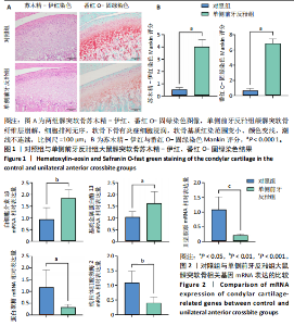
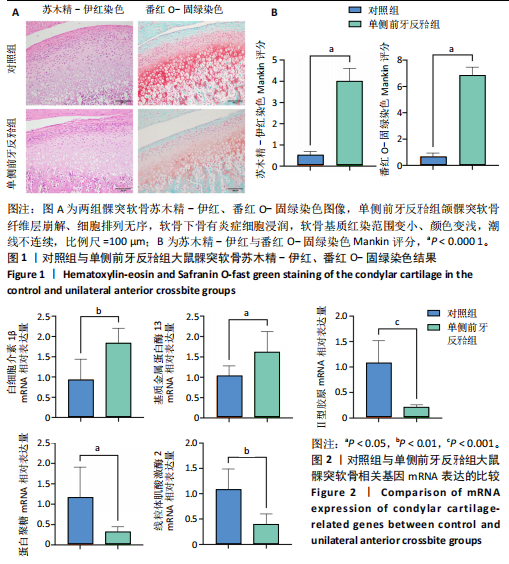
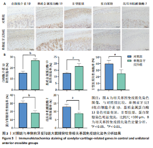
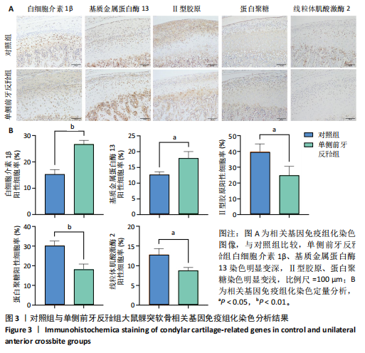
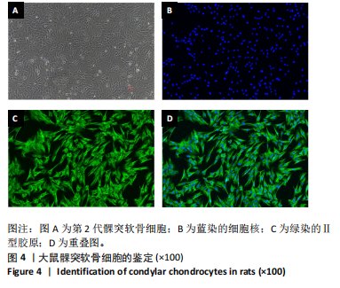
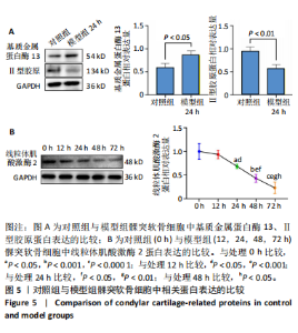
2.1 动物实验结果 2.1.1 实验动物数量分析 20只SD大鼠全部进入结果分析。 2.1.2 两组大鼠髁突软骨炎症情况 苏木精-伊红染色结果显示,对照组髁突软骨表面平整,不存在裂隙,分为纤维层、增殖层、肥大层、钙化软骨层4层;单侧前牙反牙合组髁突软骨纤维层崩解、细胞排列无序,增殖层细胞密集,细胞簇集明显,软骨下骨有炎症细胞浸润,见图1。番红O-固绿染色结果显示,相对于对照组,单侧前牙反牙合组软骨基质红染范围变小、颜色变浅,潮线不连续,见图1。单侧前牙反牙合组髁突软骨苏木精-伊红染色与番红O-固绿染色Mankin评分均高于对照组(P < 0.000 1),见图1。 2.1.3 两组大鼠髁突软骨中炎症基因及骨分解代谢基因表达比较 qRT-PCR检测结果显示,与对照组比较,单侧前牙反牙合组大鼠髁突软骨中白细胞介素1β、基质金属蛋白酶13 mRNA表达升高(P < 0.01,P < 0.05),Ⅱ型胶原、蛋白聚糖mRNA表达降低(P < 0.001,P < 0.05),见图2。免疫组化染色结果显示,与对照组比较,单侧前牙反牙合组大鼠髁突软骨中白细胞介素1β、基质金属蛋白酶13染色明显变深,Ⅱ型胶原、蛋白聚糖染色明显变浅,定量分析结果显示单侧前牙反牙合组髁突软骨中白细胞介素1β、基质金属蛋白酶13阳性细胞率均高于对照组(P < 0.01,P < 0.05),Ⅱ型胶原、蛋白聚糖阳性细胞率均低于对照组(P < 0.05,P < 0.01),见图3。 2.1.4 两组大鼠髁突软骨中线粒体肌酸激酶2表达比较 qRT-PCR检测结果显示,单侧前牙反牙合组大鼠髁突软骨中线粒体肌酸激酶2 mRNA表达低于对照组(P < 0.01),见图2。免疫组化染色结果显示,单侧前牙反牙合组大鼠髁突软骨中肥大浅层的线粒体肌酸激酶2着色不如对照组,定量分析结果显示单侧前牙反牙合组大鼠髁突软骨中线粒体肌酸激酶2阳性细胞率低于对照组(P < 0.05),见图3。 2.2 细胞实验结果 2.2.1 髁突软骨细胞的鉴定 经过四五天培养后,髁突软骨细胞进入对数生长期,待培养至第2代时细胞多呈圆形、多边形或星形,像铺路石样结构,细胞之间接触紧密,与正常的髁突软骨细胞形态相同。免疫荧光染色结果显示,超90%的细胞Ⅱ型胶原免疫荧光染色呈阳性(图4)。 2.2.2 髁突软骨细胞炎症模型鉴定 Western blot检测结果显示,模型组软骨细胞中基质金属蛋白酶13蛋白表达高于对照组(P < 0.05),Ⅱ型胶原蛋白表达低于对照组(P < 0.01),见图5A。由此确定软骨细胞炎症模型建立成功。 2.2.3 模型组髁突软骨细胞中线粒体肌酸激酶2蛋白表达变化 Western blot检测结果显示,随着白细胞介素1β处理时间的延长,髁突软骨细胞中线粒体肌酸激酶2蛋白表达逐渐降低,与处理0 h比较,处理24,48,72 h的线粒体肌酸激酶2蛋白表达降低(P < 0.05,P < 0.001,P < 0.000 1);与处理12 h比较,处理24,48,72 h的线粒体肌酸激酶2蛋白表达降低(P < 0.05,P < 0.001);与处理24 h比较,处理48,72 h的线粒体肌酸激酶2蛋白表达降低(P < 0.05,P < 0.01);与处理48 h比较,处理72 h的线粒体肌酸激酶2蛋白表达降低(P < 0.05),见图5B。"
"

"
"

| [1] TRINDADE D, CORDEIRO R, JOSÉ HC, et al. Biological Treatments for Temporomandibular Joint Disc Disorders: Strategies in Tissue Engineering. Biomolecules. 2021;11(7):933. [2] CARDONEANU A, MACOVEI LA, BURLUI AM, et al. Temporomandibular Joint Osteoarthritis: Pathogenic Mechanisms Involving the Cartilage and Subchondral Bone, and Potential Therapeutic Strategies for Joint Regeneration. Int J Mol Sci. 2022;24(1):171. [3] MÉLOU C, PELLEN-MUSSI P, JEANNE S, et al. Osteoarthritis of the Temporomandibular Joint: A Narrative Overview. Medicina. 2022; 59(1):8. [4] WANG GW, WANG MQ, WANG XJ, et al. Changes in the expression of MMP-3, MMP-9, TIMP-1 and aggrecan in the condylar cartilage of rats induced by experimentally created disordered occlusion. Arch Oral Biol. 2010;55(11):887-895. [5] CHU M, CHEN G, CHEN K, et al. Heme oxygenase 1 linked to inactivation of subchondral osteoclasts in osteoarthritis. Zhejiang Univ Sci B. 2024;25(6):513-528. [6] BRAZ MA, FREITAS PORTELLA F, SEEHABER KA, et al. Association between oxidative stress and temporomandibular joint dysfunction: A narrative review. J Oral Rehabil. 2020;47(4):536-546. [7] MA T, WU CB, SHEN QX, et al. TRIM52 knockdown inhibits proliferation, inflammatory responses and oxidative stress in IL-1β-induced synovial fibroblasts to alleviate temporomandibular joint osteoarthritis. J Cell Mol Med. 2024;28(8):e18244. [8] CHEN BY, PATHAK JL, LIN HY, et al. Inflammation Triggers Chondrocyte Ferroptosis in TMJOA via HIF-1α/TFRC. J Dent Res. 2024;103(7):712-722.
[9] GE XP, GAN YH , ZHANG CG, et al. Requirement of the NF-κB pathway for induction of Wnt-5A by interleukin-1β in condylar chondrocytes of the temporomandibular joint: functional crosstalk between the Wnt-5A and NF-κB signaling pathways. Osteoarthritis Cartilage. 2011; 19(1):111-117. [10] CHEN X, HE F, ZHANG H, et al. Syndecan-4 inhibition attenuates cartilage degeneration in temporomandibular joint osteoarthritis. J Oral Rehabil. 2024;51(11):2324-2335. [11] Tabeian H, Betti BF, Dos C, et al. IL-1β Damages Fibrocartilage and Upregulates MMP-13 Expression in Fibrochondrocytes in the Condyle of the Temporomandibular Joint. Int J Mol Sci. 2019;20(9):2260. [12] YI H, ZHANG W, CUI ZM, et al. Resveratrol alleviates the interleukin-1β-induced chondrocytes injury through the NF-κB signaling pathway. J Orthop Surg Res. 2020;15(1):424. [13] JUAN Z, XING-TONG M, XU Z, et al. Potential pathological and molecular mechanisms of temporomandibular joint osteoarthritis. J Dent Sci. 2023;18(3):959-971. [14] ZHOU Y, LIN S, HUANG Z, et al. Receptor-interacting protein 1 inhibition prevents mechanical stress-induced temporomandibular joint osteoarthritis by regulating apoptosis and later-stage necroptosis of chondrocytes. Arch Oral Biol. 2023;147:105612 [15] HE Y, ZHOU M, JIAN Z, et al. C-Reactive Protein Knockout Attenuates Temporomandibular Joint Inflammation in Rats. J Immunol Res. 2022; 2022(1):8613986. [16] 胡敏.颞下颌关节骨关节炎:认识与挑战[J].中华口腔医学杂志, 2022,57(7):665-673. [17] LIANG Y, CHEN Y, TIAN T, et al. Molecular mechanism of the ESRRG-PERM1-CKMT2 signal axis in ovariectomized female rats with OSAHS. Transpl Immunol. 2022;74:101631. [18] RICHARD I, DEVAUD C, CHERIF D, et al.The gene for creatine kinase, mitochondrial 2 (sarcomeric;CKMT2), maps to chromosome 5q13.3. Genomics. 1993;18(1):134-136. [19] ZHUANG B, NI X, MIN Z, et al. Long Non-Coding RNA CKMT2-AS1 Reduces the Viability of Colorectal Cancer Cells by Targeting AKT/mTOR Signaling Pathway. Iran J Public Health. 2022;51(2):327-335. [20] RIZO-ROCA D, GUIMARÃES DSPSF, PENDERGRAST LA, et al. Decreased mitochondrial creatine kinase 2 impairs skeletal muscle mitochondrial function independently of insulin in type 2 diabetes. Sci Transl Med. 2024;16(768):eado3022. [21] LIN W, ZHOU J, MA Y, et al. Prognostic value of mitochondrial CKMT2 in Pan-cancer and its tumor immune correlation analysis. Sci Rep. 2024;14(1):342. [22] 孙诗雨,李金岚,邢佳妮,等.简州大耳羊CKMT2基因克隆、生物信息学分析及时空表达研究[J].中国畜牧兽医,2024,51(6):2342-2353. [23] 刘金华,于晟曼,薛菲,等.CKMT2在结直肠癌中的表达及意义[J].中国实验诊断学,2022,26(12):1811-1813. [24] STAAL J, BLANCO LP, PERL A. Editorial: Mitochondrial dysfunction in inflammation and autoimmunity. Front Immunol. 2023;14:1304315. [25] DOLDER M, WALZEL B, SPEER O, et al. Inhibition of the Mitochondrial Permeability Transition by Creatine Kinase Substrates: REQUIREMENT FOR MICROCOMPARTMENTATION. J Biol Chem. 2003;278(20): 17760-17766. [26] GRZYB K, SKORKOWSKI EF. Izoenzymy kinazy kreatynowej-charakterystyka i funkcje w komórce. Postepy Biochem. 2008;54(3): 274-283. [27] LIU S, WU C, ZHANG Y. Transcriptomics analyses of IL-1β-stimulated rat chondrocytes in temporomandibular joint condyles and effect of platelet-rich plasma. Heliyon. 2024;10(4):e26739. [28] FUNATO S, YASUHARA R, YOSHIMURA K, et al. Extracellular matrix loss in chondrocytes after exposure to interleukin-1β in NADPH oxidase-dependent manner. Cell Tissue Res. 2017;368(1):135-144. [29] ZHANG M, YANG H, LU L, et al. Matrix replenishing by BMSCs is beneficial for osteoarthritic temporomandibular joint cartilage. Osteoarthritis Cartilage. 2017;25(9):1551-1562. [30] ZHANG HY, LIU YD, YANG HX, et al. Installing and thereafter removing an aberrant prosthesis elicited opposite remodelling responses in growing mouse temporomandibular joints. J Oral Rehabil. 2015;42(9):685-692. [31] MA S, ZHANG A, LI X, et al. MiR-21-5p regulates extracellular matrix degradation and angiogenesis in TMJOA by targeting Spry1. Arthritis Res Ther. 2020;22(1):99-116. [32] WANG YL, ZHANG J, ZHANG M, et al. Cartilage degradation in temporomandibular joint induced by unilateral anterior crossbite prosthesis. Oral Dis. 2014;20(3):301-306. [33] 基于网络药理学和分子对接研究高山金莲花素对颞下颌关节骨关节炎细胞模型的作用机制[J].口腔疾病防治,2024,32(8):578-588. [34] ZHANG C, WANG P, JIANG P, et al. Upregulation of lncRNA HOTAIR contributes to IL-1β-induced MMP overexpression and chondrocytes apoptosis in temporomandibular joint osteoarthritis. Gene. 2016; 586(2):248-253. [35] TUERXUN P, NG T, SUN J, et al. Lipoxin A4 modulates the PKA/CREB and NF-κB signaling pathway to mitigate chondrocyte catabolism and apoptosis in temporomandibular joint osteoarthritis. Exp Cell Res. 2024;442(2):114249. [36] CHEUNG C, TU S, FENG Y, et al. Mitochondrial quality control dysfunction in osteoarthritis: Mechanisms, therapeutic strategies & future prospects. Arch Gerontol Geriatr. 2024;125:105522. [37] LI Q, GAO Z, CHEN Y, et al. The role of mitochondria in osteogenic, adipogenic and chondrogenic differentiation of mesenchymal stem cells. Protein Cell. 2017;8(6):439-445. [38] ZHENG CX, SUI BD, QIU XY, et al. Mitochondrial Regulation of Stem Cells in Bone Homeostasis. Trends Mol Med. 2020;26(1):89-104. [39] CHEN Q, WU L, DAWA C, et al. [Research progress on the role of chondrocyte mitochondrial homeostasis imbalance in the pathogenesis of osteoarthritis]. Zhongguo Xiu Fu Chong Jian Wai Ke Za Zhi. 2023; 37(6):748-757. [40] CAO H, ZHOU X, BOWEN XU, et al. Advances in the study of mitophagy in osteoarthritis. J Zhejiang Univ Sci B. 2024;25(3):197-211. [41] PRIESNITZ C, BECKER T. Pathways to balance mitochondrial translation and protein import. Genes Dev. 2018;32(19-20):1285-1296. [42] RUIZ-ROMERO C, CALAMIA V, MATEOS J, et al. Mitochondrial dysregulation of osteoarthritic human articular chondrocytes analyzed by proteomics: a decrease in mitochondrial superoxide dismutase points to a redox imbalance. Mol Cell Proteomics. 2009;8(1):172-189. [43] BLANCO FJ, LÓPEZ-ARMADA MJ, MANEIRO E. Mitochondrial dysfunction in osteoarthritis. Mitochondrion. 2024;4(5-6):715-728. |
| [1] | Ma Chi, Wang Ning, Chen Yong, Wei Zhihan, Liu Fengji, Piao Chengzhe. Application of 3D-printing patient-specific instruments combined with customized locking plate in opening wedge high tibial osteotomy [J]. Chinese Journal of Tissue Engineering Research, 2025, 29(9): 1863-1869. |
| [2] | Yu Shuai, Liu Jiawei, Zhu Bin, Pan Tan, Li Xinglong, Sun Guangfeng, Yu Haiyang, Ding Ya, Wang Hongliang. Hot issues and application prospects of small molecule drugs in treatment of osteoarthritis [J]. Chinese Journal of Tissue Engineering Research, 2025, 29(9): 1913-1922. |
| [3] | Zhao Jiyu, Wang Shaowei. Forkhead box transcription factor O1 signaling pathway in bone metabolism [J]. Chinese Journal of Tissue Engineering Research, 2025, 29(9): 1923-1930. |
| [4] | Sun Yundi, Cheng Lulu, Wan Haili, Chang Ying, Xiong Wenjuan, Xia Yuan. Effect of neuromuscular exercise for knee osteoarthritis pain and function: a meta-analysis [J]. Chinese Journal of Tissue Engineering Research, 2025, 29(9): 1945-1952. |
| [5] | Deng Keqi, Li Guangdi, Goswami Ashutosh, Liu Xingyu, He Xiaoyong. Screening and validation of Hub genes for iron overload in osteoarthritis based on bioinformatics [J]. Chinese Journal of Tissue Engineering Research, 2025, 29(9): 1972-1980. |
| [6] | Wang Qiuyue, Jin Pan, Pu Rui . Exercise intervention and the role of pyroptosis in osteoarthritis [J]. Chinese Journal of Tissue Engineering Research, 2025, 29(8): 1667-1675. |
| [7] | Chen Yueping, Chen Feng, Peng Qinglin, Chen Huiyi, Dong Panfeng . Based on UHPLC-QE-MS, network pharmacology, and molecular dynamics simulation to explore the mechanism of Panax notoginseng in treating osteoarthritis [J]. Chinese Journal of Tissue Engineering Research, 2025, 29(8): 1751-1760. |
| [8] | Yin Lu, Jiang Chuanfeng, Chen Junjie, Yi Ming, Wang Zihe, Shi Houyin, Wang Guoyou, Shen Huarui. Effect of Complanatoside A on the apoptosis of articular chondrocytes [J]. Chinese Journal of Tissue Engineering Research, 2025, 29(8): 1541-1547. |
| [9] | Wang Peiguang, Zhang Xiaowen, Mai Meisi, Li Luqian, Huang Hao. Generalized equation estimation of the therapeutic effect of floating needle therapy combined with acupoint embedding on different stages of human knee osteoarthritis [J]. Chinese Journal of Tissue Engineering Research, 2025, 29(8): 1565-1571. |
| [10] | Yang Zhihang, Sun Zuyan, Huang Wenliang, Wan Yu, Chen Shida, Deng Jiang. Nerve growth factor promotes chondrogenic differentiation and inhibits hypertrophic differentiation of rabbit bone marrow mesenchymal stem cells [J]. Chinese Journal of Tissue Engineering Research, 2025, 29(7): 1336-1342. |
| [11] | He Guanghui, Yuan Jie, Ke Yanqin, Qiu Xiaoting, Zhang Xiaoling. Hemin regulates mitochondrial pathway of oxidative stress in mouse chondrocytes [J]. Chinese Journal of Tissue Engineering Research, 2025, 29(6): 1183-1191. |
| [12] | Qian Kun, Li Ziqing, Sun Shui . Endoplasmic reticulum stress in the occurrence and development of common degenerative bone diseases [J]. Chinese Journal of Tissue Engineering Research, 2025, 29(6): 1285-1295. |
| [13] | Ma Haoyu, Qiao Hongchao, Hao Qianqian, Shi Dongbo. Causal effects of different exercise intensities on the risk of osteoarthritis [J]. Chinese Journal of Tissue Engineering Research, 2025, 29(6): 1305-1311. |
| [14] | Xu Tianjie, Fan Jiaxin, Guo Xiaoling, Jia Xiang, Zhao Xingwang, Liu kainan, Wang Qian. Metformin exerts a protective effect on articular cartilage in osteoarthritis rats by inhibiting the PI3K/AKT/mTOR pathway [J]. Chinese Journal of Tissue Engineering Research, 2025, 29(5): 1003-1012. |
| [15] | Wu Guangtao, Qin Gang, He Kaiyi, Fan Yidong, Li Weicai, Zhu Baogang, Cao Ying . Causal relationship between immune cells and knee osteoarthritis: a two-sample bi-directional Mendelian randomization analysis [J]. Chinese Journal of Tissue Engineering Research, 2025, 29(5): 1081-1090. |
| Viewed | ||||||
|
Full text |
|
|||||
|
Abstract |
|
|||||