[1] HARRINGTON DH, STUEBEN F, LENAHAN CM. ST-Elevation Myocardial Infarction and Non-ST-Elevation Myocardial Infarction: Medical and Surgical Interventions. Crit Care Nurs Clin North Am. 2019;31(1):49-64.
[2] 苏健婷,王晶,刘庆萍,等.2010年至2019年北京市居民冠心病病死率及住院病死率变化趋势[J].心肺血管病杂志,2022,41(1):7-12.
[3] TAKAHASHI K, YAMANAKA S. Induction of pluripotent stem cells from mouse embryonic and adult fibroblast cultures by defined factors. Cell. 2006;126(4):663-676.
[4] WNOROWSKI A, SHARMA A, CHEN H, et al. Effects of Spaceflight on Human Induced Pluripotent Stem Cell-Derived Cardiomyocyte Structure and Function. Stem Cell Reports. 2019;13(6):960-969.
[5] GNECCHI M, STEFANELLO M, MURA M. Induced pluripotent stem cell technology: Toward the future of cardiac arrhythmias. Int J Cardiol. 2017;237:49-52.
[6] THOMSON JA, ITSKOVITZ-ELDOR J, SHAPIRO SS, et al. Embryonic stem cell lines derived from human blastocysts. Science. 1998; 282(5391):1145-1147.
[7] LI D, SUN J, ZHONG TP. Wnt Signaling in Heart Development and Regeneration. Curr Cardiol Rep. 2022;24(10):1425-1438.
[8] GEIGER F, ZEITLMAYR S, STAAB-WEIJNITZ CA, et al. An Inhibitory Function of TRPA1 Channels in TGF-β1-driven Fibroblast-to-Myofibroblast Differentiation. Am J Respir Cell Mol Biol. 2023;68(3): 314-325.
[9] LI J, LV Y, WANG H, et al. Cardiomyocyte-like cell differentiation by FGF-2 transfection and induction of rat bone marrow mesenchymal stem cells. Tissue Cell. 2021;73:101665.
[10] AHMED RE, CHANTHRA N, ANZAI T, et al. Sarcomere Shortening of Pluripotent Stem Cell-Derived Cardiomyocytes using Fluorescent-Tagged Sarcomere Proteins. J Vis Exp. 2021;(169). doi: 10.3791/62129.
[11] ABOUL-SOUD MAM, ALZAHRANI AJ, MAHMOUD A. Induced Pluripotent Stem Cells (iPSCs)-Roles in Regenerative Therapies, Disease Modelling and Drug Screening. Cells. 2021;10(9):2319.
[12] GAO Y, SU L, WEI Y, et al. Ascorbic acid induces MLC2v protein expression and promotes ventricular-like cardiomyocyte subtype in human induced pluripotent stem cells derived cardiomyocytes. Theranostics. 2023;13(11):3872-3896.
[13] LLUCIÀ-VALLDEPERAS A, SANCHEZ B, SOLER-BOTIJA C, et al. Physiological conditioning by electric field stimulation promotes cardiomyogenic gene expression in human cardiomyocyte progenitor cells. Stem Cell Res Ther. 2014;5(4):93.
[14] YANG X, RODRIGUEZ ML, LEONARD A, et al. Fatty Acids Enhance the Maturation of Cardiomyocytes Derived from Human Pluripotent Stem Cells. Stem Cell Reports. 2019;13(4):657-668.
[15] PARIKH SS, BLACKWELL DJ, GOMEZ-HURTADO N, et al. Thyroid and Glucocorticoid Hormones Promote Functional T-Tubule Development in Human-Induced Pluripotent Stem Cell-Derived Cardiomyocytes. Circ Res. 2017;121(12):1323-1330.
[16] OU D, WANG Q, HUANG Y, et al. Co-culture with neonatal cardiomyocytes enhances the proliferation of iPSC-derived cardiomyocytes via FAK/JNK signaling. BMC Dev Biol. 2016;16:11.
[17] LIU F, FANG Y, HOU X, et al. Enrichment differentiation of human induced pluripotent stem cells into sinoatrial node-like cells by combined modulation of BMP, FGF, and RA signaling pathways. Stem Cell Res Ther. 2020;11(1):284.
[18] GARBERN JC, HELMAN A, SEREDA R, et al. Inhibition of mTOR Signaling Enhances Maturation of Cardiomyocytes Derived From Human-Induced Pluripotent Stem Cells via p53-Induced Quiescence. Circulation. 2020; 141(4):285-300.
[19] AREFIN A, MENDOZA M, DAME K, et al. Reproducibility of drug-induced effects on the contractility of an engineered heart tissue derived from human pluripotent stem cells. Front Pharmacol. 2023;14:1212092.
[20] PAIGE SL, GALDOS FX, LEE S, et al. Patient-Specific Induced Pluripotent Stem Cells Implicate Intrinsic Impaired Contractility in Hypoplastic Left Heart Syndrome. Circulation. 2020;142(16):1605-1608.
[21] LUNDY SD, ZHU WZ, REGNIER M, et al. Structural and functional maturation of cardiomyocytes derived from human pluripotent stem cells. Stem Cells Dev. 2013;22(14):1991-2002.
[22] LOUCH WE, SHEEHAN KA, WOLSKA BM. Methods in cardiomyocyte isolation, culture, and gene transfer. J Mol Cell Cardiol. 2011;51(3):288-298.
[23] RODRIGUEZ ML, GRAHAM BT, PABON LM, et al. Measuring the contractile forces of human induced pluripotent stem cell-derived cardiomyocytes with arrays of microposts. J Biomech Eng. 2014;136(5): 051005.
[24] ZHOU J, CUI B, WANG X, et al. Overexpression of KCNJ2 enhances maturation of human-induced pluripotent stem cell-derived cardiomyocytes. Stem Cell Res Ther. 2023;14(1):92.
[25] KAMAKURA T, MAKIYAMA T, SASAKI K, et al. Ultrastructural maturation of human-induced pluripotent stem cell-derived cardiomyocytes in a long-term culture. Circ J. 2013;77(5):1307-1314.
[26] OCK S, LEE WS, KIM HM, et al. Connexin43 and zonula occludens-1 are targets of Akt in cardiomyocytes that correlate with cardiac contractile dysfunction in Akt deficient hearts. Biochim Biophys Acta Mol Basis Dis. 2018;1864(4 Pt A):1183-1191.
[27] GWIZDALA A, ROZWADOWSKA N, KOLANOWSKI TJ, et al. Safety, feasibility and effectiveness of first in-human administration of muscle-derived stem/progenitor cells modified with connexin-43 gene for treatment of advanced chronic heart failure. Eur J Heart Fail. 2017;19(1):148-157.
[28] ENG G, LEE BW, PROTAS L, et al. Autonomous beating rate adaptation in human stem cell-derived cardiomyocytes. Nat Commun. 2016;7:10312.
[29] TULLOCH NL, MUSKHELI V, RAZUMOVA MV, et al. Growth of engineered human myocardium with mechanical loading and vascular coculture. Circ Res. 2011;109(1):47-59.
[30] DAHLMANN J, AWAD G, DOLNY C, et al. Generation of functional cardiomyocytes from rat embryonic and induced pluripotent stem cells using feeder-free expansion and differentiation in suspension culture. PLoS One. 2018;13(3):e0192652.
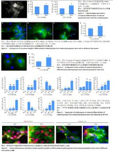
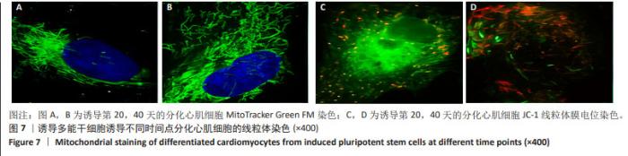
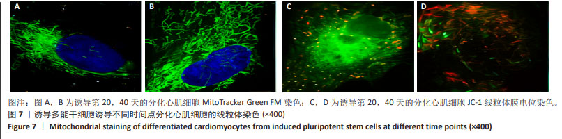
[31] KUZNETSOV AV, TROPPMAIR J, SUCHER R, et al. Mitochondrial subpopulations and heterogeneity revealed by confocal imaging: possible physiological role? Biochim Biophys Acta. 2006;1757(5-6):686-691. |