Chinese Journal of Tissue Engineering Research ›› 2025, Vol. 29 ›› Issue (7): 1408-1413.doi: 10.12307/2025.008
Previous Articles Next Articles
Verbascoside inhibits Erastin-induced ferroptosis of dopaminergic nerve cell line MN9D cells
Zhang Mingyang, Yang Xinling
- Second Affiliated Hospital of Xinjiang Medical University, Urumqi 830028, Xinjiang Uygur Autonomous Region, China
-
Received:2023-11-02Accepted:2024-01-10Online:2025-03-08Published:2024-06-27 -
Contact:Yang Xinling, MD, Doctoral supervisor, Chief physician, Second Affiliated Hospital of Xinjiang Medical University, Urumqi 830028, Xinjiang Uygur Autonomous Region, China -
About author:Zhang Mingyang, Master candidate, Physician, Second Affiliated Hospital of Xinjiang Medical University, Urumqi 830028, Xinjiang Uygur Autonomous Region, China -
Supported by:Central-Guided Local Funding Project for Scientific and Technological Development, No. ZYYD2022C17 (to YXL); Graduate Student Innovation Project of Xinjiang Uygur Autonomous Region, No. XJ2023G178 (to ZMY); Key Laboratory of Nervous System Diseases of Xinjiang Uygur Autonomous Region, No. XJDX1711 (to YXL)
CLC Number:
Cite this article
Zhang Mingyang, Yang Xinling. Verbascoside inhibits Erastin-induced ferroptosis of dopaminergic nerve cell line MN9D cells[J]. Chinese Journal of Tissue Engineering Research, 2025, 29(7): 1408-1413.
share this article
Add to citation manager EndNote|Reference Manager|ProCite|BibTeX|RefWorks

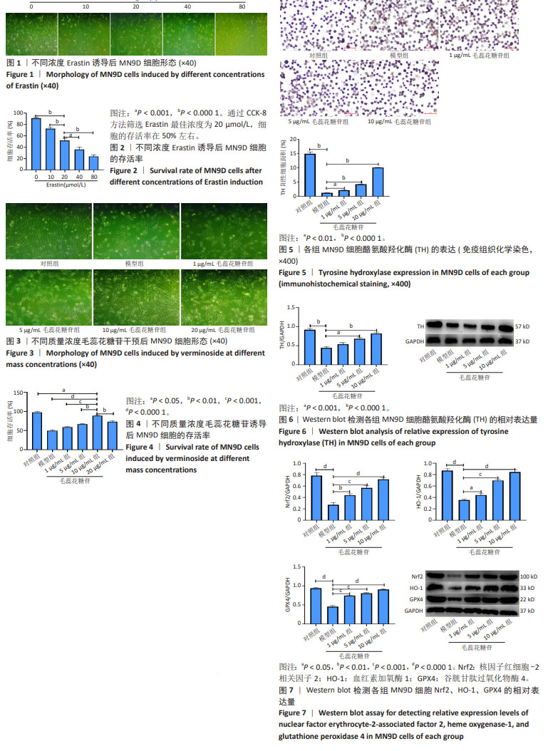
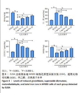
2.1 Erastin诱导铁死亡细胞模型的建立 在0,10,20,40,80 μmol/L不同浓度Erastin干预下,MN9D细胞存活率分别为(91.0±2.0)%,(73.0±2.6)%,(52.0±4.4)%,(36.0±2.6)%,(24.0±3.0)%,在20 μmol/L浓度下细胞的存活率在50%左右,故后续实验以此浓度来建立帕金森病铁死亡细胞模型,见图1,2。 2.2 不同质量浓度毛蕊花糖苷干预后MN9D细胞的存活情况 CCK-8方法检测对照组、模型组、1,5,10,20 μg/mL毛蕊花糖苷组细胞存活率分别为(98.66±1.15)%,(50.33±1.53)%,(59.00±3.60)%,(66.33±3.06)%,(89.00±3.61)%,(73.00±3.00)%,见图3,4,后续选择1,5,10 μg/mL毛蕊花糖苷进行相关实验。 2.3 免疫组织化学检测各组MN9D细胞酪氨酸羟化酶的表达 与对照组相比,模型组酪氨酸羟化酶阳性细胞面积明显减少(P < 0.01);与模型组相比,毛蕊花糖苷1,5,10 μg/mL组酪氨酸羟化酶阳性细胞面积明显增加,但仍低于对照组(P < 0.05),见图5。 2.4 Western blot法检测各组MN9D细胞酪氨酸羟化酶、Nrf2、血红素加氧酶1、GPX4的表达 与对照组相比,模型组酪氨酸羟化酶、Nrf2、血红素加氧酶1、GPX4的表达明显减少;与模型组相比,毛蕊花糖苷1,5,10 μg/mL组酪氨酸羟化酶、Nrf2、血红素加氧酶1、GPX4的表达明显增加,但仍少于对照组,差异有显著性意义(P < 0.05),见图6,7。 2.5 各组MN9D细胞还原型谷胱甘肽、丙二醛、超氧化物歧化酶、总铁离子水平 与对照组相比,模型组还原型谷胱甘肽、超氧化物歧化酶水平明显减少,丙二醛和总铁离子水平明显增加;与模型组相比,毛蕊花糖苷1,5,10 μg/mL组还原型谷胱甘肽、超氧化物歧化酶水平明显增加,但仍少于对照组,丙二醛和总铁离子水平明减少,但仍高于对照组,差异有显著性意义(P < 0.05),见图8。 "
"
| [1] NAIR AT, RAMACHANDRAN V, JOGHEE NM, et al. Gut Microbiota Dysfunction as Reliable Non-invasive Early Diagnostic Biomarkers in the Pathophysiology of Parkinson’s Disease: A Critical Review. J Neurogastroenterol Motil. 2018;24(1):30-42. [2] JANKOVIC J. Parkinson’s disease: clinical features and diagnosis. J Neurol Neurosurg Psychiatry. 2008;79(4):368-376. [3] 张辉,王运良.帕金森病的发病机制及治疗进展[J].中国实用神经疾病杂志,2021,24(15):1371-1380. [4] CHOI HK, WON LA, KONTUR PJ, et al. Immortalization of embryonic mesencephalic dopaminergic neurons by somatic cell fusion. Brain Res. 1991;552(1):67-76. [5] HELLER A, PRICE S, WON L. Glial-derived neurotrophic factor (GDNF) induced morphological differentiation of an immortalized monoclonal hybrid dopaminergic cell line of mesencephalic neuronal origin. Brain Res. 1996;725(1):132-136. [6] HAN BS, HONG HS, CHOI WS, et al. Caspase-dependent and -independent cell death pathways in primary cultures of mesencephalic dopaminergic neurons after neurotoxin treatment. J Neurosci. 2003; 23(12):5069-5078. [7] RICK CE, EBERT A, VIRAG T, et al. Differentiated dopaminergic MN9D cells only partially recapitulate the electrophysiological properties of midbrain dopaminergic neurons. Dev Neurosci. 2006;28(6):528-537. [8] DIXON SJ, LEMBERG KM, LAMPRECHT MR, et al. Ferroptosis: an iron-dependent form of nonapoptotic cell death. Cell. 2012;149(5): 1060-1072. [9] HE Y, THONG PS, LEE T, et al. Increased iron in the substantia nigra of 6-OHDA induced parkinsonian rats: a nuclear microscopy study. Brain Res. 1996;735(1):149-153. [10] SUN Y, PHAM AN, WAITE TD. Elucidation of the interplay between Fe(II), Fe(III), and dopamine with relevance to iron solubilization and reactive oxygen species generation by catecholamines. J Neurochem. 2016;137(6):955-968. [11] ANGELOVA PR, CHOI ML, BEREZHNOV AV, et al. Alpha synuclein aggregation drives ferroptosis: an interplay of iron, calcium and lipid peroxidation. Cell Death Differ. 2020;27(10):2781-2796. [12] ISLAM MT. Oxidative stress and mitochondrial dysfunction-linked neurodegenerative disorders. Neurol Res. 2017;39(1):73-82. [13] DOLMA S, LESSNICK SL, HAHN WC, et al. Identification of genotype-selective antitumor agents using synthetic lethal chemical screening in engineered human tumor cells. Cancer Cell. 2003;3(3):285-296. [14] LI M, WANG X, LU S, et al. Erastin triggers autophagic death of breast cancer cells by increasing intracellular iron levels. Oncol Lett. 2020;20(4):57. [15] YAGODA N, VON RECHENBERG M, ZAGANJOR E, et al. RAS-RAF-MEK-dependent oxidative cell death involving voltage-dependent anion channels. Nature. 2007;447(7146):864-868. [16] FRIEDMANN ANGELI JP, SCHNEIDER M, PRONETH B, et al. Inactivation of the ferroptosis regulator Gpx4 triggers acute renal failure in mice. Nat Cell Biol. 2014;16(12):1180-1191. [17] BRIGELIUS-FLOHÉ R. Tissue-specific functions of individual glutathione peroxidases. Free Radic Biol Med. 1999;27(9-10):951-965. [18] HAMBRIGHT WS, FONSECA RS, CHEN L, et al. Ablation of ferroptosis regulator glutathione peroxidase 4 in forebrain neurons promotes cognitive impairment and neurodegeneration. Redox Biol. 2017;12: 8-17. [19] 朱开梅,唐丽霞,赵文鹏,等.槲皮素脂质体对糖尿病肾病氧化应激和TGF-β1/Smad7通路的影响[J].安徽医科大学学报,2017,52(3): 319-323. [20] IMAI H, MATSUOKA M, KUMAGAI T, et al. Lipid Peroxidation-Dependent Cell Death Regulated by GPx4 and Ferroptosis. Curr Top Microbiol Immunol. 2017;403:143-170. [21] WU JR, TUO QZ, LEI P. Ferroptosis, a Recent Defined Form of Critical Cell Death in Neurological Disorders. J Mol Neurosci. 2018;66(2): 197-206. [22] BARTZOKIS G, CUMMINGS JL, MARKHAM CH, et al. MRI evaluation of brain iron in earlier- and later-onset Parkinson’s disease and normal subjects. Magn Reson Imaging. 1999;17(2):213-222. [23] LEWIS MM, DU G, BACCON J, et al. Susceptibility MRI captures nigral pathology in patients with parkinsonian syndromes. Mov Disord. 2018;33(9):1432-1439. [24] 姚辛敏,周晓洁,周妍妍.肉苁蓉化学成分及药理作用研究进展[J].中医药学报,2021,49(2):93-97. [25] HUANG DF, TANG YF, NIE SP, et al. Effect of phenylethanoid glycosides and polysaccharides from the seed of Plantago asiatica L. on the maturation of murine bone marrow-derived dendritic cells. Eur J Pharmacol. 2009;620(1-3):105-111. [26] ZHANG F, YANG YN, FENG ZM, et al. Four new phenylethanoid and flavonoid glycoside dimers from the fruit of Forsythia suspensa and their neuroprotective activities. RSC Advances. 2017;7(40): 24963-24969. [27] 陈珊珊.毛蕊花糖苷通过抑制神经炎症改善阿尔茨海默症的研究[D].长春:吉林大学,2023. [28] 芦芳,张悦,邱琦,等.毛蕊花糖苷对皮质酮损伤HT22细胞保护作用研究[J].中国临床药理学杂志,2023,39(17):2482-2486. [29] 邓敏,鞠晓东,樊东升,等.毛蕊花苷对MPP+诱导的SHSY5Y细胞凋亡的保护作用[J].中国药理学通报,2008,24(10):1297-1302. [30] PARK TJ, PARK JH, LEE GS, et al. Quantitative proteomic analyses reveal that GPX4 downregulation during myocardial infarction contributes to ferroptosis in cardiomyocytes. Cell Death Dis. 2019;10(11):835. [31] JIN M, SHI C, LI T, et al. Solasonine promotes ferroptosis of hepatoma carcinoma cells via glutathione peroxidase 4-induced destruction of the glutathione redox system. Biomed Pharmacother. 2020;129:110282. [32] DEVOS D, MOREAU C, DEVEDJIAN JC, et al. Targeting chelatable iron as a therapeutic modality in Parkinson’s disease. Antioxid Redox Signal. 2014;21(2):195-210. [33] FENG L, SUN J, XIA L, et al. Ferroptosis mechanism and Alzheimer’s disease. Neural Regen Res. 2024;19(8):1741-1750. [34] 李晶,于海波,薛晴,等.毛蕊花糖苷对戊四氮致癫痫大鼠脑保护作用研究[J].海南医学院学报,2020,26(24):1841-1844,1850. [35] HARTMANN A, HUNOT S, MICHEL PP, et al. Caspase-3: A vulnerability factor and final effector in apoptotic death of dopaminergic neurons in Parkinson’s disease. Proc Natl Acad Sci U S A. 2000;97(6):2875-2880. [36] PARZYCH KR, KLIONSKY DJ. An overview of autophagy: morphology, mechanism, and regulation. Antioxid Redox Signal. 2014;20(3):460-473. [37] ANGLADE P, VYAS S, JAVOY-AGID F, et al. Apoptosis and autophagy in nigral neurons of patients with Parkinson’s disease. Histol Histopathol. 1997;12(1):25-31. [38] DODSON M, DE LA VEGA MR, CHOLANIANS AB, et al. Modulating NRF2 in Disease: Timing Is Everything. Annu Rev Pharmacol Toxicol. 2019;59:555-575. [39] LI M, ZHOU F, XU T, et al. Acteoside protects against 6-OHDA-induced dopaminergic neuron damage via Nrf2-ARE signaling pathway. Food Chem Toxicol. 2018;119:6-13. [40] 平勇,张倩,冯雪梅.毛蕊花糖苷对缺氧/复氧诱导心肌细胞损伤的影响[J].中西医结合心脑血管病杂志,2022,20(20):3698-3703. |
| [1] | Zhao Nannan, Li Yanjie, Qin Hewei, Zhu Bochao, Ding Huimin, Xu Zhenhua. Changes in ferroptosis in hippocampal neurons of vascular dementia model rats treated with Tongmai Kaiqiao Pill [J]. Chinese Journal of Tissue Engineering Research, 2025, 29(7): 1401-1407. |
| [2] | Wang Mi, Ma Shujie, Liu Yang, Qi Rui. Identification and validation of characterized gene NFE2L2 for ferroptosis in ischemic stroke [J]. Chinese Journal of Tissue Engineering Research, 2025, 29(7): 1466-1474. |
| [3] | Gao Yang, Qin Hewei, Liu Dandan. ACSL4 mediates ferroptosis and its potential role in atherosclerotic cardiovascular disease [J]. Chinese Journal of Tissue Engineering Research, 2025, 29(6): 1239-1247. |
| [4] | Wang Chen, Zhang Weinan, Shen Jining, Liu Fan, Yuan Jishan, Liu Yake. Inhibitory effect of ferroptosis inhibitor toxicity induced by cobalt nanoparticles through reactive oxygen species [J]. Chinese Journal of Tissue Engineering Research, 2025, 29(34): 7310-7317. |
| [5] | Chen Chao, Hu Yaoquan, Lyu Zhengpin, He Qicong, Yangyang Zijiu, Luo Haoyan, Wu Guishuai, Zuo Qianlin, Wang Xuenan, Zhang Fan. tert-Butyl hydroperoxide can induce ferroptosis in nucleus pulposus cells [J]. Chinese Journal of Tissue Engineering Research, 2025, 29(32): 6858-6865. |
| [6] | Pan Yu, Zhao Renfeng, Li Xingping, Zhang Chengdong, Shi Feng, Pu Chao, Luo Xuwei, Xiao Dongqin. Iron overload induces ferroptosis in osteoblast precursor cells and inhibits osteogenic differentiation [J]. Chinese Journal of Tissue Engineering Research, 2025, 29(30): 6381-6390. |
| [7] | Fan Jiaxin, Jia Xiang, Xu Tianjie, Liu Kainan, Guo Xiaoling, Zhang Hui, Wang Qian . Metformin inhibits ferroptosis and improves cartilage damage in osteoarthritis model rats [J]. Chinese Journal of Tissue Engineering Research, 2025, 29(30): 6398-6408. |
| [8] | Yang Cheng, Li Weimin, Ran Dongcheng, Xu Jiamu, Wu Wangxiang, Xu Jiafu, Chen Jingjing, Jiang Guangfu, Wang Chunqing. Ferroptosis and osteoporosis [J]. Chinese Journal of Tissue Engineering Research, 2025, 29(3): 554-562. |
| [9] | Sun Rongyan, Xu Luchun, Jiang Guozheng, Song Jiawei, Ma Yukun, Fan Jiaojiao, Wang Guanlong, Yang Yongdong, Yu Xing. Du Meridian electroacupuncture inhibits ferroptosis and promotes neurorepair in rats with acute cervical spinal cord injury [J]. Chinese Journal of Tissue Engineering Research, 2025, 29(29): 6228-6238. |
| [10] | Tan Mingyue, Jin Yifeng, Zhang Jun, Li Hongxia. Aloin mitigates hypoxic injury in rat cardiomyocytes: inhibiting oxidative stress and ferroptosis [J]. Chinese Journal of Tissue Engineering Research, 2025, 29(25): 5335-5344. |
| [11] | Yu Qinghe, Cai Ziming, Tian He, Li Pian, Ruan Ye, Liang Jinzhu, Lin Shuhui, Lin Wenping. Heme oxygenase 1 promotes differentiation of neural stem cells into neurons under oxidative stress condition [J]. Chinese Journal of Tissue Engineering Research, 2025, 29(23): 4931-4938. |
| [12] | Shen Xiaoqiu, Wang Zhentao, Qiu Yueqing, Song Chenghao. Traditional Chinese medicine monomers regulate ferroptosis to combat myocardial ischemia-reperfusion injury [J]. Chinese Journal of Tissue Engineering Research, 2025, 29(20): 4333-4340. |
| [13] | Tao Hongcheng, Zeng Ping, Liu Jinfu, Tian Zhao, Ding Qiang, Li Chaohui, Wei Jianjie, Li Hao. Panax notoginseng saponins regulate differential miRNA expression in osteoclast exosomes and inhibit ferroptosis in osteoblasts [J]. Chinese Journal of Tissue Engineering Research, 2025, 29(19): 4011-4021. |
| [14] | Liu Dandan, Qin Hewei. Mechanism of action and progress of mitophagy, ferroptosis, cuproptosis, and disulfidptosis in Alzheimer’s disease [J]. Chinese Journal of Tissue Engineering Research, 2025, 29(19): 4132-4144. |
| [15] | Wei Jingxian, Meng Lian, Sun Hao, Zhang Tiantian, Liu Chunxia. Analysis of regulation of prognosis, immune infiltration, and ferroptosis in sarcoma based on stemness index model [J]. Chinese Journal of Tissue Engineering Research, 2025, 29(19): 4151-4160. |
| Viewed | ||||||
|
Full text |
|
|||||
|
Abstract |
|
|||||