Chinese Journal of Tissue Engineering Research ›› 2025, Vol. 29 ›› Issue (7): 1371-1379.doi: 10.12307/2025.013
Previous Articles Next Articles
Icariin-containing serum promotes chondrocyte proliferation and chondrogenic differentiation of stem cells in the co-culture system of three kinds of cells
Liu Qi1, 2, Li Linzhen1, 2, Li Yusheng1, 2, Jiao Hongzhuo1, 2, Yang Cheng1, 2, Zhang Juntao1, 2
- 1First Teaching Hospital of Tianjin University of Traditional Chinese Medicine, Tianjin 300381, China; 2National Clinical Research Center for Traditional Chinese Medicine and Acupuncture, Tianjin 300381, China
-
Received:2023-10-24Accepted:2024-01-15Online:2025-03-08Published:2024-06-27 -
Contact:Zhang Juntao, MD, Chief physician, Master’s supervisor, First Teaching Hospital of Tianjin University of Traditional Chinese Medicine, Tianjin 300381, China; National Clinical Research Center for Traditional Chinese Medicine and Acupuncture, Tianjin 300381, China -
About author:Liu Qi, Master candidate, First Teaching Hospital of Tianjin University of Traditional Chinese Medicine, Tianjin 300381, China; National Clinical Research Center for Traditional Chinese Medicine and Acupuncture, Tianjin 300381, China -
Supported by:National Natural Science Foundation of China, No. 82074470, 51573137 (to ZJT)
CLC Number:
Cite this article
Liu Qi, Li Linzhen, Li Yusheng, Jiao Hongzhuo, Yang Cheng, Zhang Juntao. Icariin-containing serum promotes chondrocyte proliferation and chondrogenic differentiation of stem cells in the co-culture system of three kinds of cells[J]. Chinese Journal of Tissue Engineering Research, 2025, 29(7): 1371-1379.
share this article
Add to citation manager EndNote|Reference Manager|ProCite|BibTeX|RefWorks

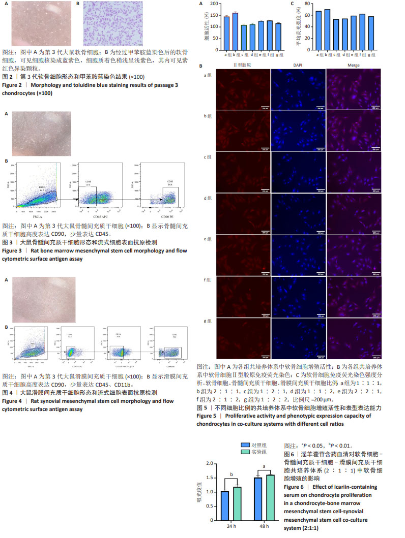
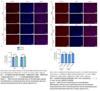
2.1 大鼠软骨细胞鉴定结果 经传代培养至第3代,倒置显微镜下可见软骨细胞呈现多角形、三角形或不规则梭形,细胞核大而圆,位于胞体中心,细胞质分布均匀,细胞密集生长后呈现“铺路石”样改变。经甲苯胺蓝染色后,培养的细胞核呈现蓝紫色,核仁明显,细胞质着色稍浅呈浅紫色,其内可见紫红色异染颗粒,细胞周围有少许蓝紫色异染颗粒出现。根据形态学观察和甲苯胺蓝染色阳性,结合取材部位,结果表明所提取的细胞为关节软骨细胞,见图2。 2.2 BMSC表面抗原鉴定结果 分离培养约48 h可出现贴壁生长,细胞呈圆形、三角形或梭形,生长缓慢,换液后细胞增殖速度加快,呈克隆性生长,出现以梭形细胞为主且大小不一的细胞集落。2周左右细胞融合度为80%-90%时达到传代要求,传代后细胞形态单一,似成纤维细胞样长梭形,第3代BMSC纯度可达90%以上,细胞增殖融合时呈漩涡状和放射状排列,表现出较强的细胞极性特征,见图3A。流式细胞表型鉴定显示,表达CD45为阴性,表达CD90为阳性,见图3B,基本符合BMSC的特征,证实了从大鼠骨髓中提取的细胞为BMSC。 2.3 SMSC表面抗原鉴定结果 SMSC在细胞形态和免疫表型方面与BMSC相似,但具有比BMSC更强的集落形成能力和成软骨分化能力。分离培养约24 h即可出现贴壁生长,细胞呈不规则梭形,换液后细胞生长速度加快,7-10 d细胞可达80%-90%融合,传代后3-5 d可铺满瓶底,传至第3代时,细胞生长速度和分化能力都处于最佳状态,此时细胞呈现长梭形的成纤维状,为间充质干细胞的标准状态,见图4A。流式细胞表型鉴定显示,表达CD45和CD11b为阴性,CD45阴性率为92.6%,CD11b阴性率为78.9%,表达CD90为阳性,阳性率为78.2%,见图4B,符合国际细胞治疗协会鉴定SMSC的标准。 2.4 不同细胞比例的共培养体系中软骨细胞增殖和表型能力 培养72 h后,软骨细胞、BMSC和SMSC的接种比例为2∶1∶1时软骨细胞增殖活性最强,见图5A;软骨细胞Ⅱ型胶原免疫荧光染色结果,见图5B;免疫荧光强度分析显示软骨细胞、BMSC和SMSC接种比例为2∶1∶1时软骨细胞表型能力最佳,Ⅱ型胶原表达量最高,平均荧光强度为70%,见图5C,因此后续实验研究将采用这一细胞比例。 2.5 淫羊藿苷含药血清对共培养体系中软骨细胞增殖的影响 从图6可以看出,淫羊藿苷含药血清干预共培养体系24,48 h后,实验组吸光度值均明显增高,表明软骨细胞增殖数量明显增加,淫羊藿苷含药血清干预24 h后两组之间差异有显著性意义(P < 0.01),48 h后两组相比差异仍有显著性意义(P < 0.05),说明淫羊藿苷含药血清能够促进软骨细胞的增殖,且以作用24 h效果最佳,推测淫羊藿苷含药血清在短时间内起效明显。 2.6 淫羊藿苷含药血清对共培养体系中软骨细胞表型能力的影响 Ⅱ型胶原是软骨标志性蛋白,其合成和分泌是维持软骨细胞功能的特征性指标。软骨细胞中Ⅱ型胶原的表达量越高,表明软骨细胞分化表型活性越强[24]。两组软骨细胞Ⅱ型胶原免疫荧光染色结果,见图7A,同一时间下两组荧光强度分析,见图7B,与对照组相比,淫羊藿苷含药血清干预24 h后实验组荧光强度明显增高(P < 0.01),48 h时两组荧光强度仍有显著差异(P < 0.05),表明淫羊藿苷含药血清能够提高共培养体系中软骨细胞Ⅱ型胶原的分泌量,促进其表型表达,且这种促进作用在干预24 h时最明显。 2.7 淫羊藿苷含药血清对共培养体系中BMSC和SMSC成软骨分化的影响 成软骨分化的BMSC和SMSC可以被Ⅱ型胶原免疫荧光染色,荧光越强,表明BMSC和SMSC转化为软骨细胞的数量越多[25]。图8A,B分别为BMSC和SMSC的Ⅱ型胶原免疫荧光染色图,均可见部分细胞免疫荧光染色呈阳性,对照组整体荧光强度均较弱,而实验组荧光强度均较强,图8C为BMSC和SMSC的荧光强度分析,实验组荧光强度明显高于对照组(P < 0.01),表明淫羊藿苷含药血清能够促进共培养体系中BMSC和SMSC的成软骨分化。 "
"

| [1] KACPRZAK B, ROSIŃSKA K. Rehabilitation of Soccer Players’ Knee Injuries: Cartilage Reconstruction, Anterior Cruciate Ligament Surgery, and Intensive Recovery-A Pilot Study. J Clin Med. 2023; 12(21):6893. [2] HOUSMANS BAC, NEEFJES M, SURTEL DAM, et al. Synovial fluid from end-stage osteoarthritis induces proliferation and fibrosis of articular chondrocytes via MAPK and RhoGTPase signaling. Osteoarthritis Cartilage. 2022;30(6):862-874. [3] GUO X, XI L, YU M, et al. Regeneration of articular cartilage defects: Therapeutic strategies and perspectives. J Tissue Eng. 2023;14: 20417314231164765. [4] CHAU M, DOU Z, BARONCELLI M, et al. The synovial microenvironment suppresses chondrocyte hypertrophy and promotes articular chondrocyte differentiation. NPJ Regen Med. 2022;7(1):51. [5] MARCHAN J, WITTIG O, DIAZ-SOLANO D, et al. Enhanced chondrogenesis from chondrocytes co-cultured on mesenchymal stromal cells: Implication for cartilage repair. Injury. 2022;53(2):399-407. [6] ZHANG J, QIN X, DENG Y, et al. Transforming Growth Factor-β1 Enhances Mesenchymal Characteristics of Buffalo (Bubalus bubalis) Bone Marrow-Derived Mesenchymal Stem Cells. Cell Reprogram. 2021;23(2):127-138. [7] OKAMURA G, EBINA K, HIRAO M, et al. Promoting Effect of Basic Fibroblast Growth Factor in Synovial Mesenchymal Stem Cell-Based Cartilage Regeneration. Int J Mol Sci. 2020;22(1):300. [8] LIU X, LIU Y, HE H, et al. Human adipose and synovial mesenchymal stem cells improve osteoarthritis in rats by reducing chondrocyte reactive oxygen species and inhibiting inflammatory response. J Clin Lab Anal. 2022;36(5):e24353. [9] TANG Z, LU Y, ZHANG S, et al. Chondrocyte secretome enriched microparticles encapsulated with the chondrocyte membrane to facilitate the chondrogenesis of BMSCs and reduce hypertrophy. J Mater Chem B. 2021;9(48):9989-10002. [10] WEI W, DAI H. Articular cartilage and osteochondral tissue engineering techniques: Recent advances and challenges. Bioact Mater. 2021; 6(12):4830-4855. [11] STAMPOULTZIS T, KARAMI P, PIOLETTI DP. Thoughts on cartilage tissue engineering: A 21st century perspective. Curr Res Transl Med. 2021; 69(3):103299. [12] 张熙南,张君涛,靳博.骨关节炎软骨缺损修复的动物实验研究[J].中国中医基础医学杂志,2021,27(10):1685-1690. [13] GAO ZR, FENG YZ, ZHAO YQ, et al. Traditional Chinese medicine promotes bone regeneration in bone tissue engineering. Chin Med. 2022;17(1):86. [14] SEYEDI Z, AMIRI MS, MOHAMMADZADEH V, et al. Icariin: A Promising Natural Product in Biomedicine and Tissue Engineering. J Funct Biomater. 2023;14(1):44. [15] BAHRAMI M, VALIANI A, AMIRPOUR N, et al. Cartilage Tissue Engineering Via Icariin and Adipose-derived Stem Cells in Fibrin Scaffold. Adv Biomed Res. 2018;7:36. [16] 罗云梅,李铭铭,熊乙林,等.基于CX3CL1介导的炎症反应研究淫羊藿苷对缺氧诱导的肺动脉高压小鼠的作用[J].中草药,2022,53(4): 1068-1075. [17] 张君涛,王平,穆刚,等.淫羊藿苷含药血清对兔软骨细胞增殖及糖胺聚糖分泌的影响[J].中华中医药杂志,2018,33(4):1601-1604. [18] BEDNARCZYK E. Chondrocytes In Vitro Systems Allowing Study of OA. Int J Mol Sci. 2022;23(18):10308. [19] ZHANG B, HUANG J, LIU J, et al. Injectable composite hydrogel promotes osteogenesis and angiogenesis in spinal fusion by optimizing the bone marrow mesenchymal stem cell microenvironment and exosomes secretion. Mater Sci Eng C Mater Biol Appl. 2021;123:111782. [20] LI F, TANG Y, SONG B, et al. Nomenclature clarification: synovial fibroblasts and synovial mesenchymal stem cells. Stem Cell Res Ther. 2019;10(1):260. [21] ILAS DC, CHURCHMAN SM, MCGONAGLE D, et al. Targeting subchondral bone mesenchymal stem cell activities for intrinsic joint repair in osteoarthritis. Future Sci OA. 2017;3(4):FSO228. [22] LE H, XU W, ZHUANG X, et al. Mesenchymal stem cells for cartilage regeneration. J Tissue Eng. 2020;11:2041731420943839. [23] XU X, XU L, XIA J, et al. Harnessing knee joint resident mesenchymal stem cells in cartilage tissue engineering. Acta Biomater. 2023;168:372-387. [24] SZUSTAK M, GENDASZEWSKA-DARMACH E. Extracellular Nucleotides Selectively Induce Migration of Chondrocytes and Expression of Type II Collagen. Int J Mol Sci. 2020;21(15):5227. [25] YANG K, SUN J, WEI D, et al . Photo-crosslinked mono-component type II collagen hydrogel as a matrix to induce chondrogenic differentiation of bone marrow mesenchymal stem cells. J Mater Chem B. 2017;5(44): 8707-8718. [26] HAGHWERDI F, KHOZAEI RAVARI M, TAGHIYAR L, et al. Application of bone and cartilage extracellular matrices in articular cartilage regeneration. Biomed Mater. 2021;16(4):042014. [27] RAPP AE, ZAUCKE F. Cartilage extracellular matrix-derived matrikines in osteoarthritis. Am J Physiol Cell Physiol. 2023;324(2):C377-C394. [28] CAO X, LUO P, HUANG J, et al. Intraarticular senescent chondrocytes impair the cartilage regeneration capacity of mesenchymal stem cells. Stem Cell Res Ther. 2019;10(1):86. [29] CHEN M, WEN H, ZHOU S, et al. Patchouli Alcohol Inhibits D-Gal Induced Oxidative Stress and Ameliorates the Quality of Aging Cartilage via Activating the Nrf2/HO-1 Pathway in Mice. Oxid Med Cell Longev. 2022;2022:6821170. [30] MATA-MIRANDA MM, MARTINEZ-MARTINEZ CM, NORIEGA-GONZALEZ JE, et al. Morphological, genetic and phenotypic comparison between human articular chondrocytes and cultured chondrocytes. Histochem Cell Biol. 2016;146(2):183-189. [31] DUAN A, SHEN K, LI B, et al. Extracellular vesicles derived from LPS-preconditioned human synovial mesenchymal stem cells inhibit extracellular matrix degradation and prevent osteoarthritis of the knee in a mouse model. Stem Cell Res Ther. 2021;12(1):427. [32] EPANOMERITAKIS IE, LEE E, LU V, et al. The Use of Autologous Chondrocyte and Mesenchymal Stem Cell Implants for the Treatment of Focal Chondral Defects in Human Knee Joints-A Systematic Review and Meta-Analysis. Int J Mol Sci. 2022;23(7):4065. [33] ROTHDIENER M, UYNUK-OOL T, SÜDKAMP N, et al. Human osteoarthritic chondrons outnumber patient- and joint-matched chondrocytes in hydrogel culture-Future application in autologous cell-based OA cartilage repair? J Tissue Eng Regen Med. 2018;12(2): e1206-e1220. [34] ZHAO Z, ZHOU X, GUAN J, et al. Co-implantation of bone marrow mesenchymal stem cells and chondrocytes increase the viability of chondrocytes in rat osteo-chondral defects. Oncol Lett. 2018;15(5): 7021-7027. [35] 刘朝政,李克文,牛兴邦,等.骨髓间充质干细胞基于Rho/ROCK信号通路对膝骨性关节炎模型大鼠软骨细胞凋亡的影响[J].中国老年学杂志,2023,43(13):3255-3259. [36] QIONG J, XIA Z, JING L, et al. Synovial mesenchymal stem cells effectively alleviate osteoarthritis through promoting the proliferation and differentiation of meniscus chondrocytes. Eur Rev Med Pharmacol Sci. 2020;24(4):1645-1655. [37] 马春辉,阎作勤,郭常安,等.Ⅱ型胶原与Bcl-2在骨关节炎软骨细胞中的表达[J].中国矫形外科杂志,2012,20(19):1786-1789. [38] 张松,王鹏程,胡海清.关节软骨细胞与CD105阳性滑膜间充质干细胞共培养并促进其成软骨分化[J].华中科技大学学报(医学版), 2020,49(4):408-413. [39] 范帅,吴春飞,梁祖建,等.补肾调肝方含药血清促进大鼠软骨细胞自噬治疗关节炎[J].中国组织工程研究,2022,26(20):3178-3183. [40] 张成龙,刘爱峰,张超,等.基于文献计量学的淫羊藿研究现状及热点分析[J].药物评价研究,2021,44(10):2242-2251. [41] XU S, ZHAO S, JIAN Y, et al. Icariin-loaded hydrogel with concurrent chondrogenesis and anti-inflammatory properties for promoting cartilage regeneration in a large animal model. Front Cell Dev Biol. 2022;10:1011260. [42] TANG W, ZHANG H, LIU D, et al. Icariin accelerates cartilage defect repair by promoting chondrogenic differentiation of BMSCs under conditions of oxygen-glucose deprivation. J Cell Mol Med. 2022; 26(1):202-215. [43] ZHANG J, FAN F, ZHANG C, et al. Icariin-conditioned serum combined with chitosan attenuates cartilage injury in rabbit knees with osteochondral defect. J Orthop Surg Res. 2023; 18(1):125. [44] CHENG T, ZHANG Y, ZHANG T, et al. Comparative Pharmacokinetics Study of Icariin and Icariside II in Rats. Molecules. 2015;20(12): 21274-21286. |
| [1] | Zhao Jiyu, Wang Shaowei. Forkhead box transcription factor O1 signaling pathway in bone metabolism [J]. Chinese Journal of Tissue Engineering Research, 2025, 29(9): 1923-1930. |
| [2] | Yin Lu, Jiang Chuanfeng, Chen Junjie, Yi Ming, Wang Zihe, Shi Houyin, Wang Guoyou, Shen Huarui. Effect of Complanatoside A on the apoptosis of articular chondrocytes [J]. Chinese Journal of Tissue Engineering Research, 2025, 29(8): 1541-1547. |
| [3] | Hu Taotao, Liu Bing, Chen Cheng, Yin Zongyin, Kan Daohong, Ni Jie, Ye Lingxiao, Zheng Xiangbing, Yan Min, Zou Yong. Human amniotic mesenchymal stem cells overexpressing neuregulin-1 promote skin wound healing in mice [J]. Chinese Journal of Tissue Engineering Research, 2025, 29(7): 1343-1349. |
| [4] | Zhang Zhenyu, Liang Qiujian, Yang Jun, Wei Xiangyu, Jiang Jie, Huang Linke, Tan Zhen. Target of neohesperidin in treatment of osteoporosis and its effect on osteogenic differentiation of bone marrow mesenchymal stem cells [J]. Chinese Journal of Tissue Engineering Research, 2025, 29(7): 1437-1447. |
| [5] | Chi Wenxin, Zhang Cunxin, Gao Kai, Lyu Chaoliang, Zhang Kefeng. Mechanism by which nobiletin inhibits inflammatory response of BV2 microglia [J]. Chinese Journal of Tissue Engineering Research, 2025, 29(7): 1321-1327. |
| [6] | Yu Ting, Lyu Dongmei, Deng Hao, Sun Tao, Cheng Qian. Icariin pretreatment enhances effect of human periodontal stem cells on M1-type macrophages [J]. Chinese Journal of Tissue Engineering Research, 2025, 29(7): 1328-1335. |
| [7] | Yang Zhihang, Sun Zuyan, Huang Wenliang, Wan Yu, Chen Shida, Deng Jiang. Nerve growth factor promotes chondrogenic differentiation and inhibits hypertrophic differentiation of rabbit bone marrow mesenchymal stem cells [J]. Chinese Journal of Tissue Engineering Research, 2025, 29(7): 1336-1342. |
| [8] | Xiang Pan, Che Yanjun, Luo Zongping. Compressive stress induces degeneration of cartilaginous endplate cells through the SOST/Wnt/beta-catenin pathway [J]. Chinese Journal of Tissue Engineering Research, 2025, 29(5): 951-957. |
| [9] | Sun Xianjuan, Wang Qiuhua, Zhang Jinyi, Yang Yangyang, Wang Wenshuang, Zhang Xiaoqing. Adhesion, proliferation, and vascular smooth muscle differentiation of bone marrow mesenchymal stem cells on different electrospinning membranes [J]. Chinese Journal of Tissue Engineering Research, 2025, 29(4): 661-669. |
| [10] | Ge Xiao, Zhao Zhuangzhuang, Guo Shuyu, Xu Rongyao. HOXA10 gene-modified bone marrow mesenchymal stem cells promote bone regeneration [J]. Chinese Journal of Tissue Engineering Research, 2025, 29(36): 7701-7708. |
| [11] | Zhang Xiongjinfu, Chen Yida, Cheng Xinyi, Liu Daihui, Shi Qin . Exosomes derived from bone marrow mesenchymal stem cells of young rats to reverse senescence in aged rat bone marrow mesenchymal stem cells [J]. Chinese Journal of Tissue Engineering Research, 2025, 29(36): 7709-7718. |
| [12] | Sima Xinli, Liu Danping, Qi Hui. Effect and mechanism of metformin-modified bone marrow mesenchymal stem cell exosomes on regulating chondrocytes [J]. Chinese Journal of Tissue Engineering Research, 2025, 29(36): 7728-7734. |
| [13] | Ma Weibang, Xu Zhe, Yu Qiao, Ouyang Dong, Zhang Ruguo, Luo Wei, Xie Yangjiang, Liu Chen. Screening and cytological validation of cartilage degeneration-related genes in exosomes from osteoarthritis synovial fluid [J]. Chinese Journal of Tissue Engineering Research, 2025, 29(36): 7783-7789. |
| [14] | Liu Chengyuan, Guo Qianping. Differential effects of kartogenin on chondrogenic and osteogenic differentiation of rat and rabbit bone marrow mesenchymal stem cells [J]. Chinese Journal of Tissue Engineering Research, 2025, 29(35): 7490-7498. |
| [15] | Fang Yuan, Qian Zhiyong, He Yuanhada, Wang Haiyan, Sha Lirong, Li Xiaohe, Liu Jing, He Yachao, Zhang Kai, Temribagen. Mechanism of Mongolian medicine Echinops sphaerocephalus L. in proliferation and angiogenesis of vascular endothelial cells [J]. Chinese Journal of Tissue Engineering Research, 2025, 29(35): 7519-7528. |
| Viewed | ||||||
|
Full text |
|
|||||
|
Abstract |
|
|||||