Chinese Journal of Tissue Engineering Research ›› 2024, Vol. 28 ›› Issue (14): 2166-2172.doi: 10.12307/2024.332
Previous Articles Next Articles
Bioinformatics analysis and validation of differentially expressed genes and small molecule drug prediction in proliferative scar
Zuo Jun, Ma Shaolin
- Department of Plastic and Aesthetic Surgery, The First Affiliated Hospital of Xinjiang Medical University, Urumqi 830000, Xinjiang Uygur Autonomous Region, China
-
Received:2023-03-21Accepted:2023-05-25Online:2024-05-18Published:2023-07-28 -
Contact:Ma Shaolin, Master, Chief physician, Professor, Doctoral supervisor, Department of Plastic and Aesthetic Surgery, The First Affiliated Hospital of Xinjiang Medical University, Urumqi 830000, Xinjiang Uygur Autonomous Region, China -
About author:Zuo Jun, MD candidate, Attending physician, Department of Plastic and Aesthetic Surgery, The First Affiliated Hospital of Xinjiang Medical University, Urumqi 830000, Xinjiang Uygur Autonomous Region, China -
Supported by:National Natural Science Foundation of China (Regional Program), No. 81760345 (to MSL); Youth Program of Natural Science Foundation of Hunan Province, No. 2021JJ40487 (to ZJ)
CLC Number:
Cite this article
Zuo Jun, Ma Shaolin. Bioinformatics analysis and validation of differentially expressed genes and small molecule drug prediction in proliferative scar[J]. Chinese Journal of Tissue Engineering Research, 2024, 28(14): 2166-2172.
share this article
Add to citation manager EndNote|Reference Manager|ProCite|BibTeX|RefWorks

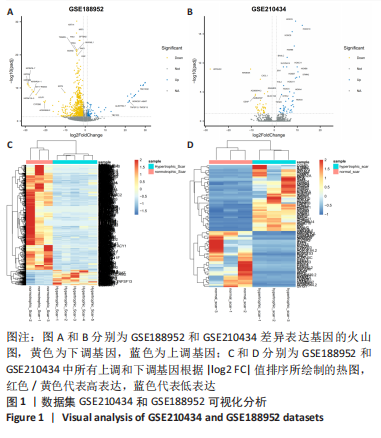
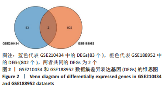
2.1 差异表达基因的筛选 此次研究纳入两组基因芯片,其中GSE188952数据集包括3例正常瘢痕样本,5例增生性瘢痕样本;GSE210434数据集包括3例正常瘢痕样本,3例增生性瘢痕样本。两组芯片预处理后中位数位于同一水平线,提示样本的基因表达谱数据一致性良好,可用于正常和增生性瘢痕患者差异表达基因的后续分析。GSE188952数据集共筛选出804个差异基因,其中上调基因102个,下调基因702个;GSE210434数据集共筛选出85个差异基因,其中上调基因46个,下调基因39个。进一步采用火山图(图1A,B)和热图(图1C,D)进行可视化处理。取交集后仅有2个共同DEGs(图2),故选取DEGs数量较多的GSE188952数据集进一步分析。"
"
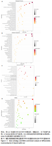
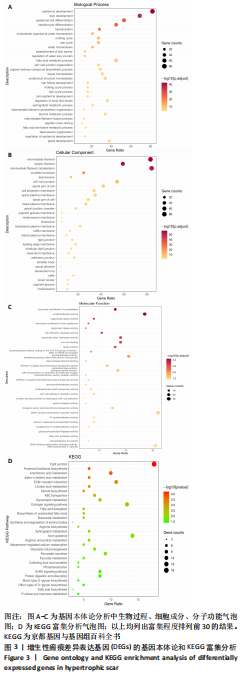
2.2 DEGs的生物学功能及通路富集分析 基因本体论富集分析中生物学过程结果显示,DEGs主要在表皮发育(epidermis development)、表皮细胞分化(epidermal cell differentiation)、角质形成细胞分化(keratinocyte differentiation)、角质化(keratinization)等子集中富集(图3A);细胞组分结果显示,DEGs主要在中间丝(intermediate filament)、角蛋白纤维(keratin filament)、中间丝细胞骨架(intermediate filament cytoskeleton)和细胞桥粒(desmosome)等子集中富集(图3B);分子功能结果显示,DEGs主要在细胞骨架的结构组成(structural constituent of cytoskeleton)、酰基转移酶活性(acyltransferase activity)、三酰甘油脂肪酶活性(Triglyceride lipase activity)和表皮的结构组成(structural constituent of skin epidermis)(图3C)。KEGG信号通路富集分析显示主要参与的信号通路有紧密连接(Tight junction)、萜类骨架生物合成(Terpenoid backbone biosynthesis)、花生四烯酸代谢(Arachidonic acid metabolism)和细胞外基质受体交互(ECM-receptor interaction)等(图3D)。"

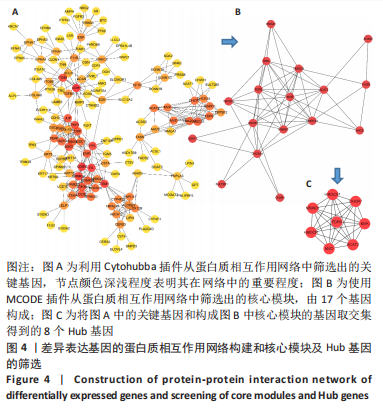
2.3 蛋白质相互作用网络的构建与Hub基因的筛选 利用Cytoscape中的Cytohubba插件找出关键基因,见图4A,节点颜色越深代表分值越高。再根据MCODE插件分析模块的重要程度,进一步筛选核心模块,包括HMGA1、MVK、ACSS、HMGCR、FASN、ACAT2、MVD、FDPS、TM7SF2、DHCR7、MSMO1、HMGCS1、AACS、SOAT1、DHCR24、SULT2B1和CH25H共17个基因(图4B)。为了进一步缩小Hub基因的范围,将Degree排名前20的关键基因与构成核心模块的基因求交集,筛选出HMGCS1、DHCR7、MSMO1、FDPS、MVK、HMGCR、MVD、ACAT2 共8 个Hub基因,均为下调基因(图4C)。"
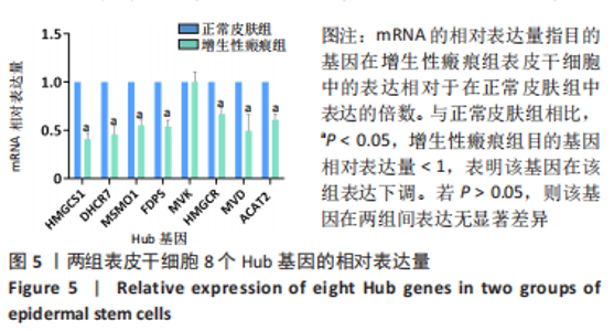
2.4 荧光定量PCR验证Hub基因mRNA的表达 如图5所示,对比正常皮肤组,增生性瘢痕表皮干细胞中Hub基因HMGCS1、DHCR7、MSMO1、FDPS、HMGCR、MVD、ACAT2的mRNA的表达均显著下降(P < 0.05) ,而MVK mRNA的表达无明显变化(P > 0.05)。"


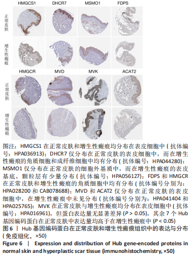
2.5 Hub基因编码蛋白在正常皮肤和增生性瘢痕组织中的表达差异 人类蛋白图谱数据库免疫组织化学法检测数据显示,除MVK外,其余Hub基因编码蛋白在正常皮肤组织中表达水平均高于增生性瘢痕组织(P < 0.05)。见图6。"
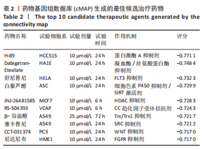
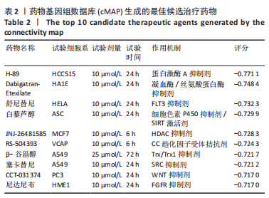
2.6 根据Hub基因预测小分子药物 由于cMAP数据库将上调基因的上传数目限定在10-150 个,此次研究将上述8 个下调的Hub基因和全部102 个上调的DEGs上传到cMAP,以原始评分0.7为界值,共筛选出47 个潜在药物,包括蛋白激酶A抑制剂(H-89)、凝血酶/丝氨酸蛋白酶抑制剂(Dabigatran- Etexilate)、FLT3抑制剂(舒尼替尼)、细胞色素P450抑制剂/SIRT激活剂(白藜芦醇)、HDAC抑制剂(JNJ-26481585)、SRC抑制剂(塞卡替尼)、TRX/TRX1抑制剂(β-谷甾醇)、CC趋化因子受体拮抗剂(RS-504393和AMD-11 070)、FGFR抑制剂(尼达尼布)、5-羟色胺受体拮抗剂(甲麦角林)、固醇去甲基酶抑制剂(酮康唑)等。去除药物列表中作用机制或靶点名称不详的化合物,评分排序前10的增生性瘢痕候选药物详见表2。"
| [1] WEI J, WANG Z, ZHONG C, et al. LncRNA MIR503HG promotes hypertrophic scar progression via miR-143-3p-mediated Smad3 expression. Wound Repair Regen. 2021;29(5):792-800. [2] ZHANG H, WANG HY, WANG DL, et al. Effect of pressure therapy for treatment of hypertrophic scar. Med. Baltim. 2019;98(26):e16263. [3] TAN J, ZHOU J, HUANG L, et al. Hypertrophic Scar Improvement by Early Intervention With Ablative Fractional Carbon Dioxide Laser Treatment. Lasers Surg Med. 2021;53(4):450-457. [4] TUNCA M, GAMSIZKAN M, YÜREKLI A, et al. Cryosurgery to remove perichondrium for the rabbit ear hypertrophic scar model: a simplified method. Acta Dermatovenerol Alp Pannonica Adriat. 2019;28(2):57-59. [5] BI M, SUN P, LI D, et al. Intralesional Injection of Botulinum Toxin Type A Compared with Intralesional Injection of Corticosteroid for the Treatment of Hypertrophic Scar and Keloid: A Systematic Review and Meta-Analysis. Med Sci Monit. 2019;25:2950-2958. [6] ELSAIE ML. Update on management of keloid and hypertrophic scars: A systemic review. J Cosmet Dermatol. 2021;20(9):2729-2738. [7] OGAWA R. The Most Current Algorithms for the Treatment and Prevention of Hypertrophic Scars and Keloids: A 2020 Update of the Algorithms Published 10 Years Ago. Plast Reconstr Surg. 2022;149(1):79e-94e. [8] MCGINTY S, SIDDIQUI WJ. Keloid. London: BTI-StatPearls: StatPearls Publishing, 2021. [9] KLIFTO KM, ASIF M, HULTMAN CS. Laser management of hypertrophic burn scars: a comprehensive review. Burns Trauma. 2020;8:tkz002. [10] ANDERSON JB, FOGLIO A, HARRANT AB, et al. Scoping Review of Therapeutic Strategies for Keloids and Hypertrophic Scars. Plast Reconstr Surg Glob Open. 2021;9(3):e3469. [11] 贠张君,王慧静,俞仪萱,等. 基于生物信息学技术筛选结直肠癌的差异基因和中药预测研究[J]. 中国中药杂志,2022,47(6):1666-1676. [12] EGAWA G, KABASHIMA K. Barrier dysfunction in the skin allergy. Allergol Int. 2018;67(1):3-11. [13] XIAO Y, ZHANG B, CLOYD JM, et al. Gene signature and connectivity mapping to assist with drug prediction for pancreatic ductal adenocarcinoma. Surg Oncol. 2022;44:101849. [14] LEE HJ, JANG YJ. Recent Understandings of Biology, Prophylaxis and Treatment Strategies for Hypertrophic Scars and Keloids. Int J Mol Sci. 2018;19(3):711. [15] LESZCZYNSKI R, DA SILVA CA, PINTO ACPN, et al. Laser therapy for treating hypertrophic and keloid scars. Cochrane Database Syst Rev. 2022;9(9):CD011642. [16] DENG X, ZHAO F, ZHAO D, et al. Oxymatrine promotes hypertrophic scar repair through reduced human scar fibroblast viability, collagen and induced apoptosis via autophagy inhibition. Int Wound J. 2022;19(5):1221-1231. [17] BUTZELAAR L, ULRICH MM, MINK VAN DER MOLEN AB, et al. Currently known risk factors for hypertrophic skin scarring: A review. J Plast Reconstr Aesthet Surg. 2016;69(2):163-169. [18] MOUSAVIZADEH SM, TORBATI PM, DARYANI A. The effects of kiwifruit dressing on hypertrophic scars in a rabbit ear model. J Wound Care. 2021;30(Sup9a):XVi-XVvii. [19] LI W, WANG D, LI M, et al. Emodin inhibits the proliferation of papillary thyroid carcinoma by activating AMPK. Exp Ther Med. 2021;22(4):1075. [20] JIANG N, LI Z, LUO Y, et al. Emodin ameliorates acute pancreatitis-induced lung injury by suppressing NLRP3 inflammasome-mediated neutrophil recruitment. Exp Ther Med. 2021;22(2):857. [21] RU Z, HU Y, HUANG S, et al.Bioflavonoid GalanginSuppresses Hypertrophic Scar Formation by the TGF-β/Smad Signaling Pathway. Evid Based Complement Alternat Med. 2021;2021:2444839. [22] CHEN D, LI Q, ZHANG H, et al. Traditional Chinese medicine for hypertrophic scars-A review of the therapeutic methods and potential effects. Front Pharmacol. 2022;13:1025602. [23] BÄSLER K, BERGMANN S, HEISIG M, et al. The role of tight junctions in skin barrier function and dermal absorption. J Control Release. 2016;242:105-118. [24] 席榕,朱慧婷,李伯华,等. 皮肤屏障紧密连接在特应性皮炎发病机制中的作用[J]. 中华临床免疫和变态反应杂志,2022,16(2):172-177. [25] FERNÁNDEZ-MAYOLA M, BETANCOURT L, MOLINA-KAUTZMAN A, et al. Growth hormone-releasing peptide 6 prevents cutaneous hypertrophic scarring: early mechanistic data from a proteome study. Int Wound J. 2018;15(4):538-546. [26] HUANG C, OGAWA R. Roles of lipid metabolism in keloid development. Lipids Health Dis. 2013;12:60. [27] 郑宇锟,王光忠,吴骁伟,等. 基于甲羟戊酸途径的盐酸小檗碱体外抗肺癌细胞药效及机制[J]. 中国实验方剂学杂志,2022,28(13):92-101. [28] KWON J, BAKHOUM SF. The Cytosolic DNA-Sensing cGAS-STING Pathway in Cancer. Cancer Discov. 2020;10(1):26-39. [29] MARTINS ÁM, RAMOS CC, FREITAS D, et al. Glycosylation of Cancer Extracellular Vesicles: Capture Strategies, Functional Roles and Potential Clinical Applications. Cells. 2021;10(1):109. [30] WANG IH, HUANG TT, CHEN JL, et al. Mevalonate Pathway Enzyme HMGCS1 Contributes to Gastric Cancer Progression. Cancers (Basel). 2020;12(5):1088. [31] 蔡硕,陈思敏,王媛媛,等. 基于高通量测序技术探讨补骨脂宁治疗肺癌的潜在机制[J]. 中国实验方剂学杂志,2021,27(14):183 -192 . [32] HASHEMI M, HOSHYAR R, ANDE SR, et al. Mevalonate Cascade and its Regulation in Cholesterol Metabolism in Different Tissues in Health and Disease. Curr Mol Pharmacol. 2017;10(1):13-26. [33] HA NT, LEE CH. Roles of Farnesyl-Diphosphate Farnesyltransferase 1 in Tumour and Tumour Microenvironments. Cells. 2020;9(11):2352. [34] ERSHOV P, KALUZHSKIY L, MEZENTSEV Y, et al. Enzymes in the Cholesterol Synthesis Pathway: Interactomics in the Cancer Context. Biomedicines. 2021; 9(8):895. [35] PRABHU AV, LUU W, LI D, et al. DHCR7: A vital enzyme switch between cholesterol and vitamin D production. Prog Lipid Res. 2016;64:138-151. [36] 赵路,周洋,黄君富,等. DHCR7基因在膀胱癌中的表达及其与临床特征及预后关系的生物信息学分析[J]. 国际检验医学杂志,2022,43(21):2649-2654. [37] SU L, FU L, LI Y, et al. Disruption of the association between drug transporter and actin cytoskeleton abolishes drug resistance in hypertrophic scar. Oncotarget. 2017;8(2):2617-2627. [38] SHAIKH G, CRONSTEIN B. Signaling pathways involving adenosine A2A and A2B receptors in wound healing and fibrosis. Purinergic Signal. 2016;12(2):191-197. [39] VORSTANDLECHNER V, LAGGNER M, COPIC D, et al. The serine proteases dipeptidyl-peptidase 4 and urokinase are key molecules in human and mouse scar formation. Nat Commun. 2021;12(1):6242. [40] HU M, YANG T, YANG L, et al. Preclinical studies of Flonoltinib Maleate, a novel JAK2/FLT3 inhibitor, in treatment of JAK2V617F-induced myeloproliferative neoplasms. Blood Cancer J. 2022;12(3):37. [41] BAI XZ, LIU JQ, YANG LL, et al. Identification of sirtuin 1 as a promising therapeutic target for hypertrophic scars. Br J Pharmacol. 2016;173(10):1589-1601. [42] DEVARAJ E, ROY A, ROYAPURAM VEERARAGAVAN G, et al. β-Sitosterol attenuates carbon tetrachloride-induced oxidative stress and chronic liver injury in rats.Naunyn Schmiedebergs Arch Pharmacol. 2020;393(6):1067-1075. [43] PARK YJ, BANG IJ, JEONG MH, et al. Effects of β-Sitosterol from Corn Silk on TGF-β1-Induced Epithelial-Mesenchymal Transition in Lung Alveolar Epithelial Cells. J Agric Food Chem. 2019;67(35):9789-9795. [44] 郭京龄,马晨. 白藜芦醇调节脂质代谢研究进展[J]. 中国食品学报,2022,22(6): 390-402. [45] CUI H, ZHANG M, YANG Q, et al. The Prediction of Drug-Disease Correlation Based on Gene Expression Data. Biomed Res Int. 2018;2018:4028473. |
| [1] | Liu Lin, Liu Shixuan, Lu Xinyue, Wang Kan. Metabolomic analysis of urine in a rat model of chronic myofascial trigger points [J]. Chinese Journal of Tissue Engineering Research, 2025, 29(8): 1585-1592. |
| [2] | Li Jun, Gong Jingjing, Sun Guobin, Guo Rui, Ding Yang, Qiang Lijuan, Zhang Xiaoli, Fang Zhanhai . miR-27a-3p promotes the proliferation of human hypertrophic scar fibroblasts by regulating mitogen-activated protein kinase signaling pathway [J]. Chinese Journal of Tissue Engineering Research, 2025, 29(8): 1609-1617. |
| [3] | Zhao Jiacheng, Ren Shiqi, Zhu Qin, Liu Jiajia, Zhu Xiang, Yang Yang. Bioinformatics analysis of potential biomarkers for primary osteoporosis [J]. Chinese Journal of Tissue Engineering Research, 2025, 29(8): 1741-1750. |
| [4] | Zhang Zhenyu, Liang Qiujian, Yang Jun, Wei Xiangyu, Jiang Jie, Huang Linke, Tan Zhen. Target of neohesperidin in treatment of osteoporosis and its effect on osteogenic differentiation of bone marrow mesenchymal stem cells [J]. Chinese Journal of Tissue Engineering Research, 2025, 29(7): 1437-1447. |
| [5] | Zhang Haojun, Li Hongyi, Zhang Hui, Chen Haoran, Zhang Lizhong, Geng Jie, Hou Chuandong, Yu Qi, He Peifeng, Jia Jinpeng, Lu Xuechun. Identification and drug sensitivity analysis of key molecular markers in mesenchymal cell-derived osteosarcoma [J]. Chinese Journal of Tissue Engineering Research, 2025, 29(7): 1448-1456. |
| [6] | Wang Mi, Ma Shujie, Liu Yang, Qi Rui. Identification and validation of characterized gene NFE2L2 for ferroptosis in ischemic stroke [J]. Chinese Journal of Tissue Engineering Research, 2025, 29(7): 1466-1474. |
| [7] | Chen Yuning, Jiang Ying, Liao Xiangyu, Chen Qiongjun, Xiong Liang, Liu Yue, Liu Tong. Buqi Huoxue Compounds intervene with the expression of related factors and autophagy related proteins in a rat model of cerebral ischemia/reperfusion [J]. Chinese Journal of Tissue Engineering Research, 2025, 29(6): 1152-1158. |
| [8] | Zhang Debao, Wang Peng, Li Kun, Zhang Shaojie, Li Zhijun, Li Shuwen, Wu Yimin. Epidural fibrous scar formation in rabbits following autologous ligamentum flavum intervention [J]. Chinese Journal of Tissue Engineering Research, 2025, 29(6): 1168-1175. |
| [9] | Wang Yuru, Li Siyuan, Xu Ye, Zhang Yumeng, Liu Yang, Hao Huiqin. Effects of wogonin on joint inflammation in collagen-induced arthritis rats via the endoplasmic reticulum stress pathway [J]. Chinese Journal of Tissue Engineering Research, 2025, 29(5): 1026-1035. |
| [10] | Liu Yani, Yang Jinghuan, Lu Huihui, Yi Yufang, Li Zhixiang, Ou Yangfu, Wu Jingli, Wei Bing . Screening of biomarkers for fibromyalgia syndrome and analysis of immune infiltration [J]. Chinese Journal of Tissue Engineering Research, 2025, 29(5): 1091-1100. |
| [11] | Han Haihui, Meng Xiaohu, Xu Bo, Ran Le, Shi Qi, Xiao Lianbo. Effect of fibroblast growth factor receptor 1 inhibitor on bone destruction in rats with collagen-induced arthritis [J]. Chinese Journal of Tissue Engineering Research, 2025, 29(5): 968-977. |
| [12] | Dong Meilin, Du Haiyu, Liu Yuan. Quercetin-loaded carboxymethyl chitosan hydrogel promotes wound healing in diabetic rats [J]. Chinese Journal of Tissue Engineering Research, 2025, 29(4): 692-699. |
| [13] | Zhu Xuekun, Liu Heng, Feng Hui, Gao Yunlong, Wen Lei, Cai Xiaosong, Zhao Ben, Zhong Min. Identification of core genes of osteoarthritis by bioinformatics [J]. Chinese Journal of Tissue Engineering Research, 2025, 29(3): 637-644. |
| [14] | Wu Ao, Yu Peng, Teng Jiawen, Kong Peng, Bian Sishan. Analysis of oxidative stress-related genes and immune infiltration in osteoarthritis [J]. Chinese Journal of Tissue Engineering Research, 2025, 29(2): 302-311. |
| [15] | Zhang Yanfeng, Zhang Huimin, He Xiang, Zheng Yuping. Effects of long non-coding RNA nuclear enriched abundant transcript 1 on the proliferation, apoptosis and migration of keloid fibroblasts [J]. Chinese Journal of Tissue Engineering Research, 2025, 29(2): 347-354. |
| Viewed | ||||||
|
Full text |
|
|||||
|
Abstract |
|
|||||