Chinese Journal of Tissue Engineering Research ›› 2015, Vol. 19 ›› Issue (49): 8015-8020.doi: 10.3969/j.issn.2095-4344.2015.49.025
Previous Articles Next Articles
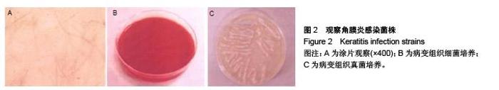
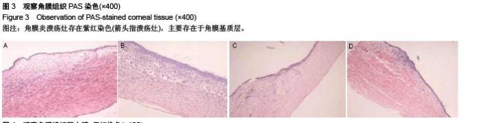
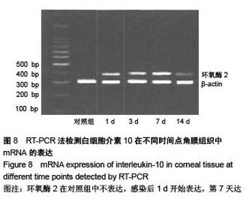
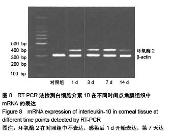
Expression of interleukin-10 and cyclooxygenase 2 in keratitis rat models
Lu Xia1, Pan Xu-bin1, Xie Yu-tao2, Ji Qiu-di3
- 1Department of Ophthalmology, 2Operating Room, 3Department of Dermatology, the Fourth People’s Hospital of Wuxi, Wuxi 214062, Jiangsu Province, China
-
Received:2015-09-23Online:2015-11-30Published:2015-11-30 -
About author:Lu Xia, Nurse-in-charge, Department of Ophthalmology, the Fourth People’s Hospital of Wuxi, Wuxi 214062, Jiangsu Province, China
CLC Number:
Cite this article
Lu Xia, Pan Xu-bin, Xie Yu-tao, Ji Qiu-di. Expression of interleukin-10 and cyclooxygenase 2 in keratitis rat models[J]. Chinese Journal of Tissue Engineering Research, 2015, 19(49): 8015-8020.
share this article

"

"
"
"

"

"


"
"
| [1] 李洪霞,张宏波,王宜强.T 和 B 细胞缺陷对真菌性角膜炎发病过程的影响[J].国际眼科杂志,2012,12(12):2247-2252.[2] 苏晶,崔红平.烟曲霉菌性角膜炎大鼠角膜IL-6和IL-10的表达[J].眼科研究,2008,26(8):579-582. [3] 王晓雪.IL-L与IL-10在大鼠真菌性角膜炎固有免疫阶段角膜上皮组织中的表达[D]. 青岛:青岛大学,2012. [4] 苏晶.大鼠烟曲霉菌性角膜炎动物模型的建立及其免疫病理的研究[D].上海:同济大学医学院,2006. [5] 刘筱楠.CXCL1在大鼠烟曲霉菌角膜炎早期的表达[D]. 青岛:青岛大学, 2012.[6] Sellers RS, Silverman L, Khan KN. Cyclooxygenase-2 expression in the cornea of dogs with keratitis.Vet Pathol. 2004; 41(2):116-121.[7] Li N, Che CY, Hu LT, et al.Effects of COX-2 inhibitor NS-398 on IL-10 expression in rat fungal keratitis.Int J Ophthalmol. 2011; 4(2):165-169.[8] 张明昌,王勇.角膜碱烧伤中白介素-1与核转录因子κB表达的相关性研究[J].眼科研究,2007,25(1):33-36.[9] 梁丽娜,庄曾渊.带状疱疹病毒性角膜内皮炎合并瞳孔损害 2 例[J].中国中医眼科杂志,2013,2(1):30-31.[10] 夏丽坤,张劲松,陈晓隆,等.HSV-1上调白细胞介素-18 mRNA在小鼠角膜组织中表达的研究[J].眼科研究,2005,23(4):393-396.[11] 夏丽坤,高殿文,濮伟,等.白细胞介素10对小鼠单纯疱疹性角膜基质炎作用的实验研究[J].中华眼科杂志,2003,39(10):592-596.[12] 林秀丽.兔棘阿米巴性角膜炎IL-1β和MCP-1的表达[D].福州:福建医科大学,2010.[13] 曾宗圣,韩晓丽,胡建章,等.白细胞介素-17和维甲酸相关核孤儿受体γt在小鼠真菌性角膜炎中的表达[J].中华实验眼科杂志, 2013,31(7):653-658.[14] Hazlett LD, Jiang X, McClellan SA.IL-10 function, regulation, and in bacterial keratitis.J Ocul Pharmacol Ther.2014;30(5): 373-380.[15] Biswas PS, Banerjee K, Kim B,et al. Role of inflammatory cytokine-induced cyclooxygenase 2 in the ocular immunopathologic disease herpetic stromal karatitis. J Virol.2005;79(16):10589-10600. |
| [1] | Chen Ziyang, Pu Rui, Deng Shuang, Yuan Lingyan. Regulatory effect of exosomes on exercise-mediated insulin resistance diseases [J]. Chinese Journal of Tissue Engineering Research, 2021, 25(25): 4089-4094. |
| [2] | Chen Yang, Huang Denggao, Gao Yuanhui, Wang Shunlan, Cao Hui, Zheng Linlin, He Haowei, Luo Siqin, Xiao Jingchuan, Zhang Yingai, Zhang Shufang. Low-intensity pulsed ultrasound promotes the proliferation and adhesion of human adipose-derived mesenchymal stem cells [J]. Chinese Journal of Tissue Engineering Research, 2021, 25(25): 3949-3955. |
| [3] | Yang Junhui, Luo Jinli, Yuan Xiaoping. Effects of human growth hormone on proliferation and osteogenic differentiation of human periodontal ligament stem cells [J]. Chinese Journal of Tissue Engineering Research, 2021, 25(25): 3956-3961. |
| [4] | Sun Jianwei, Yang Xinming, Zhang Ying. Effect of montelukast combined with bone marrow mesenchymal stem cell transplantation on spinal cord injury in rat models [J]. Chinese Journal of Tissue Engineering Research, 2021, 25(25): 3962-3969. |
| [5] | Gao Shan, Huang Dongjing, Hong Haiman, Jia Jingqiao, Meng Fei. Comparison on the curative effect of human placenta-derived mesenchymal stem cells and induced islet-like cells in gestational diabetes mellitus rats [J]. Chinese Journal of Tissue Engineering Research, 2021, 25(25): 3981-3987. |
| [6] | Hao Xiaona, Zhang Yingjie, Li Yuyun, Xu Tao. Bone marrow mesenchymal stem cells overexpressing prolyl oligopeptidase on the repair of liver fibrosis in rat models [J]. Chinese Journal of Tissue Engineering Research, 2021, 25(25): 3988-3993. |
| [7] | Liu Jianyou, Jia Zhongwei, Niu Jiawei, Cao Xinjie, Zhang Dong, Wei Jie. A new method for measuring the anteversion angle of the femoral neck by constructing the three-dimensional digital model of the femur [J]. Chinese Journal of Tissue Engineering Research, 2021, 25(24): 3779-3783. |
| [8] | Meng Lingjie, Qian Hui, Sheng Xiaolei, Lu Jianfeng, Huang Jianping, Qi Liangang, Liu Zongbao. Application of three-dimensional printing technology combined with bone cement in minimally invasive treatment of the collapsed Sanders III type of calcaneal fractures [J]. Chinese Journal of Tissue Engineering Research, 2021, 25(24): 3784-3789. |
| [9] | Qian Xuankun, Huang Hefei, Wu Chengcong, Liu Keting, Ou Hua, Zhang Jinpeng, Ren Jing, Wan Jianshan. Computer-assisted navigation combined with minimally invasive transforaminal lumbar interbody fusion for lumbar spondylolisthesis [J]. Chinese Journal of Tissue Engineering Research, 2021, 25(24): 3790-3795. |
| [10] | Hu Jing, Xiang Yang, Ye Chuan, Han Ziji. Three-dimensional printing assisted screw placement and freehand pedicle screw fixation in the treatment of thoracolumbar fractures: 1-year follow-up [J]. Chinese Journal of Tissue Engineering Research, 2021, 25(24): 3804-3809. |
| [11] | Shu Qihang, Liao Yijia, Xue Jingbo, Yan Yiguo, Wang Cheng. Three-dimensional finite element analysis of a new three-dimensional printed porous fusion cage for cervical vertebra [J]. Chinese Journal of Tissue Engineering Research, 2021, 25(24): 3810-3815. |
| [12] | Wang Yihan, Li Yang, Zhang Ling, Zhang Rui, Xu Ruida, Han Xiaofeng, Cheng Guangqi, Wang Weil. Application of three-dimensional visualization technology for digital orthopedics in the reduction and fixation of intertrochanteric fracture [J]. Chinese Journal of Tissue Engineering Research, 2021, 25(24): 3816-3820. |
| [13] | Sun Maji, Wang Qiuan, Zhang Xingchen, Guo Chong, Yuan Feng, Guo Kaijin. Development and biomechanical analysis of a new anterior cervical pedicle screw fixation system [J]. Chinese Journal of Tissue Engineering Research, 2021, 25(24): 3821-3825. |
| [14] | Lin Wang, Wang Yingying, Guo Weizhong, Yuan Cuihua, Xu Shenggui, Zhang Shenshen, Lin Chengshou. Adopting expanded lateral approach to enhance the mechanical stability and knee function for treating posterolateral column fracture of tibial plateau [J]. Chinese Journal of Tissue Engineering Research, 2021, 25(24): 3826-3827. |
| [15] | Zhu Yun, Chen Yu, Qiu Hao, Liu Dun, Jin Guorong, Chen Shimou, Weng Zheng. Finite element analysis for treatment of osteoporotic femoral fracture with far cortical locking screw [J]. Chinese Journal of Tissue Engineering Research, 2021, 25(24): 3832-3837. |
| Viewed | ||||||
|
Full text |
|
|||||
|
Abstract |
|
|||||