Chinese Journal of Tissue Engineering Research ›› 2026, Vol. 30 ›› Issue (33): 8653-8661.doi: 10.12307/2026.494
Previous Articles Next Articles
Gait kinematics and electromyography features in patients with Lenke type 5 adolescent idiopathic scoliosis
Liu Jiaqi1, Zhou Ding2, Jiang Yawen2, Ni Simin3, Li Ning1, Zhu Zhitao2, Liu Yue1, Chen Qinghua2, Mei Zhao4, Yao Yifei5, Wang Jinwu6
- 1Rehabilitation School, Shandong Second Medical University, Weifang 261053, Shandong Province, China; 2School of Exercise and Health, Shanghai University of Sport, Shanghai 200438, China; 3University College London, London WC1E 6BT, UK; 4Huazhu Medical Technology Co., Ltd. (Shanghai), Shanghai 201204, China; 5School of Biomedical Engineering, Shanghai Jiao Tong University, Shanghai 200030, China; 6Shanghai Key Laboratory of Orthopedics, Department of Orthopedic Surgery, Shanghai Ninth People’s Hospital Affiliated to Shanghai Jiao Tong University School of Medicine, Shanghai 200011, China
-
Received:2026-03-05Revised:2026-03-23Online:2026-11-28Published:2026-06-10 -
Contact:Wang Jinwu, MD, Professor, Chief physician, Shanghai Key Laboratory of Orthopedics, Department of Orthopedic Surgery, Shanghai Ninth People’s Hospital Affiliated to Shanghai Jiao Tong University School of Medicine, Shanghai 200011, China -
About author:Liu Jiaqi, MS, Rehabilitation School, Shandong Second Medical University, Weifang 261053, Shandong Province, China Zhou Ding, MS, School of Exercise and Health, Shanghai University of Sport, Shanghai 200438, China Liu Jiaqi and Zhou Ding contributed equally to this work. -
Supported by:Clinical Research-Oriented MDT Project, Ninth People’s Hospital Affiliated to Shanghai Jiao Tong University School of Medicine, No. 201914 (to WJW); Guizhou Provincial Science and Technology Support Program, No. [2023] General 196 (to WJW [project participant])

CLC Number:
Cite this article
Liu Jiaqi, Zhou Ding, Jiang Yawen, Ni Simin, Li Ning, Zhu Zhitao, Liu Yue, Chen Qinghua, Mei Zhao, Yao Yifei, Wang Jinwu. Gait kinematics and electromyography features in patients with Lenke type 5 adolescent idiopathic scoliosis[J]. Chinese Journal of Tissue Engineering Research, 2026, 30(33): 8653-8661.
share this article
Add to citation manager EndNote|Reference Manager|ProCite|BibTeX|RefWorks

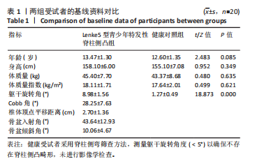
2.1 研究对象一般资料 使用G*Power软件(G*Power 3.1版)计算研究所需样本量,效应量中值设置为0.5,α概率为0.05,计算结果表明,为了达到0.8的统计检验力,需要总样本量34例。根据研究的纳排标准共计纳入受试者40例,均为女性。Lenke5型青少年特发性脊柱侧凸患者20例,同年龄段正常人群20例,试验过程无脱落,全部进入结果分析。Lenke5型青少年特发性脊柱侧凸患者统一纳入左侧为凸侧,右侧为凹侧。两组受试者在年龄、身高、体质量和体质量指数之间无显著性差异(P > 0.05)。Lenke5型特发性脊柱侧凸组的躯干旋转角、Cobb角均显著大于健康对照组(P < 0.05)。受试者的一般资料见表1。"
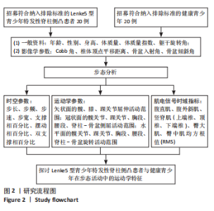
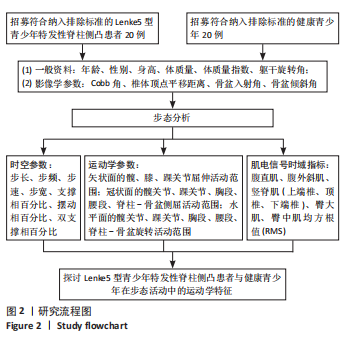
2.2 试验流程图 见图2。"
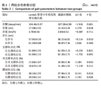
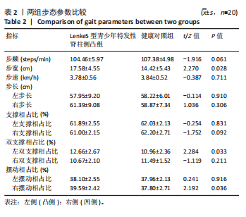
2.3 时空参数 比较 Lenke5型青少年特发性脊柱侧凸组与健康对照组在自然行走状态下的步态时空参数发现,Lenke5型青少年特发性脊柱侧凸组步宽增大,差异有显著性意义(P < 0.05);步频、步速以及双侧步长与健康对照组差异均无显著性意义 (P > 0.05)。Lenke5型青少年特发性脊柱侧凸组凸侧双支撑相、凹侧摆动相占整个步态周期的百分比增加,差异均有显著性意义 (P < 0.05);而双侧支撑相占比、凹侧双支撑相占比及凸侧摆动相占比在组间比较中亦未见显著差异(P > 0.05),见表2。"

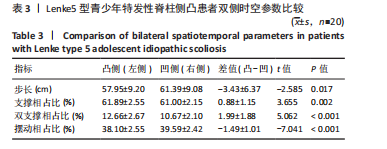
如表3所示,Lenke5型青少年特发性脊柱侧凸患者在凸侧(左侧)与凹侧(右侧)之间的步态时空参数存在显著不对称性。与凸侧相比,凹侧步长显著增加(P < 0.05)。在步态周期百分比方面,凸侧支撑相占比、双支撑相占比显著增加(P < 0.05);凸侧摆动相占比显著降低(P < 0.05)。"
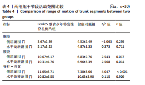
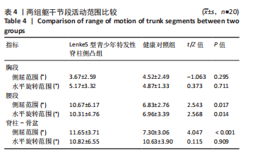
2.4 运动学参数 2.4.1 躯干运动学参数 与健康对照组相比,Lenke5型青少年特发性脊柱侧凸组在步行过程中的躯干运动以腰段活动范围增大为主要差异特征,见表4。节段分析显示,Lenke5型青少年特发性脊柱侧凸组腰段侧屈和水平旋转幅度均显著大于健康对照组(P < 0.05)。胸段侧屈与水平旋转幅度在两组间差异均未达到统计学差异(P > 0.05)。从脊柱-骨盆的相对运动来看,Lenke5型青少年特发性脊柱侧凸组侧屈幅度显著增加(P < 0.05),水平旋转在两组间无显著差异(P > 0.05)。"
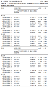
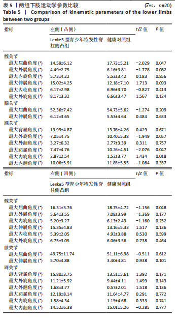
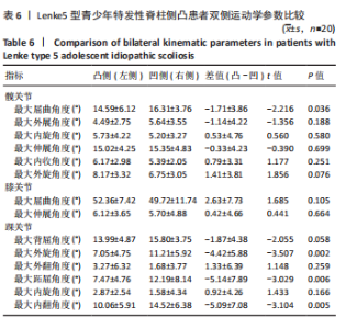
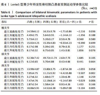
2.4.2 下肢运动学参数 比较两组在自然行走状态下的下肢运动学参数(髋、膝、踝关节的峰值角度),在髋关节方面,Lenke5型青少年特发性脊柱侧凸组双侧屈曲峰值角度均小于健康对照组(P < 0.05);伸展、外展/内收及内旋/外旋的峰值角度无统计学差异(P > 0.05)。双侧膝关节屈曲/伸展的峰值角度组间比较无显著差异(P > 0.05)。在踝关节方面,Lenke5型青少年特发性脊柱侧凸组凸侧跖屈峰值角度显著小于健康对照组(P < 0.05);而内旋峰值角度显著大于健康对照组(P < 0.05);背屈、外旋、内翻/外翻峰值角度与健康对照组差异均无显著性意义(P > 0.05)。凹侧踝关节各活动峰值角度组间比较无显著差异(P > 0.05),见表5。 在Lenke5型青少年特发性脊柱侧凸组内凸侧(左侧)与凹侧(右侧)的比较中,髋关节与踝关节部分峰值角度呈现显著差异,见表6。在髋关节方面,凸侧屈曲峰值角度显著小于凹侧(P < 0.05);伸展、内旋/外旋、内收/外展峰值角度的双侧差异均无显著性意义(P > 0.05)。膝关节屈曲/伸展峰值角度双侧比较无明显差异(P > 0.05)。在踝关节方面,凸侧外旋、跖屈和内翻峰值角度均显著小于凹侧(P < 0.05),背屈、外翻和内旋峰值角度的双侧差异均无显著性意义(P > 0.05)。"
"
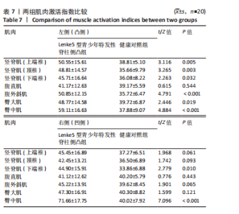
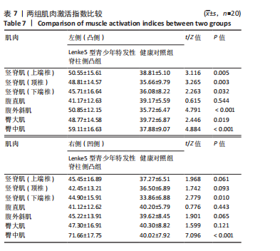
2.5 表面肌电分析 Lenke5型青少年特发性脊柱侧凸组与健康对照组在步行过程中的核心肌群肌电信号的时域指标均方根值具有一定差异,见表7。在竖脊肌方面,Lenke5型青少年特发性脊柱侧凸组的凸侧竖脊肌均方根值在上端椎、顶椎和下端椎水平均显著高于健康对照组(P < 0.05);凹侧竖脊肌均方根值在下端椎水平显著高于健康对照组(P < 0.05),在上端椎、顶椎水平与健康对照组差异无显著性意义(P > 0.05)。在腹部核心肌群方面,Lenke5型青少年特发性脊柱侧凸组凸侧腹外斜肌均方根值高于健康对照组(P < 0.05),凹侧腹外斜肌和双侧腹直肌的均方根值与健康对照组差异无显著性意义(P > 0.05)。在臀周肌群方面,Lenke5型青少年特发性脊柱侧凸组双侧臀中肌均方根值均显著高于健康对照组(P < 0.05);凸侧臀大肌均方根值显著高于健康对照组(P < 0.05);凹侧臀大肌均方根值与健康对照组差异无显著性意义(P > 0.05)。"

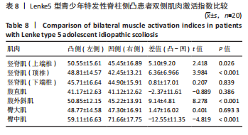
组内分析结果显示,Lenke5型青少年特发性脊柱侧凸患者在步行过程中多块肌群的均方根值存在明显不对称,见表8。竖脊肌方面,凸侧(左侧)竖脊肌均方根值在上端椎、顶椎水平均显著高于凹侧(P < 0.05);而下端椎水平两侧差异不显著(P > 0.05)。在腹部核心肌群方面,腹外斜肌均方根值在凸侧显著高于凹侧(P < 0.05),而腹直肌两侧差异无显著性意义(P > 0.05)。在臀周肌群方面,凹侧臀中肌均方根值显著高于凸侧(P < 0.05);双侧臀大肌均方根值差异无显著性意义(P > 0.05)。"
| [1] LIU X, WANG Y, LIU M, et al. The efficacy of core stabilization exercise in mild and moderate adolescent idiopathic scoliosis: a systematic review and meta-analysis. J Orthop Surg Res. 2025;20(1):214. [2] WANG H, MA Z, WU Z, et al. Biomechanical analysis of spinal range of motion and intervertebral disc loadings in normal and adolescent idiopathic scoliosis models. Front Bioeng Biotechnol. 2025;13:1473776. [3] FAZALBHOY A, MCAVINEY J, MIRENZI R. Compliance of Physiotherapeutic Scoliosis-Specific Exercise in Adolescent Idiopathic Scoliosis: A Scoping Review. J Clin Med. 2025;14(9):2950. [4] ZHANG X, WANG D, LV D, et al. Reducing the Brace Correction Stress on the Secondary Lumbar Curve Results in Excellent Muscle, Bone, and Disc Mechanical Performance: A Musculoskeletal Finite Element Simulation of AIS Patient With Rigo A3. Orthop Surg. 2025;17(2):525-539. [5] PAU M, LEBAN B, PILLONI G, et al. Trunk rotation alters postural sway but not gait in female children and early adolescents: Results from a school-based screening for scoliosis. Gait Posture. 2018;61:301-305. [6] AN JK, BERMAN D, SCHULZ J. Back pain in adolescent idiopathic scoliosis: A comprehensive review. J Child Orthop. 2023;17(2):126-140. [7] SARWARK JF, CASTELEIN RM, MAQSOOD A, et al. The Biomechanics of Induction in Adolescent Idiopathic Scoliosis: Theoretical Factors. J Bone Joint Surg Am. 2019;101(6):e22. [8] CHEN T, BIAN H, PENG X, et al. Sagittal spinopelvic parameters in adolescent idiopathic scoliosis: divergence between Lenke type 5, 6, and healthy adolescents. Eur Spine J. 2025;34(7):2669-2676. [9] XIE K, ZHU S, LIN J, et al. A novel artificial Intelligence-Based model for automated Lenke classification in adolescent idiopathic scoliosis. Eur Spine J. 2025;34(9):3929-3939. [10] LIU Y, LI X, DOU X, et al. Correlational analysis of three-dimensional spinopelvic parameters with standing balance and gait characteristics in adolescent idiopathic scoliosis: A preliminary research on Lenke V. Front Bioeng Biotechnol. 2022;10:1022376. [11] GÖNDER N, ÖZTÜRK C, TAŞDEMIR R, et al. Morphological and Morphometric Assessment of Adolescent Idiopathic Scoliosis According to Pelvic Axial Rotation-A Retrospective Cohort Study with 397 Patients. Children (Basel). 2025;12(8):991. [12] CHAN CYW, NAING KS, CHIU CK, et al. Pelvic obliquity in adolescent idiopathic scoliosis planned for posterior spinal fusion: A preoperative analysis of 311 lower limb axis films. J Orthop Surg (Hong Kong). 2019; 27(2):2309499019857250. [13] HAN SM, YANG C, WEN JX, et al. Morphology and deformity of the shoulder and pelvis in the entire spine radiographs of adolescent idiopathic scoliosis. Quant Imaging Med Surg. 2023;13(5):3266-3278. [14] 侯俞彤,黄承兰,杨云霄,等.Lenke5型青少年特发性脊柱侧凸腰椎与骨盆参数的相关性[J].中国组织工程研究,2024,28(36):5753-5758. [15] ZHU F, HONG Q, GUO X, et al. A comparison of foot posture and walking performance in patients with mild, moderate, and severe adolescent idiopathic scoliosis. PLoS One. 2021;16(5):e0251592. [16] KRAMERS-DE QUERVAIN IA, MÜLLER R, STACOFF A, et al. Gait analysis in patients with idiopathic scoliosis. Eur Spine J. 2004;13(5):449-456. [17] MAHAUDENS P, BANSE X, MOUSNY M, et al. Gait in adolescent idiopathic scoliosis: kinematics and electromyographic analysis. Eur Spine J. 2009; 18(4):512-521. [18] GAN X, LIU X, CAI D, et al. Wearable accelerometers reveal objective assessment of walking symmetry and regularity in idiopathic scoliosis patients. PeerJ. 2024;12:e17739. [19] HABER CK, SACCO M. Scoliosis: lower limb asymmetries during the gait cycle. Arch Physiother. 2015;5:4. [20] MAHAUDENS P, DETREMBLEUR C, MOUSNY M, et al. Gait in adolescent idiopathic scoliosis: energy cost analysis. Eur Spine J. 2009;18(8):1160-1168. [21] MAHAUDENS P, THONNARD JL, DETREMBLEUR C. Influence of structural pelvic disorders during standing and walking in adolescents with idiopathic scoliosis. Spine J. 2005;5(4):427-433. [22] PARK YS, LIM YT, KOH K, et al. Association of spinal deformity and pelvic tilt with gait asymmetry in adolescent idiopathic scoliosis patients: Investigation of ground reaction force. Clin Biomech (Bristol). 2016;36:52-57. [23] YANG X, HU B, SONG Y, et al. Coronal and sagittal balance in Lenke 5 AIS patients following posterior fusion: important role of the lowest instrument vertebrae selection. BMC Musculoskelet Disord. 2018;19(1):212. [24] 刘静,徐纯鑫,陆洋阳,等.青少年特发性脊柱侧凸患者步态参数、肌肉参数与影像学的相关性[J].中国组织工程研究,2025,29(21): 4477-4485. [25] FARAHPOUR N, YOUNESIAN H, BAHRPEYMA F. Electromyographic activity of erector spinae and external oblique muscles during trunk lateral bending and axial rotation in patients with adolescent idiopathic scoliosis and healthy subjects. Clin Biomech (Bristol). 2015;30(5):411-417. [26] 贺银川,王军,刘佳男,等.表面肌电图在青少年特发性脊柱侧凸评估中应用现状的研究进展[J].中国脊柱脊髓杂志,2025,35(2):200-204. [27] 张珊珊,张振发,许轶,等.中度S型青少年特发性脊柱侧凸胸腰背部肌电变化特征[J].中国康复医学杂志,2025,40(10):1482-1489. [28] TU Q, XU W, FENG Y, et al. Prevalence and determinants of adolescent idiopathic scoliosis from school screening in Xiaoshan District, Hangzhou, China. Front Public Health. 2025;13:1595793. [29] BAUMGARTEN A, KIM JK, ROBISON J, et al. Analysis of surgeon biometrics during open and robotic radical cystectomy with electromyography and motion capture analysis. Int Braz J Urol. 2020;46(1):138. [30] WILCZYŃSKI J, KAROLAK P. Relationship Between Electromyographic Frequency of the Erector Spinae and Location, Direction, and Number of Spinal Curvatures in Children with Scoliotic Changes. Risk Manag Healthc Policy. 2021;14:1881-1896. [31] PARK HJ, SIM T, SUH SW, et al. Analysis of coordination between thoracic and pelvic kinematic movements during gait in adolescents with idiopathic scoliosis. Eur Spine J. 2016;25(2):385-393. [32] GARG B, GUPTA M, MEHTA N, et al. Influence of Etiology and Onset of Deformity on Spatiotemporal, Kinematic, Kinetic, and Electromyography Gait Variables in Patients with Scoliosis-A Prospective, Comparative Study. Spine (Phila Pa 1976). 2021;46(6):374-382. [33] XU J, CHEN M, WANG X, et al. Biomechanical changes in adolescent idiopathic scoliosis during walking: A protocol for systematic review and meta-analysis. Medicine (Baltimore). 2023;102(49):e36528. [34] KARAM MR, ASMAR M, RACHKIDI R, et al. Different kinematic strategies are adopted by AIS patients during walking depending on their Lenke type. Eur Spine J. 2025. doi: 10.1007/s00586-025-09531-3. [35] DA SILVEIRA GE, ANDRADE RM, GUILHERMINO GG, et al. The Effects of Short- and Long-Term Spinal Brace Use with and without Exercise on Spine, Balance, and Gait in Adolescents with Idiopathic Scoliosis. Medicina (Kaunas). 2022;58(8):1024. [36] YANG JH, SUH SW, SUNG PS, et al. Asymmetrical gait in adolescents with idiopathic scoliosis. Eur Spine J. 2013;22(11):2407-2413. [37] STRUBER L, NOUGIER V, GRIFFET J, et al. Comparison of Trunk Motion between Moderate AIS and Healthy Children. Children (Basel). 2022; 9(5):738. [38] SHENG Z, KUAI S, TONG S, et al. The asymmetry spinal kinematics in C-shaped adolescent idiopathic scoliosis during level walking. J Biomech. 2025;193:113022. [39] 宋云雅,郭锦丽,曹策.青少年特发性脊柱侧弯患者的三维步态特征研究[J].实用骨科杂志,2023,29(5):417-421. [40] KIM DS, PARK SH, GOH TS, et al. A meta-analysis of gait in adolescent idiopathic scoliosis. J Clin Neurosci. 2020;81:196-200. [41] EUN IS, CHO YJ, GOH TS, et al. Association between gait profile and spinal alignment in patients with adolescent idiopathic scoliosis. J Clin Neurosci. 2024;130:110915. [42] CAȚAN L, CERBU S, AMARICAI E, et al. Assessment of Static Plantar Pressure, Stabilometry, Vitamin D and Bone Mineral Density in Female Adolescents with Moderate Idiopathic Scoliosis. Int J Environ Res Public Health. 2020; 17(6):2167. [43] NORIEGA-GONZALEZ DC, CRESPO J, ARDURA F, et al. Cerebral White Matter Connectivity in Adolescent Idiopathic Scoliosis: A Diffusion Magnetic Resonance Imaging Study. Children (Basel). 2022;9(7):1023. [44] MAHAUDENS P, RAISON M, BANSE X, et al. Effect of long-term orthotic treatment on gait biomechanics in adolescent idiopathic scoliosis. Spine J. 2014;14(8):1510-1509. [45] KUO FC, WANG NH, HONG CZ. Impact of visual and somatosensory deprivation on dynamic balance in adolescent idiopathic scoliosis. Spine (Phila Pa 1976). 2010;35(23):2084-2090. [46] PARAMENTO M, PASSAROTTO E, MACCARONE MC, et al. Neurophysiological, balance and motion evidence in adolescent idiopathic scoliosis: A systematic review. PLoS One. 2024;19(5):e0303086. [47] SHAHIDI B, YOO A, FARNSWORTH C, et al. Paraspinal muscle morphology and composition in adolescent idiopathic scoliosis: A histological analysis. JOR Spine. 2021;4(3):e1169. [48] STETKAROVA I, ZAMECNIK J, BOCEK V, et al. Electrophysiological and histological changes of paraspinal muscles in adolescent idiopathic scoliosis. Eur Spine J. 2016;25(10):3146-3153. [49] MARIN L, KAWCZYŃSKI A, CARNEVALE PELLINO V, et al. Displacement of Centre of Pressure during Rehabilitation Exercise in Adolescent Idiopathic Scoliosis Patients. J Clin Med. 2021;10(13):2837. [50] CHWAŁA W, KOZIANA A, KASPERCZYK T, et al. Electromyographic assessment of functional symmetry of paraspinal muscles during static exercises in adolescents with idiopathic scoliosis. Biomed Res Int. 2014;2014:573276. |
| [1] | Li Zhifei, Han Bin, Liu Qiuli, Zhang Zhanming, Wei Haokai, Zuo Kuangshi, Zhang Yisheng. Cervical motion characteristics in patients with cervical spondylotic radiculopathy based on motion capture technology [J]. Chinese Journal of Tissue Engineering Research, 2026, 30(9): 2286-2293. |
| [2] | Chen Long, Wang Xiaozhen, Xi Jintao, Lu Qilin. Biomechanical performance of short-segment screw fixation combined with expandable polyetheretherketone vertebral body replacement in osteoporotic vertebrae [J]. Chinese Journal of Tissue Engineering Research, 2026, 30(9): 2226-2235. |
| [3] | Xinjiang Branch of China Trauma Rescue & Treatment Association. Expert consensus on diagnosis and treatment of brucellar osteoarthritis [J]. Chinese Journal of Tissue Engineering Research, 2026, 30(9): 2403-2412. |
| [4] | Fu Lyupeng, Yu Peng, Liang Guoyan, Chang Yunbing. Electroactive materials applied in spinal surgery [J]. Chinese Journal of Tissue Engineering Research, 2026, 30(8): 2113-2123. |
| [5] | Li Hanyue, Li Yini, Xiang Linmei, Li Sen. Effects of resistance exercise therapy on pain and function in patients with cervical spondylotic radiculopathy: a meta-analysis [J]. Chinese Journal of Tissue Engineering Research, 2026, 30(4): 987-996. |
| [6] | Hao Yunteng, Shi Jun, Zhang Shaojie, Li Zhijun, Yang Yang, Wang Chaoqun, Ma Yuan, Zhao Hailong, Chen Jie, Dong Chongyang, Zhang Zhifeng, Li Kun, Wang Xing. Comparative finite element analysis of cervical intervertebral discs in adult humans and macaques [J]. Chinese Journal of Tissue Engineering Research, 2026, 30(36): 9488-9496. |
| [7] | Guo Zicheng, Meng Jingyuan, Zhang Jiechao, Ding Li, Tang Xiaoye, Tian Lichao, Wang Yilin, He Yong. Differences in scapular kinematics between healthy individuals and rotator cuff tear patients based on biplane X-ray [J]. Chinese Journal of Tissue Engineering Research, 2026, 30(3): 652-660. |
| [8] | Sun Kui, Huang Hailun, Wei Yatao, Liu Yongai, Gao Heng. Bibliometric analysis of exercise therapy interventions for adolescent idiopathic scoliosis [J]. Chinese Journal of Tissue Engineering Research, 2026, 30(29): 7755-7763. |
| [9] | Fu Guangliang, Bao Chunyu, Meng Qinghua, Wang Baochen, Cao Jiaxing, Sun Jiawei. Biomechanical analysis during non-anticipated stop-jump cutting before and after exercise fatigue in functional ankle instability and healthy populations [J]. Chinese Journal of Tissue Engineering Research, 2026, 30(29): 7572-7580. |
| [10] | Bai Xue, Tian Yukui, Guo Lei, Shi Mengni, Cui Xiaofeng, Wang Cheng, Li Jingxian, Zhu Qingguang, Liu Junchang. Construction of an early knee osteoarthritis rat model: CatWalk-based gait analysis and evaluation [J]. Chinese Journal of Tissue Engineering Research, 2026, 30(28): 7280-7286. |
| [11] | Zhang Le, Julaiti·Maitirouzi, Xie Xuechen, Li Chunchao, Wang Yixi, Parhat·Rexiti. Finite element analysis of biomechanical performance of a novel double-screw technique in lumbar revision of the original fixed segment [J]. Chinese Journal of Tissue Engineering Research, 2026, 30(27): 6985-6994. |
| [12] | He Zhuoqun, Wang Haiyan, Li Xiaohe, Zhang Ruofan, Shi Guopeng, Bai Lili, Wang Xiaolu. Correlation between sacro-femoro-pubic angle and spine-pelvic parameters in patients with Lenke types 1, 5, and 6 adolescent idiopathic scoliosis [J]. Chinese Journal of Tissue Engineering Research, 2026, 30(27): 7030-7036. |
| [13] | Liu Yakun, Lu Guangqi, Liang Long, Li Jing, Sun Xinyue, Liu Guangwei, Zhou Shuaiqi, Mao Hanze, Ma Mingming, Hu Jiaming, Zhu Liguo, Zhuang Minghui, Yu Jie. Highly sensitive indicators of neck muscle fatigue derived from multimodal electrophysiological and metabolic coupling analysis [J]. Chinese Journal of Tissue Engineering Research, 2026, 30(25): 6506-6511. |
| [14] | Shao Yunbo, Guo Jiayi, Li Feng. Characteristics of lower limb muscle motor activation in patients with unilateral knee osteoarthritis [J]. Chinese Journal of Tissue Engineering Research, 2026, 30(24): 6174-6181. |
| [15] | Gu Jiangpeng, Guo Wei, Chen Xujing, Liu Xiaomin, Liu Hongbo, Sun Peng, Ye Chaoqun, Feng Wei, Wang Fei. Feng’s spinal manipulation for cervical spondylosis: kinematic changes [J]. Chinese Journal of Tissue Engineering Research, 2026, 30(23): 6081-6091. |
| Viewed | ||||||
|
Full text |
|
|||||
|
Abstract |
|
|||||