Chinese Journal of Tissue Engineering Research ›› 2025, Vol. 29 ›› Issue (36): 7872-7879.doi: 10.12307/2025.555
Previous Articles Next Articles
Role of exosomes derived from mesenchymal stem cells in treatment of colorectal cancer
Guo Zhao1, Zhuang Haoyan1, Shi Xuewen2
- 1First Clinical Medical College, Shandong University of Traditional Chinese Medicine, Jinan 250000, Shandong Province, China; 2Department of Anorectal Surgery, Affiliated Hospital of Shandong University of Traditional Chinese Medicine, Jinan 250000, Shandong Province, China
-
Received:2024-04-22Accepted:2024-09-21Online:2025-12-28Published:2025-03-24 -
Contact:Shi Xuewen, MD, Chief physician, Department of Anorectal Surgery, Affiliated Hospital of Shandong University of Traditional Chinese Medicine, Jinan 250000, Shandong Province, China -
About author:Guo Zhao, Master candidate, First Clinical Medical College, Shandong University of Traditional Chinese Medicine, Jinan 250000, Shandong Province, China -
Supported by:Shandong Province Traditional Chinese Medicine Science and Technology Development Project, No. 2019-0187 (to SXW); Qilu Traditional Chinese Medicine Advantage Specialty Cluster Construction Project, No. YWC2022ZKJQ0003 (to SXW)
CLC Number:
Cite this article
Guo Zhao, Zhuang Haoyan, Shi Xuewen. Role of exosomes derived from mesenchymal stem cells in treatment of colorectal cancer[J]. Chinese Journal of Tissue Engineering Research, 2025, 29(36): 7872-7879.
share this article
Add to citation manager EndNote|Reference Manager|ProCite|BibTeX|RefWorks
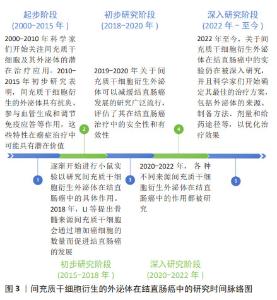
2.1 MSC-Exos在结直肠癌中的研究时间脉络图 见图3。 "
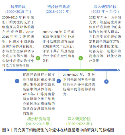
2.2 MSC-Exos 2.2.1 不同来源MSC-Exos 1983年,科学家在绵羊网织红细胞中首次发现了外泌体。外泌体是由多种细胞类型分泌的脂质双层细胞外囊泡。细胞外囊泡可分为3种类型:外泌体(直径30-150 nm)、微囊泡(100-1 000 nm)和凋亡小体(直径50-2 000 nm)。外泌体是通过细胞质膜向内出芽和随后形成的多囊泡体形成的。相反,微囊泡是通过质膜向外出芽产生的。细胞凋亡期间释放凋亡小体。许多传统标志物(CD9、CD63、CD81、TSG101、Alix、Flotillin-1、HSC70、肌动蛋白、MHCⅠ和MHCⅡ)在细胞外囊泡的多种亚型中错综分布,所以很难根据表面标志物将它们进行划分,目前主要通过直径大小进行划分,见图4。 "

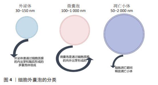
外泌体富含蛋白质、脂质和RNA,包括mRNA和miRNA,可以介导其功能发挥[22]。外泌体由多种细胞分泌,存在于几乎所有体液中[23],包括血液、唾液、尿液、脑脊液和乳汁等。但是根据大量研究证实,不同来源的MSC-Exos存在着各自的独特优势,见表1。"
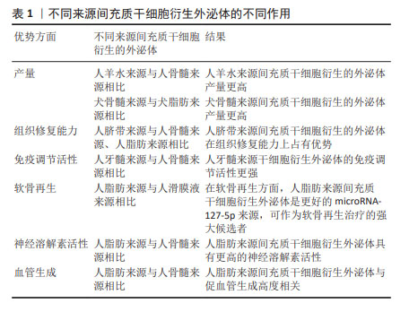
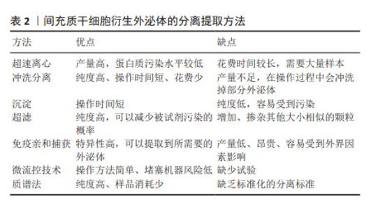
在产量方面,与人骨髓来源MSC-Exos相比,人羊水来源MSC-Exos产量更高[24];与犬脂肪来源MSC-Exos相比,犬骨髓来源MSC-Exos产量更高[25]。在组织修复能力方面,与人骨髓来源、人脂肪来源MSC-Exos相比,人脐带来源MSC-Exos更具优势[26]。在免疫调节活性方面,与人骨髓来源MSC-Exos相比,人牙髓来源MSC-Exos免疫调节活性更强[27]。研究表明,外泌体microRNA-127-5p的产量越高,软骨再生能力越好。人脂肪来源MSC-Exos比人滑膜液来源MSC-Exos表达更多的microRNA-127-5p,可以作为软骨再生的强大候选者[28]。在一项针对阿尔茨海默病的研究中发现,人脂肪来源MSC-Exos具有更高的神经溶解素活性[29]。在血管生成方面,与人骨髓来源MSC-Exos相比,人脂肪来源MSC-Exos与血管生成更具相关性[30]。 2.2.2 MSC-Exos的分离与提取 MSC-Exos的分离和提取一直是研究者所热衷的方向,为了使提取的产量更高、浓度更纯、结构稳定、污染低、操作时间短、花费少,目前科学家主要提出了以下几种分离方法:超速离心、冲洗分离、沉淀、超滤、免疫亲和捕获、微流控技术、质谱法等,见表2。"
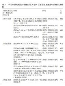
超速离心仍然是目前应用最广泛的方法,提取方案简单,提取的外泌体纯度也较高[31]。冲洗分离的方法相比较超速离心操作时间短,但是在操作过程中不可避免地会冲洗掉部分外泌体,导致提取到的产量不足[32]。沉淀方法最大的优点在于步骤简单、操作时间短[33-35]。超滤方法最大的局限性在于无法过滤出与外泌体大小相似的颗粒[36-38]。免疫亲和捕获可以通过细胞外囊泡的表面标记物很好地区分各种外泌体,从而提取到所需的外泌体类型,但是这种方法具有产量低的缺点[39-41]。此外,最近新兴的提取方法,包括微流控技术和质谱法等[42-47],都还缺少充分的分离标准,其效果还没有得到充分的评估。尽管目前尚未就外泌体分离的“金标准”达成共识,但建议结合使用多种技术以获得最佳外泌体分离结果[43]。 2.2.3 MSC-Exos的生物学特性 2010年,MSC-Exos首次在心肌缺血再灌注损伤中进行了研究[48],随后开展了大量研究探讨MSC-Exos在各类疾病中的作用。自从2012年,ZHU等[49]首次报道MSC-Exos可以促进体内肿瘤生长以来,关于MSC-Exos在肿瘤中的作用就被积极讨论。MSC-Exos在肿瘤中的作用与间充质干细胞类似,参与血管生成、抑制肿瘤细胞增殖和促进肿瘤细胞凋亡,这在李佳林等[50]的研究中也被证实。 (1)参与血管生成。在结肠癌异种移植的小鼠模型中,人骨髓来源MSC-Exos可以通过激活ERK1/2和p38 MAPK通路增加肿瘤细胞中血管内皮生长因子和趋化因子受体4的表达,从而促进血管生成,进而促进结直肠癌的发展[49]。 然而在乳腺癌中科学家们发现了相反的结果,LEE等[51]报道人骨髓来源MSC-Exos可以通过miR-16靶向血管内皮生长因子在乳腺癌细胞中的表达,从而抑制血管生成和肿瘤进展。PAKRAVAN等[52]支持这一观点,他们发现人骨髓来源间充质干细胞携带miR-100,通过下调血管内皮生长因子的表达,抑制乳腺癌血管生成。人月经血来源MSC-Exos通过抑制活性氧通路,下调促血管生成因子和NF-κB转录因子的分泌,抑制前列腺癌的进展[53]。 (2)抑制肿瘤细胞增殖和促进肿瘤细胞凋亡。人脐带来源MSC-Exos可以通过对肿瘤细胞发挥抗增殖和促凋亡作用,从而抑制膀胱癌的进展[54]。REZA等[55]观察到人脂肪来源MSC-Exos可以抑制A2780和SKOV-3癌细胞的增殖,从而抑制卵巢癌的生长与转移。人脂肪来源MSC-Exos通过Caspase-3/7通路诱导细胞凋亡,抑制前列腺癌的发展[56]。人骨髓来源MSC-Exos通过KDM3A/DCLK1/FXYD3轴抑制肿瘤细胞增殖,抑制肺癌的进展[57]。 综上,MSC-Exos在治疗肿瘤方面具有积极性,这毋庸置疑,但是也有部分科学家提出了MSC-Exos对肿瘤具有促进作用,在将来的研究中,应该侧重于研究其促进与抑制作用的不同机制,从而为肿瘤患者提供更完善的方案。 2.3 MSC-Exos治疗结直肠癌 2.3.1 MSC-Exos作为结直肠癌的治疗工具 越来越多的证据表明,从不同来源间充质干细胞中分离出的外泌体可以抑制结直肠癌的进展。此文章主要就人脐带来源、人骨髓来源、人脂肪来源MSC-Exos对结直肠癌的作用展开研究,见表3。"
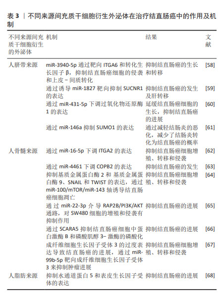
人脐带来源MSC-Exos可以携带miR-3940-5p通过靶向ITGA6和转化生长因子β,抑制结直肠癌细胞的侵袭,并抑制上皮-间质转化,从而抑制了结直肠癌的生长与转移[58]。CHEN等[59]研究也证实了人脐带来源MSC-Exos可以抑制结直肠癌的发生并且对肝转移具有抑制作用,其作用机制是通过诱导miR-1827靶向抑制SUCNR1而发挥作用。QU等[60]研究证实人脐带来源MSC-Exos携带的miR-431-5p 可以通过下调过氧化物还原酶1的表达,从而延缓结直肠癌细胞的生长,抑制结直肠癌的进展。WANG等[61]研究发现人脐带来源MSC-Exos通过miR-146a抑制SUMO1的表达,延缓结肠炎的恶化,避免了结肠炎长期恶化后发生结直肠癌的不良结局。多项实验证明,人骨髓来源MSC-Exos对结直肠癌也具有抑制作用,主要体现在以下几个机制:通过miR-16-5p下调ITGA2的表达[62];通过miR-4461下调 COPB2的表达,抑制HCT116和SW480细胞的迁移和侵袭[63],抑制基质金属蛋白酶2、基质金属蛋白酶9、SNAIL和TWIST的表达;通过miR-100/mTOR/miR-143轴诱导结直肠癌细胞凋亡[64];通过miR-22-3p介导 RAP2B/PI3K/AKT通路,对SW480细胞的增殖和侵袭有抑制作用[65];通过SCARA5抑制结直肠癌细胞中蛋白激酶B和磷酸肌醇3-激酶的磷酸化[66];通过miR-99b-5p靶向 FGFR3来抑制肿瘤进展[67]。在最近的一项研究中,发现人脂肪来源MSC-Exos可以通过抑制水通道蛋白5和表皮生长因子受体的表达,进而抑制结直肠癌的发展[68]。 综上所述,间充质干细胞对结直肠癌的治疗作用可能归因于富含肿瘤抑制性miRNA的外泌体,这可能会成为未来临床实践中治疗结直肠癌的一种有前途的方法。 2.3.2 MSC-Exos作为结直肠癌的潜在治疗靶点 尽管有大量的实验证据已经表明MSC-Exos对结直肠癌具有抑制作用,但仍有研究显示其在结直肠癌的发展中发挥促进作用,这表明MSC-Exos可以作为结直肠癌的潜在治疗靶点,为治疗结直肠癌提供更有价值的方法,见表4。"
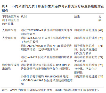
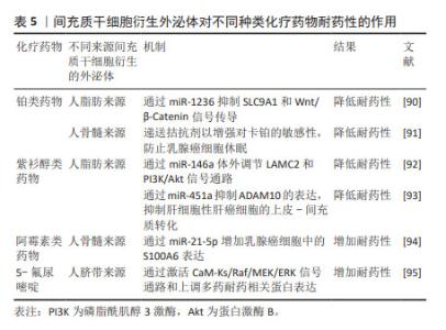
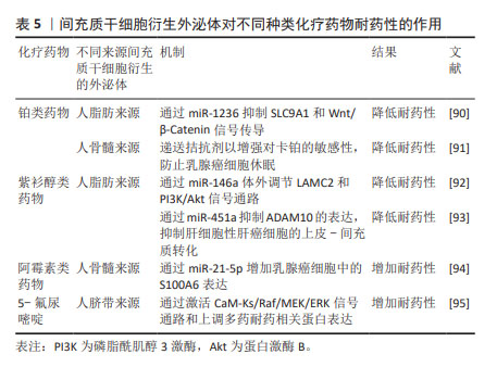
XUE等[69]研究发现,人脂肪来源MSC-Exos可以通过TRPC3/NF-KB轴诱导肿瘤相关成纤维细胞的表型,从而加速结肠癌的进展。人骨髓来源MSC-Exos可以通过携带的miR-142-3p增加结肠癌中肿瘤干细胞的数量,从而加速结肠癌的发生与进展[70]。一项大鼠模型研究中也证实了人骨髓来源间充质干细胞可以通过腺苷单磷酸活化蛋白激酶/哺乳动物雷帕霉素靶蛋白(AMP-activated protein kinase/mammalian target of rapamycin,AMPK/mTOR)介导的NF-κB激活促进癌细胞的增殖、迁移和集落形成,促进结直肠癌的发生与转移[71]。另外,最近的一项体外研究表明,人骨髓来源MSC-Exos中携带的miR-424具有诱导结直肠癌发生的作用[72]。REN等[73]研究发现,癌症相关成纤维细胞通过转移外泌体长链非编码RNA H19促进结肠癌干细胞增殖和增加化疗耐药性,从而加速结直肠癌的进展。同样的结果在ZHOU等 [74]的研究中也被证实,癌症相关成纤维细胞分泌的外泌体长链非编码RNA LINC00659通过miR-342-3p/ANXA2轴促进结直肠癌细胞增殖。通过以上研究可以发现,抑制这些促进结直肠癌进展的靶点可以为治疗结直肠癌提供一种新思路。 2.3.3 MSC-Exos可以作为治疗结直肠癌的载体 外泌体具有高稳定性、高生物相容性的特点,这使其作为药物输送的载体成为可能[75]。同时,外泌体还具有生物利用度高的特点,可以更顺利地通过血脑屏障等生物屏障[76]。有研究报道,MSC-Exos可以通过细胞间连接、P-糖蛋白的高表达、隧道纳米管和胞吐方式与癌细胞相互作用[77-79]。 另外,可以将对癌症具有良好治疗潜力的小干扰 RNA (siRNA)或microRNA (miRNA)装载到MSC-Exos上,使其作为合适的递送系统[80-82]。HAN等[83]研究发现,人脐带来源MSC-Exos可以作为抗miRNA寡核苷酸的载体,被肿瘤细胞特异性摄取,以发挥抗肿瘤作用。在另外一项研究中发现,人骨髓来源MSC-Exos可以作为阿霉素的载体,包封率高达35%,与单纯的游离阿霉素相比,它表现出更显著的抑制结直肠癌的作用[84]。结直肠癌干细胞衍生外泌体携带的miR-1270选择性地增强了对大肠癌细胞的抑制作用[85]。最近的一项研究也证实了MSC-Exos可以作为良好的抗结直肠癌药物的载体,在结直肠癌中发挥着重要作用[86]。以上所有研究都证明了MSC-Exos可以作为一种创新的药物递送策略,其特点是靶向更精确、免疫原性更低,以实现更好的靶向治疗[87]。 2.3.4 MSC-Exos在化疗耐药性中的作用 化疗贯穿着结直肠癌的整个治疗过程,早期手术治疗后,一般需要化疗巩固治疗,晚期无法进行手术时,更需要化疗维持生命。铂类药物、紫杉醇、阿霉素和5-氟尿嘧啶等多种具有不同特性和靶点的化疗药物已被有效应用于晚期癌症患者[88]。然而,长期化疗耐药性成为化疗成功与否的重要挑战[89]。很多研究已经证明MSC-Exos与癌症化疗耐药性密切相关,因为它们可以直接递送功能蛋白和RNA,避免了细胞凋亡,这充分表明MSC-Exos可以通过调节细胞凋亡蛋白来介导化疗耐药性。但是也有很多研究表明MSC-Exos可以通过介导不同的信号通路而增加化疗药物耐药性,见表5。"
| [1] XI Y, XU P. Global colorectal cancer burden in 2020 and projections to 2040. Transl Oncol. 2021;14(10):101174. [2] ALZAHRANI SM, AL DOGHAITHER HA, AL-GHAFARI AB. General insight into cancer: An overview of colorectal cancer (Review). Mol Clin Oncol. 2021;15(6):271. [3] BILLER LH, SCHRAG D. Diagnosis and Treatment of Metastatic Colorectal Cancer: A Review. JAMA. 2021;325(7):669-685. [4] SHAHAR N, LARISCH S. Inhibiting the inhibitors: Targeting anti-apoptotic proteins in cancer and therapy resistance. Drug Resist Updat. 2020;52: 100712. [5] GATENBY RA, BROWN JS. Integrating evolutionary dynamics into cancer therapy. Nat Rev Clin Oncol. 2020;17(11):675-686. [6] WYLD L, AUDISIO RA, POSTON GJ. The evolution of cancer surgery and future perspectives. Nat Rev Clin Oncol. 2015;12(2):115-124. [7] CREMOLINI C, ANTONIOTTI C, ROSSINI D, et al. Upfront FOLFOXIRI plus bevacizumab and reintroduction after progression versus mFOLFOX6 plus bevacizumab followed by FOLFIRI plus bevacizumab in the treatment of patients with metastatic colorectal cancer (TRIBE2): a multicentre, open-label, phase 3, randomised, controlled trial. Lancet Oncol. 2020;21(4):497-507. [8] SALEM HK, THIEMERMANN C. Mesenchymal stromal cells: current understanding and clinical status. Stem Cells. 2010;28(3):585-596. [9] TEIXEIRA FG, PANCHALINGAM KM, ANJO SI, et al. Do hypoxia/normoxia culturing conditions change the neuroregulatory profile of Wharton Jelly mesenchymal stem cell secretome? Stem Cell Res Ther. 2015;6(1):133. [10] KISIEL AH, MCDUFFEE LA, MASAOUD E, et al. Isolation, characterization, and in vitro proliferation of canine mesenchymal stem cells derived from bone marrow, adipose tissue, muscle, and periosteum. Am J Vet Res. 2012; 73(8):1305-1317. [11] MURPHY MB, MONCIVAIS K, CAPLAN AI. Mesenchymal stem cells: environmentally responsive therapeutics for regenerative medicine. Exp Mol Med. 2013;45(11):e54. [12] RIDGE SM, SULLIVAN FJ, GLYNN SA. Mesenchymal stem cells: key players in cancer progression. Mol Cancer. 2017;16(1):31. [13] MENG L, ZHAO Y, BU W, et al. Bone mesenchymal stem cells are recruited via CXCL8-CXCR2 and promote EMT through TGF-β signal pathways in oral squamous carcinoma. Cell Prolif. 2020;53(8):e12859. [14] XUNIAN Z, KALLURI R. Biology and therapeutic potential of mesenchymal stem cell-derived exosomes. Cancer Sci. 2020;111(9):3100-3110. [15] BIGONI-ORDÓÑEZ GD, CZARNOWSKI D, PARSONS T, et al. Integrin α6 (CD49f), The Microenvironment and Cancer Stem Cells. Curr Stem Cell Res Ther. 2019;14(5):428-436. [16] BAEK G, CHOI H, KIM Y, et al. Mesenchymal Stem Cell-Derived Extracellular Vesicles as Therapeutics and as a Drug Delivery Platform. Stem Cells Transl Med. 2019;8(9):880-886. [17] LIU H, DENG S, HAN L, et al. Mesenchymal stem cells, exosomes and exosome-mimics as smart drug carriers for targeted cancer therapy. Colloids Surf B Biointerfaces. 2022;209(Pt 1):112163. [18] YANG Y, BUCAN V, BAEHRE H, et al. Acquisition of new tumor cell properties by MSC-derived exosomes. Int J Oncol. 2015;47(1):244-252. [19] PESSINA A, SISTO F, COCCÈ V, et al. A mesenchymal stromal cell line resistant to paclitaxel that spontaneously differentiates into osteoblast-like cells. Cell Biol Toxicol. 2011;27(3):169-180. [20] DOS SANTOS GGL, OLIVEIRA ALL, SANTOS DS, et al. Mesenchymal stem cells reduce the oxaliplatin-induced sensory neuropathy through the reestablishment of redox homeostasis in the spinal cord. Life Sci. 2021; 265:118755. [21] ALTANEROVA U, JAKUBECHOVA J, BENEJOVA K, et al. Intracellular prodrug gene therapy for cancer mediated by tumor cell suicide gene exosomes. Int J Cancer. 2021;148(1):128-139. [22] ALZHRANI GN, ALANAZI ST, ALSHARIF SY, et al. Exosomes: Isolation, characterization, and biomedical applications. Cell Biol Int. 2021;45(9): 1807-1831. [23] SIMPSON RJ, KALRA H, MATHIVANAN S. ExoCarta as a resource for exosomal research. J Extracell Vesicles. 2012;1(1):18374. [24] TRACY SA, AHMED A, TIGGES JC, et al. A comparison of clinically relevant sources of mesenchymal stem cell-derived exosomes: Bone marrow and amniotic fluid. J Pediatr Surg. 2019;54(1):86-90. [25] VILLATORO AJ, ALCOHOLADO C, MARTÍN-ASTORGA MC, et al. Comparative analysis and characterization of soluble factors and exosomes from cultured adipose tissue and bone marrow mesenchymal stem cells in canine species. Vet Immunol Immunopathol. 2019;208:6-15. [26] WANG ZG, HE ZY, LIANG S, et al. Comprehensive proteomic analysis of exosomes derived from human bone marrow, adipose tissue, and umbilical cord mesenchymal stem cells. Stem Cell Res Ther. 2020; 11(1):511. [27] JI L, BAO L, GU Z, et al. Comparison of immunomodulatory properties of exosomes derived from bone marrow mesenchymal stem cells and dental pulp stem cells. Immunol Res. 2019;67(4-5):432-442. [28] SEMERCI SEVIMLI T, SEVIMLI M, QOMI EKENEL E, et al. Comparison of exosomes secreted by synovial fluid-derived mesenchymal stem cells and adipose tissue-derived mesenchymal stem cells in culture for microRNA-127-5p expression during chondrogenesis. Gene. 2023;865:147337. [29] KATSUDA T, TSUCHIYA R, KOSAKA N, et al. Human adipose tissue-derived mesenchymal stem cells secrete functional neprilysin-bound exosomes. Sci Rep. 2013;3:1197. [30] POMATTO M, GAI C, NEGRO F, et al. Differential Therapeutic Effect of Extracellular Vesicles Derived by Bone Marrow and Adipose Mesenchymal Stem Cells on Wound Healing of Diabetic Ulcers and Correlation to Their Cargoes. Int J Mol Sci. 2021;22(8):3851. [31] GARDINER C, DI VIZIO D, SAHOO S, et al. Techniques used for the isolation and characterization of extracellular vesicles: results of a worldwide survey. J Extracell Vesicles. 2016;5:32945. [32] CHENG H, FANG H, XU RD, et al. Development of a rinsing separation method for exosome isolation and comparison to conventional methods. Eur Rev Med Pharmacol Sci. 2019;23(12):5074-5083. [33] SOARES MARTINS T, CATITA J, MARTINS ROSA I, et al. Exosome isolation from distinct biofluids using precipitation and column-based approaches. PLoS One. 2018;13(6):e0198820. [34] BUSCHMANN D, KIRCHNER B, HERMANN S, et al. Evaluation of serum extracellular vesicle isolation methods for profiling miRNAs by next-generation sequencing. J Extracell Vesicles. 2018;7(1):1481321. [35] ZHAO L, YU J, WANG J, et al. Isolation and Identification of miRNAs in exosomes derived from serum of colon cancer patients. J Cancer. 2017;8(7): 1145-1152. [36] VAN DEUN J, MESTDAGH P, SORMUNEN R, et al. The impact of disparate isolation methods for extracellular vesicles on downstream RNA profiling. J Extracell Vesicles. 2014;3(1):24858. [37] LAMPARSKI HG, METHA-DAMANI A, YAO JY, et al. Production and characterization of clinical grade exosomes derived from dendritic cells. J Immunol Methods. 2002;270(2):211-226. [38] LOBB RJ, BECKER M, WEN SW, et al. Optimized exosome isolation protocol for cell culture supernatant and human plasma. J Extracell Vesicles. 2015; 4:27031. [39] POPOVIC M, MAZZEGA E, TOFFOLETTO B, et al. Isolation of anti-extra-cellular vesicle single-domain antibodies by direct panning on vesicle-enriched fractions. Microb Cell Fact. 2018;17(1):6. [40] GREENING DW, XU R, JI H, et al. A protocol for exosome isolation and characterization: evaluation of ultracentrifugation, density-gradient separation, and immunoaffinity capture methods. Methods Mol Biol. 2015;1295:179-209. [41] ZHU L, SUN HT, WANG S, et al. Isolation and characterization of exosomes for cancer research. J Hematol Oncol. 2020;13(1):152. [42] ABRAMOWICZ A, MARCZAK L, WOJAKOWSKA A, et al. Harmonization of exosome isolation from culture supernatants for optimized proteomics analysis. PLoS One. 2018;13(10):e0205496. [43] THÉRY C, WITWER KW, AIKAWA E, et al. Minimal information for studies of extracellular vesicles 2018 (MISEV2018): a position statement of the International Society for Extracellular Vesicles and update of the MISEV2014 guidelines. J Extracell Vesicles. 2018;7(1):1535750. [44] LANGEVIN SM, KUHNELL D, ORR-ASMAN MA, et al. Balancing yield, purity and practicality: a modified differential ultracentrifugation protocol for efficient isolation of small extracellular vesicles from human serum. RNA Biol. 2019;16(1):5-12. [45] KANWAR SS, DUNLAY CJ, SIMEONE DM, et al. Microfluidic device (ExoChip) for on-chip isolation, quantification and characterization of circulating exosomes. Lab Chip. 2014;14(11):1891-1900. [46] ILIESCU FS, VRTAČNIK D, NEUZIL P, et al. Microfluidic Technology for Clinical Applications of Exosomes. Micromachines (Basel). 2019; 10(6):392. [47] ZHANG P, ZHOU X, HE M, et al. Ultrasensitive detection of circulating exosomes with a 3D-nanopatterned microfluidic chip. Nat Biomed Eng. 2019;3(6):438-451. [48] LAI RC, ARSLAN F, LEE MM, et al. Exosome secreted by MSC reduces myocardial ischemia/reperfusion injury. Stem Cell Res. 2010;4(3):214-222. [49] ZHU W, HUANG L, LI Y, et al. Exosomes derived from human bone marrow mesenchymal stem cells promote tumor growth in vivo. Cancer Lett. 2012; 315(1):28-37. [50] 李佳林,张耀东,娄艳茹,等.间充质干细胞分泌组发挥作用的分子机制[J].中国组织工程研究,2025,29(7):1512-1522. [51] LEE JK, PARK SR, JUNG BK, et al. Exosomes derived from mesenchymal stem cells suppress angiogenesis by down-regulating VEGF expression in breast cancer cells. PLoS One. 2013;8(12):e84256. [52] PAKRAVAN K, BABASHAH S, SADEGHIZADEH M, et al. MicroRNA-100 shuttled by mesenchymal stem cell-derived exosomes suppresses in vitro angiogenesis through modulating the mTOR/HIF-1α/VEGF signaling axis in breast cancer cells. Cell Oncol (Dordr). 2017;40(5):457-470. [53] ALCAYAGA-MIRANDA F, GONZÁLEZ PL, LOPEZ-VERRILLI A, et al. Prostate tumor-induced angiogenesis is blocked by exosomes derived from menstrual stem cells through the inhibition of reactive oxygen species. Oncotarget. 2016;7(28):44462-44477. [54] WU S, JU GQ, DU T, et al. Microvesicles derived from human umbilical cord Wharton’s jelly mesenchymal stem cells attenuate bladder tumor cell growth in vitro and in vivo. PLoS One. 2013;8(4):e61366. [55] REZA AMMT, CHOI YJ, YASUDA H, et al. Human adipose mesenchymal stem cell-derived exosomal-miRNAs are critical factors for inducing anti-proliferation signalling to A2780 and SKOV-3 ovarian cancer cells. Sci Rep. 2016;6:38498. [56] TAKAHARA K, II M, INAMOTO T, et al. microRNA-145 Mediates the Inhibitory Effect of Adipose Tissue-Derived Stromal Cells on Prostate Cancer. Stem Cells Dev. 2016;25(17):1290-1298. [57] LIU J, FENG Y, ZENG X, et al. Extracellular vesicles-encapsulated let-7i shed from bone mesenchymal stem cells suppress lung cancer via KDM3A/DCLK1/FXYD3 axis. J Cell Mol Med. 2021;25(4):1911-1926. [58] LI T, WAN Y, SU Z, et al. Mesenchymal Stem Cell-Derived Exosomal microRNA-3940-5p Inhibits Colorectal Cancer Metastasis by Targeting Integrin α6. Dig Dis Sci. 2021;66(6):1916-1927. [59] CHEN J, LI Z, YUE C, et al. Human umbilical cord mesenchymal stem cell-derived exosomes carrying miR-1827 downregulate SUCNR1 to inhibit macrophage M2 polarization and prevent colorectal liver metastasis. Apoptosis. 2023;28(3-4):549-565. [60] QU M, LI J, HONG Z, et al. The role of human umbilical cord mesenchymal stem cells-derived exosomal microRNA-431-5p in survival and prognosis of colorectal cancer patients. Mutagenesis. 2022;37(2):164-171. [61] WANG J, PEI B, YAN J, et al. hucMSC-Derived Exosomes Alleviate the Deterioration of Colitis via the miR-146a/SUMO1 Axis. Mol Pharm. 2022; 19(2):484-493. [62] XU Y, SHEN L, LI F, et al. microRNA-16-5p-containing exosomes derived from bone marrow-derived mesenchymal stem cells inhibit proliferation, migration, and invasion, while promoting apoptosis of colorectal cancer cells by downregulating ITGA2. J Cell Physiol. 2019;234(11):21380-21394. [63] CHEN HL, LI JJ, JIANG F, et al. MicroRNA-4461 derived from bone marrow mesenchymal stem cell exosomes inhibits tumorigenesis by downregulating COPB2 expression in colorectal cancer. Biosci Biotechnol Biochem. 2020; 84(2):338-346. [64] JAHANGIRI B, KHALAJ-KONDORI M, ASADOLLAHI E, et al. MSC-Derived exosomes suppress colorectal cancer cell proliferation and metastasis via miR-100/mTOR/miR-143 pathway. Int J Pharm. 2022;627:122214. [65] WANG Y, LIN C. Exosomes miR-22-3p Derived from Mesenchymal Stem Cells Suppress Colorectal Cancer Cell Proliferation and Invasion by Regulating RAP2B and PI3K/AKT Pathway. J Oncol. 2021;2021:3874478. [66] FANG Y, WU F, SHANG G, et al. SCARA5 in bone marrow stromal cell-derived exosomes inhibits colorectal cancer progression by inactivating the PI3K/Akt pathway. Genomics. 2023;115(4):110636. [67] NING S, CHEN Y, LI S, et al. Exosomal miR-99b-5p Secreted from Mesenchymal Stem Cells Can Retard the Progression of Colorectal Cancer by Targeting FGFR3. Stem Cell Rev Rep. 2023;19(8):2901-2917. [68] MANSOURABADI AH, AGHAMAJIDI A, FARAJI F, et al. Mesenchymal stem cells- derived exosomes inhibit the expression of Aquaporin-5 and EGFR in HCT-116 human colorectal carcinoma cell line. BMC Mol Cell Biol. 2022;23(1):40. [69] XUE C, GAO Y, LI X, et al. Mesenchymal stem cells derived from adipose accelerate the progression of colon cancer by inducing a MT-CAFs phenotype via TRPC3/NF-KB axis. Stem Cell Res Ther. 2022;13(1):335. [70] LI H, LI F. Exosomes from BM-MSCs increase the population of CSCs via transfer of miR-142-3p. Br J Cancer. 2018;119(6):744-755. [71] WU XB, LIU Y, WANG GH, et al. Mesenchymal stem cells promote colorectal cancer progression through AMPK/mTOR-mediated NF-κB activation. Sci Rep. 2016;6:21420. [72] ZHANG N, LI L, LUO J, et al. Inhibiting microRNA-424 in bone marrow mesenchymal stem cells-derived exosomes suppresses tumor growth in colorectal cancer by upregulating TGFBR3. Arch Biochem Biophys. 2021;709:108965. [73] REN J, DING L, ZHANG D, et al. Carcinoma-associated fibroblasts promote the stemness and chemoresistance of colorectal cancer by transferring exosomal lncRNA H19. Theranostics. 2018;8(14):3932-3948. [74] ZHOU L, LI J, TANG Y, et al. Exosomal LncRNA LINC00659 transferred from cancer-associated fibroblasts promotes colorectal cancer cell progression via miR-342-3p/ANXA2 axis. J Transl Med. 2021;19(1):8. [75] LI YJ, WU JY, WANG JM, et al. Gemcitabine loaded autologous exosomes for effective and safe chemotherapy of pancreatic cancer. Acta Biomater. 2020;101:519-530. [76] 马宝南,孙丽娜,韩美华.肿瘤治疗的细胞药物递送系统的研究进展[J].现代药物与临床,2024,39(2):503-513. [77] PESSINA A, BONOMI A, COCCÈ V, et al. Mesenchymal stromal cells primed with paclitaxel provide a new approach for cancer therapy. PLoS One. 2011;6(12):e28321. [78] PESSINA A, COCCÈ V, PASCUCCI L, et al. Mesenchymal stromal cells primed with Paclitaxel attract and kill leukaemia cells, inhibit angiogenesis and improve survival of leukaemia-bearing mice. Br J Haematol. 2013; 160(6):766-778. [79] GOODARZI A, VALIKHANI M, AMIRI F, et al. The mechanisms of mutual relationship between malignant hematologic cells and mesenchymal stem cells: Does it contradict the nursing role of mesenchymal stem cells? Cell Commun Signal. 2022;20(1):21. [80] LOU G, SONG X, YANG F, et al. Exosomes derived from miR-122-modified adipose tissue-derived MSCs increase chemosensitivity of hepatocellular carcinoma. J Hematol Oncol. 2015;8:122. [81] GRECO KA, FRANZEN CA, FOREMAN KE, et al. PLK-1 Silencing in Bladder Cancer by siRNA Delivered With Exosomes. Urology. 2016;91:241.e1-7. [82] WANG Y, CHEN X, TIAN B, et al. Nucleolin-targeted Extracellular Vesicles as a Versatile Platform for Biologics Delivery to Breast Cancer. Theranostics. 2017;7(5):1360-1372. [83] HAN S, LI G, JIA M, et al. Delivery of Anti-miRNA-221 for Colorectal Carcinoma Therapy Using Modified Cord Blood Mesenchymal Stem Cells-Derived Exosomes. Front Mol Biosci. 2021;8:743013. [84] BAGHERI E, ABNOUS K, FARZAD SA, et al. Targeted doxorubicin-loaded mesenchymal stem cells-derived exosomes as a versatile platform for fighting against colorectal cancer. Life Sci. 2020;261:118369. [85] JIN Y, SUN L, CHEN Y, et al. The homologous tumor-derived-exosomes loaded with miR-1270 selectively enhanced the suppression effect for colorectal cancer cells. Cancer Med. 2024;13(1):e6936. [86] KARAMI FATH M, ANJOMROOZ M, TAHA SR, et al. The therapeutic effect of exosomes from mesenchymal stem cells on colorectal cancer: Toward cell-free therapy. Pathol Res Pract. 2022;237:154024. [87] WANG M, LI J, WANG D, et al. The effects of mesenchymal stem cells on the chemotherapy of colorectal cancer. Biomed Pharmacother. 2023; 160:114373. [88] GHOSH S. Cisplatin: The first metal based anticancer drug. Bioorg Chem. 2019;88:102925. [89] GUERRA F, ARBINI AA, MORO L. Mitochondria and cancer chemoresistance. Biochim Biophys Acta Bioenerg. 2017;1858(8):686-699. [90] JIA Z, ZHU H, SUN H, et al. Adipose Mesenchymal Stem Cell-Derived Exosomal microRNA-1236 Reduces Resistance of Breast Cancer Cells to Cisplatin by Suppressing SLC9A1 and the Wnt/β-Catenin Signaling. Cancer Manag Res. 2020;12:8733-8744. [91] BLISS SA, SINHA G, SANDIFORD OA, et al. Mesenchymal Stem Cell-Derived Exosomes Stimulate Cycling Quiescence and Early Breast Cancer Dormancy in Bone Marrow. Cancer Res. 2016;76(19):5832-5844. [92] QIU L, WANG J, CHEN M, et al. Exosomal microRNA‑146a derived from mesenchymal stem cells increases the sensitivity of ovarian cancer cells to docetaxel and taxane via a LAMC2‑mediated PI3K/Akt axis. Int J Mol Med. 2020;46(2):609-620. [93] XU Y, LAI Y, CAO L, et al. Human umbilical cord mesenchymal stem cells-derived exosomal microRNA-451a represses epithelial-mesenchymal transition of hepatocellular carcinoma cells by inhibiting ADAM10. RNA Biol. 2021;18(10):1408-1423. [94] LUO T, LIU Q, TAN A, et al. Mesenchymal Stem Cell-Secreted Exosome Promotes Chemoresistance in Breast Cancer via Enhancing miR-21-5p-Mediated S100A6 Expression. Mol Ther Oncolytics. 2020;19:283-293. [95] JI R, ZHANG B, ZHANG X, et al. Exosomes derived from human mesenchymal stem cells confer drug resistance in gastric cancer. Cell Cycle. 2015;14(15):2473-2483. [96] GUO G, TAN Z, LIU Y, et al. The therapeutic potential of stem cell-derived exosomes in the ulcerative colitis and colorectal cancer. Stem Cell Res Ther. 2022;13(1):138. |
| [1] | Lai Pengyu, Liang Ran, Shen Shan. Tissue engineering technology for repairing temporomandibular joint: problems and challenges [J]. Chinese Journal of Tissue Engineering Research, 2025, 29(在线): 1-9. |
| [2] | Liu Lin, Liu Shixuan, Lu Xinyue, Wang Kan. Metabolomic analysis of urine in a rat model of chronic myofascial trigger points [J]. Chinese Journal of Tissue Engineering Research, 2025, 29(8): 1585-1592. |
| [3] | Su Xiaoyang, Chen Wenting, Fu Yidan, Zhao Yan, Lan Danfeng, Yang Qiuping. Correlation between Mer receptor tyrosine kinase and diabetic peripheral neuropathy in Sprague-Dawley rats [J]. Chinese Journal of Tissue Engineering Research, 2025, 29(8): 1593-1599. |
| [4] | Li Kaiying, Wei Xiaoge, Song Fei, Yang Nan, Zhao Zhenning, Wang Yan, Mu Jing, Ma Huisheng. Mechanism of Lijin manipulation regulating scar formation in skeletal muscle injury repair in rabbits [J]. Chinese Journal of Tissue Engineering Research, 2025, 29(8): 1600-1608. |
| [5] | Li Jun, Gong Jingjing, Sun Guobin, Guo Rui, Ding Yang, Qiang Lijuan, Zhang Xiaoli, Fang Zhanhai . miR-27a-3p promotes the proliferation of human hypertrophic scar fibroblasts by regulating mitogen-activated protein kinase signaling pathway [J]. Chinese Journal of Tissue Engineering Research, 2025, 29(8): 1609-1617. |
| [6] | Li Huayuan, Li Chun, Liu Junwei, Wang Ting, Li Long, Wu Yongli. Effect of warm acupuncture on PINK1/Parkin pathway in the skeletal muscle of rats with chronic fatigue syndrome [J]. Chinese Journal of Tissue Engineering Research, 2025, 29(8): 1618-1625. |
| [7] | Jing Ruyi, Chen Yingxin, Cao Lei . Prognosis of deep lamellar keratoplasty versus penetrating keratoplasty in the treatment of stromal corneal dystrophy [J]. Chinese Journal of Tissue Engineering Research, 2025, 29(8): 1626-1633. |
| [8] | Wang Xuanqiang, Zhang Wenyang, Li Yang, Kong Weiqian, Li Wei, Wang Le, Li Zhongshan, Bai Shi. Effects of chronic exposure to low-frequency pulsed magnetic fields on contractility and morphology of the quadriceps muscle in healthy adults [J]. Chinese Journal of Tissue Engineering Research, 2025, 29(8): 1634-1642. |
| [9] | Zhang Yuxin, Yu Cong, Zhang Cui, Ding Jianjun, Chen Yan. Differences in postural control ability between older adults with mild cognitive impairment and those with normal cognition under different single-task and dual-task conditions [J]. Chinese Journal of Tissue Engineering Research, 2025, 29(8): 1643-1649. |
| [10] | Zhou Panpan, Cui Yinglin, Zhang Wentao, Wang Shurui, Chen Jiahui, Yang Tong . Role of cellular autophagy in cerebral ischemic injury and the regulatory mechanism of traditional Chinese medicine [J]. Chinese Journal of Tissue Engineering Research, 2025, 29(8): 1650-1658. |
| [11] | Yu Jingbang, Wu Yayun. Regulatory effect of non-coding RNA in pulmonary fibrosis [J]. Chinese Journal of Tissue Engineering Research, 2025, 29(8): 1659-1666. |
| [12] | Wang Qiuyue, Jin Pan, Pu Rui . Exercise intervention and the role of pyroptosis in osteoarthritis [J]. Chinese Journal of Tissue Engineering Research, 2025, 29(8): 1667-1675. |
| [13] | Zhu Hanmin, Wang Song, Xiao Wenlin, Zhang Wenjing, Zhou Xi, He Ye, Li Wei, . Mitophagy regulates bone metabolism [J]. Chinese Journal of Tissue Engineering Research, 2025, 29(8): 1676-1683. |
| [14] | Yuan Weibo, Liu Chan, Yu Limei. Potential application of liver organoids in liver disease models and transplantation therapy [J]. Chinese Journal of Tissue Engineering Research, 2025, 29(8): 1684-1692. |
| [15] | Wang Yida, Liu Jun, Wang Xiaoling, Wang Liyan, Yang Chengru, Zhang Xuexiao. Effects of wearable electronic device-based interventions on physical activity and sedentary behavior in healthy adolescents: a meta-analysis [J]. Chinese Journal of Tissue Engineering Research, 2025, 29(8): 1693-1704. |
| Viewed | ||||||
|
Full text |
|
|||||
|
Abstract |
|
|||||