Chinese Journal of Tissue Engineering Research ›› 2017, Vol. 21 ›› Issue (31): 5072-5077.doi: 10.3969/j.issn.2095-4344.2017.31.026
Previous Articles Next Articles
Imaging evaluation for scaphoid fracture: which surgical approach is more effective and safer?
Feng Yan-hua1, Cui Ying-tie1, Tian Yi-ren1, Zhang Fang1, Wang Shi-bo2, He Ming-zhe1
- 1First Department of Orthopedics, Children’s Hospital of Hebei Province, Shijiazhuang 050000, Hebei Province, China; 2Department of Orthopedics, No. 101 Hospital of Chinese PLA, Wuxi 214044, Jiangsu Province, China
-
Online:2017-11-08Published:2017-12-01 -
Contact:Zhang Fang, Associate chief physician, First Department of Orthopedics, Children’s Hospital of Hebei Province, Shijiazhuang 050000, Hebei Province, China -
About author:Feng Yan-hua, Master, Attending physician, First Department of Orthopedics, Children’s Hospital of Hebei Province, Shijiazhuang 050000, Hebei Province, China
CLC Number:
Cite this article
Feng Yan-hua, Cui Ying-tie, Tian Yi-ren, Zhang Fang, Wang Shi-bo, He Ming-zhe. Imaging evaluation for scaphoid fracture: which surgical approach is more effective and safer? [J]. Chinese Journal of Tissue Engineering Research, 2017, 21(31): 5072-5077.
share this article

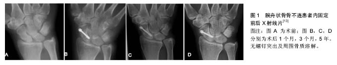
2.1 腕舟状骨骨折的影像学检查 对怀疑有腕舟状骨骨折的患者,需行影像学检查确诊。通常采用的影像学检查包括X射线平片、CT、MRI以及核素骨扫描等。 2.1.1 X射线平片 因X射线平片价格低廉且操作简便,故临床上腕关节平片是腕部外伤患者的常规检查,通常采用的体位包括后前位、侧位及腕舟状骨位。 由于腕舟状骨形态不规则,且与邻近的月骨、头状骨和大小多角骨等不规则骨有复杂的关节连接,如果投照方法不恰当,体位摆放不标准,则在X射线平片上容易出现腕舟状骨与邻近诸骨的重叠现象,可导致对腕舟状骨骨折的误诊或漏诊[2]。 有研究表明,腕舟状骨的X射线平片投照时至少要有2个与腕舟状骨相互垂直的体位,但由于腕舟状骨特殊的解剖形态及毗邻关系,很难投照出两个互相垂直体位均清晰的图像[3]。 国内丁双双等[4]的研究显示尽管握拳尺偏位(腕舟状骨位)是显示腕舟状骨的最佳位置,但仅采用此种体位评价腕舟状骨骨折仍有相当比例的骨折患者被漏诊,而腕关节旋前60°和旋后45°、斜位片及侧位这4种体位可以从不同角度显示腕舟状骨,具有较好的互补作用,可以作为评价腕舟状骨骨折较为理想的X射线检查体位,从而提高诊断的准确性。 Morin等[5]报道X射线平片对腕舟状骨骨折的发现率为75%,另外25%的腕舟状骨骨折早期难以用腕部X射线平片明确诊断,需2-4周后才能在X射线平片上表现出来,这也是腕舟状骨骨折容易误诊、漏诊从而延误临床治疗的主要因素。 有学者认为腕关节后前位、侧位、尺偏后前位、尺偏斜位、放大侧位、球管向近侧倾斜20°的尺偏后前位组合能获得对腕舟状骨较满意的影像分析。其研究显示,组合投照与矢状面CT一样,诊断腕舟状骨骨折具有很好的敏感度和特异度。 多体位组合投照虽然能了解更多信息,但检查时间、辐射剂量以及费用也相应增加,甚至高于CT,因而临床应用受到限制。可见X射线摄片对腕舟状骨骨折的诊断存在一定不足之处,这要求在诊断腕舟状骨骨折时若摄片阴性需行进一步检查,避免漏诊对患者造成不必要的伤害。 2.1.2 CT检查 常规X射线检查显示失败时,CT的薄层三螺旋体层检查对评价腕舟状骨骨折是很有效的,CT不仅等对腕舟状骨性横断扫描,而且可以直接进行冠状位、矢状位进行扫描,还可对完舟状骨轴位行1 mm层厚的三螺旋体层的切层扫描,使腕舟状骨可疑的骨折线得以充分显示,且还可以应用于后期骨折愈合的复查过程中,特别是对骨折后腕舟状骨驼背畸形(即骨折近端向背侧屈曲而骨折远端向掌测屈曲所造成的腕舟状骨背侧变尖成角)可有CT充分评价,且对体位要求包括受检者加几个胳膊举止头上,腕部用绷带固定防止活动,用层厚1 mm三螺旋体层扫描或用1.5 mm层厚重叠扫描以提高重建图像的质量便于提供较好的细节信息[6]。 有学者认为CT检查可以确诊在X射线片上不能发现的腕舟状骨骨折,并且这种差别具有统计学意义,因此可作为诊断腕部骨折的金标准[7]。 Welling等[3]报道CT在诊断腕舟状骨骨折方面的敏感性达到72%-80%,特异性为80%-100%,并且在腕舟状骨骨折早期诊断中的特异性和敏感性会更高。在随访中发现,由于早期确诊可以提早确定合适的治疗方案,从而减少并发症的发生,使患者早日回到正常的生活工作中去,故许多患者对CT检查后的疗效满意。 CT检查不仅对腕舟状骨骨折的早期诊断有帮助,而且对腕部其他诸骨的诊断也同样适用,现阶段采用CT检查最可靠也最直观。但是CT检查尚缺少一些精确的量化参数来判断腕舟状骨骨折,尤其对微小的骨折来说更是如此。 以往一些学者推荐使用腕舟状骨内角、舟状骨背侧角、皮质角、舟状骨高长比、舟状骨主轴、舟状骨长轴等参数。但这些参数在以后的许多研究中被认为其重要性与准确性都不甚理想。 随后又有学者设想评价腕舟状骨骨折可以用健侧作为标准,但并未经统计分析是否可行。国内也有一些学者在致力于这方面的研究,他们提出用腕舟状骨的最大长度、最长轴的位置端点、近端尺侧关节面与最长轴的夹角等一些参数来确定,以确定腕舟状骨的最大长轴与近端尺侧关节面的夹角是否具有性别、侧别差异,从而判断是否可以用健侧参数来评价患侧是否存在骨折。 郭阳等[8]对30例60侧腕舟状骨的长度及与尺侧关节面的夹角的研究显示,可以用健侧为依据来判断患侧是否发生骨折。但患者如果双侧腕关节都有损伤,这种判断方法是否依然可靠目前还没有进一步的研究证实。 虽然CT检查对诊断腕舟状骨骨折的帮助很大,但其辐射量较大,尤其对儿童的辐射危害更大,应重视对受检者的保护,尽量减少辐射对身体的危害。此外,由于 CT图像缺乏整体观,不能全面反映关节的受伤情况,对腕舟状骨骨折的整体情况反映也不如X线片直观,因此CT检查同样存在一定的不足。 2.1.3 MRI检查 与CT扫描相比,MRI不仅能很好地评价腕舟状骨结构的完整性,更重要的是能准确地反映腕舟状骨的血液灌注情况,这是X射线平片、CT扫描等检查所不能及的,故受到广大临床工作者的青睐。 腕舟状骨血供少,损伤后易发生缺血坏死而引起一些严重的并发症,所以及时诊断并观察腕舟状骨的血供情况,对于选择治疗方案十分重要,MRI具有这方面的优势。 当腕舟状骨骨折和缺血性坏死后,X射线平片检查结果不明确,CT扫描检查难以确诊时,MRI检查对上述评估有很大的优势,具体表现如下:常规用自旋回波T1加权像,通常不用其他的序列即可诊断骨折,如果T1加权像不能对骨折进行定论,可加用T2加权序列,必要时加上脂肪抑制序列;急性腕舟状骨骨折时在T1加权像上表现为舟状骨远端呈现低信号,在T2加权及脂肪抑制序列呈现高信号;但是大多数情况下骨折部位可以见到无论高信号还是低信号的突然改变;缺血性坏死的MRI典型改变为骨形态的不规则,且在所有序列均呈现低信号改变,Gd-DTPA增强检查能够进一步对急性或慢性腕舟状骨骨折的血运情况进行进一步的评价,主要观察静脉注射药物后血管是否存在及信号的变化程度,并且MRI有助于对急性骨折中可能出现缺血性坏死、延迟愈合或不愈合的患者进行评价,还可提供关于关节软骨和腕部韧带损伤情况进行评价。 Ng等[9]提出CT检查对骨小梁骨折的诊断率很低,而MRI可以弥补这一缺陷。但是,MRI对骨皮质骨折的诊断率不高[10]。 2.1.4 核素骨扫描 核素骨扫描属于核医学范畴,国外近年来已广泛应用于腕舟状骨骨折的诊断。曾有学者报道核素骨扫描最大的优点在于腕舟状骨骨折后24 h内即可有阳性表现。 Ezoddini等[11]报道核素骨扫描对腕舟状骨骨折的敏感性达100%,特异性达98%。Leppänen 等[12]报道对于首次X射线平片检查阴性的腕舟状骨骨折,采用核素骨扫描比反复进行X射线检查或CT检查的准确率更高。 由此可见核素骨扫描已经成为发现隐形腕舟状骨骨折较为客观的检查技术。以往曾有研究表明核素骨扫描技术对发现隐形腕舟状骨骨折有较高的敏感度,一般在受伤后1年或更长的时间仍有阳性表现,早期通常显示腕部由于外伤引起的滑膜弥漫性充血,若怀疑腕舟状骨骨折可在受伤后4 d性核素骨扫描检查,骨折时为阳性表现,若为隐形表现可排除骨折可能。 目前核素骨扫描已经用于筛选X射线和临床评估无结果的病例,最初X射线检查呈阴性的腕部外伤患者,但临床上高度怀疑存在舟状骨骨折时,核素骨扫描比反复的X射线检查更加精准有效[13]。 综合上述文献报道每种影像学检查措施均存在一定的不足之处,在临床上若怀疑腕舟状骨骨折应根据患者具体情况确定行何种检查,在减轻患者经济负担并减少辐射的基础上提高诊断率。 一般来讲对于腕部损伤患者行腕部X射线平片检查是常规,如果阳性则行进一步的相应治疗,但是一旦X射线平片检查阴性,而且临床上高度存在骨折患者需选择何种检查目前面临一定困难。 以下结合各种检查优缺点、设备的有效性及费用等情况下提供几种选择途径,希望能对广大临床工作者提供一定参考:一,如果条件允许的情况下可在患者受伤后24 h内行核素骨扫描检查,若骨折则为阳性表现,可行进一步治疗,若为阴性则可排除骨折可能,同时行核素骨扫描检查还可以发现其他骨折。MRI检查可以发现细微的隐匿性骨折,其敏感性为100%,检查效果和核素骨扫描相似,确定是费用较高;二,如果检查设备不具备但是临床上又高度怀疑存在腕舟状骨骨折是,可暂时按无移位骨折处理,定期复查,若在随访中发现骨折症状可行CT或MRI检查以便发现隐匿性骨折,尽量做到 早期治疗;三,若经济条件允许情况下应尽早行CT检查,以便确定是否存在腕舟状骨或其他腕骨出现损伤,CT尤其适用于腕部符合损伤,如经舟骨-月骨周围骨折-脱位。如果还不确定可行MRI或核素骨扫描检查。 2.2 腕舟状骨骨折的手术入路 手术治疗腕舟状骨骨折由来已久,各种内固定器材如克氏针、Herbert螺钉或空心加压螺钉、可吸收拉力螺钉等已在临床应用多年[14],采用关节镜技术也已取得良好手术效果。但如前所述,由于腕舟状骨和邻近诸骨形成关节且部分重叠,手术操作存在一定的困难。 骨折部位血液供应是影响骨折愈合的重要因素,腕舟状骨的血液供应主要依赖于附着于舟状骨结节及舟状骨腰部韧带中的滋养血管,其余部位均为关节覆盖,章莹等[15]报道腕舟状骨的主要营养血管来自桡背部,由桡动脉斜行穿过拇长展肌腱和拇短伸肌腱神深面至手背后分出2-4支恒定的细小动脉,由腕舟状骨体部沿嵴部贯穿整个纵轴。 基于上述腕舟状骨血液供应的特点何种手术入路既能达治疗的目的又能够很好的保护血液供应是骨折愈合的前提。目前文献报道的手术入路包括掌侧入路、背侧入路、桡腕侧入路等,但目前尚无一种公认的理想手术入路。 2.2.1 掌侧入路 传统的掌侧入路由Russe于1960年首次报道,在腕掌侧从腕舟状骨结节向近侧作一长约 4 cm的纵行切口,切口位于桡侧腕屈肌的桡侧,切开皮肤、皮下组织以及深筋膜,将桡侧腕屈肌牵向尺侧,小心保护桡动脉及其分支,并将桡动、静脉牵向桡侧,在桡腕关节的近端沿长轴方向将关节囊纵行切开即可显示腕舟状骨。 Huang等[16]认为传统的掌侧入路不能很好地暴露腕舟状骨,因此在对49例腕舟状骨骨不连患者的治疗中采用了改良的掌侧入路,即在桡侧腕屈肌和桡动脉之间做一纵弧形切口,切开关节囊及骨间韧带,暴露骨折的位置及腕舟状骨结节(图1)。 Martin等[17]也采用该入路并取得了满意效果,认为这是一种较理想的手术入路。 Garcia等[18]采用该入路治疗38例腕舟状骨骨折患者,认为该入路具有术后并发症少、腕关节功能恢复好、无后遗症的优点。但是掌侧入路可能破坏腕舟状骨的血 供从而影响骨折的愈合,故术中应仔细保护腕舟状骨周围的软组织及韧带。 "
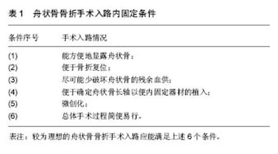
2.2.2 背侧入路 许多报道认为掌侧入路可能破坏腕舟状骨的血供从而影响骨折愈合。 Slade于2002年首次报道了背侧入路,此入路以lister结节为标志,于桡骨茎突背侧0.5 cm处沿前臂长轴作纵行切口,向两侧延长,依次切开皮肤、皮下组织及深筋膜,分离腕背侧伸肌,暴露骨折端。 近年来国内外较多文献报道采用该入路行经皮小切口加压螺钉置入术治疗腕舟状骨骨折。随着背侧入路手术的日臻成熟,近年来的研究显示背侧入路可更准确地将螺钉置于腕舟状骨的中轴线,而且对于腕舟状骨近1/3处的骨折或近极骨折,背侧入路从近极置入螺钉的方式更具有生物力学优势。 Garcia等[18]报道采用该入路治疗51例HerbertA2,B2和B3型腕舟状骨骨折,术后患者功能恢复至伤前的95%以上,认为对上述类型骨折的治疗尤为适用。 刘波等[19]对6例腕舟状骨骨折患者采用背侧入路小切口经皮加压螺钉内固定,其中包括HerbertB2型4例,B3型2例,患者术后腕关节活动度达到健侧的90%以上,且无疼痛不适等并发症。 Leppänen等[12]采用背侧入路方法治疗腕舟状骨骨不连,认为采用该入路加桡骨茎突骨移植是一种较好的选择,术后随访效果显著。 Cohen等[20]报道螺钉放置的最佳位置和长度是沿着腕舟状骨的纵轴线,并在近极和远极均位于关节面下 2 mm。目前的研究显示国人腕舟状骨长轴相对较短,而手术中螺钉生产厂家往往不能备齐全部长度型号,所以有时不得已而选择不合适长度的螺钉,往往会影响手术效果。与掌侧入路相比,背侧入路置入螺钉是一种相对较新的治疗技术,尤其是采用经皮小切口的微创方法对置入螺钉要求较高,操作难度较大。但是,背侧入路对血运的干扰是否比掌侧入路小,学术界存在不同观 点[21-23]。 2.2.3 桡腕侧入路 Ricardo等[24]报道了采用桡腕侧入路治疗腕舟状骨骨折,即在桡腕侧做与鼻烟窝长轴一致的纵行切口,并在腕横纹近侧向腕背侧弧形延长,以利于暴露腕舟状骨及桡骨茎突,切开皮肤和皮下组织,保护好桡神经浅支和桡动脉,向两侧翻开皮瓣即可暴露桡骨茎突,这种方法有利于桡骨茎突切除,并便于桡骨远端楔形截骨移植,术后随访发现无并发症发生。虽然这种手术平均减少了7°关节面的倾斜度,但这种减少并不影响腕关节的活动功能。 Kirkeby等[25]认为桡腕侧入路需和桡骨茎突切除移植联合应用,腕舟状骨在此种入路下位置较浅,暴露较容易,术野也较大,对治疗腕舟状骨骨折具有一定的优点。 2.2.4 关节镜手术入路 近年来微创技术的不断发展,尤其具有创伤小、痛苦少、操作简单、安全疗效好的特点引起人们的关注,人们对腕舟状骨骨折的微创治疗的兴趣也逐渐增加。 关节镜技术能够利用微创切口在直视下准确复位腕舟状骨骨折,可以最大限度地减少切开复位对血供的破坏和对软组织的损伤,且能够较早开始功能锻炼。 目前关节镜技术应用于腕部疾病治疗的研究重点主要集中在解剖学基础的研究、腕关节不稳及韧带损伤诊断与治疗、三角纤维软骨复合体损伤的诊断与治疗、关节软骨及滑膜疾病的诊断与治疗以及辅助与治疗桡骨远端粉碎性关节内骨折等几个方面。关节镜技术治疗腕舟状骨骨折报道目前国内外相关文献报道较少。 标准的腕关节镜入路大多为背侧入路,近年来亦有掌侧入路的文献报道[26]。 Geissler[27]利用关节镜技术联合经皮穿针技术治疗运动员腕舟状骨骨折,取得了良好疗效。 Korompilias[2]采用该技术联合空心加压螺钉治疗腕舟状骨骨折延迟愈合或不愈合,术中可使骨折处紧密贴合,在保护血供的基础上促进了骨折的愈合,临床效果明显。但关节镜手术技术含量较高,需要医师在熟练掌握切开复位手术技术和关节镜技术后方可开展。 国内肖颖锋等[28]在应用腕关节镜辅助下复位AO加压螺钉内固定治疗新鲜腕舟状骨14例骨折愈合疗效满意的报道,虽然腕关节镜技术要求较高,但相信随着目前科技的发展及微创技术的日臻完善,微创技术治疗腕舟状骨的前景是非常值得期待的,也相信会活动满意的疗效,使广大患者得到积极满意的治疗。 目前国内外学者在何种手术入路的效果更好这一方面仍存在很大争议,综合文献报道作者认为一个较为理想的舟状骨骨折手术入路应能满足6个条件(表1),可惜上述手术入路仍难以完全满足这6个条件,有待于临床上进一步探索。"

| [1] Markuszewski M, Kraus A, Studniarek M, et al NMR findings in patients after wrist trauma with a negative plain radiographs. Pol J Radiol. 2012;77(2):7-13.[2] Korompilias A, Lykissas M, Kostas-Agnantis I, et al An alternative graft fixation technique for scaphoid nonunions treated with vascular bone grafting. J Hand Surg Am. 2014;39(7):1308-1312.[3] Welling RD,Jacobson JA,Jamadar DA,et al.MDCT and radiography of wrist fractures: radiographic sensitivity and fracture patterns.AJR.2008;190(1):10-16[4] 丁双双,刘军平,卞国清,等.手腕舟状骨X 线DR投照体位探讨[J].放射学实践,2009,24(11):1262-1264. [5] Morin P, Reindl R, Berry GK, et al. Incorrect radiographic evaluation after vascularized bone grafting forscaphoid fracture or nonunion. Spring.2011;19(1):6-9.[6] (美)Greenspan A(主编). 唐光健(译). 骨放射学[M].3版.北京:中国医药科技出版社,2002:171-174.[7] Querellou S , Arnaud L, Williams T, et al. Role of SPECT/CT compared with MRI in the diagnosis and management of patients with wrist trauma occultfractures. Clin Nucl Med. 2014;39(1):8-13.[8] 郭阳,姜保国,田光磊,等.腕舟状骨螺旋CT影像最长轴的确立与测量[J].中华关节外科杂志,2010,4(2):227-232.[9] Ng A, Griffith JF, Taljanovic MS, et al. Is dynamic contrast-enhanced MRI useful for assessing proximal fragment vascularity in scaphoid fracturedelayed and non-union? Skeletal Radiol. 2013;42(7):983-992.[10] Senevirathna S, Rajeev A, Newby M. The value of delayed MRI scans in the assessment of acute wrist injuries. Acta Orthop Belg. 2013;79(3):275-279.[11] Ezoddini F, Zangoie M, Banadaki H, et al. Comparative study of the diagnostic value of panoramic and conventional radiography of the wrist in scaphoid fractures. Iran J Radiol. 2012;10(1):14-20.[12] Leppänen O, Karjalainen T. Treatment of the scaphoid fracture. Duodecim. 2013;129(21):2273-2279. [13] Freyschmidet J(主编).徐文军,刘吉华,肖德责(主译).骨放射学-正常与早期病理表现的界定[M]. 5版,济南:山东科学技术出版社,2005:159-160.[14] 洪加源,康两期,丁真奇等,逆行可吸收拉力螺钉内固定治疗腕舟骨骨折[J].中国骨伤,2009,22(1):822-823.[15] 章莹,尹庆水,许家军等,手舟骨微创内固定解剖学基础.中国临床解剖学杂志,2004,22(2):176-178.[16] Huang YC, Liu Y, Chen TH. Long-term results of scaphoid nonunion treated by intercalated bone grafting and Herbert’s screw fixation-a study of 49 patients for at least five years. Int Orthop. 2009;33(5):1295-1300. [17] Martin AG,Downing ND,Davis TR.Bone grafting of scaphoid non-unions:a simple distraction technique to optimise fracture exposure.Ann R Coll Surg Engl.2008;90(5):425-436.[18] Garcia R, Leversedge F, Aldridge J, et al. Scaphoid nonunions treated with 2 headless compression screws and bone grafting. J Hand Surg Am.2014;39(7):1301-1307.[19] 刘波,陈山林,田光磊.背侧入路经皮加压螺钉内固定治疗腕舟状骨骨折的临床研究[J].中华关节外科杂志,2010,4(2): 155-160. [20] Cohen M, Jupiter J, Fallahi K, et al. Scaphoid waist nonunion with humpback deformity treated without structural bone graft. J Hand Surg Am. 2013;38(4):701-705.[21] Adamani DC,Mikola EA,Fraser BJ.Percutaneous fixation of the scaphoid through a dorsal approach:an anatomic study.J Hand Surg Am.2008;33(3):327-331.[22] Bedi A,Jebson PJ,Hayden RJ,et al.Internal fixation of acute,nondisplaced scaphoid waist fractures via a limited dorsal approach:an assessment of radiographic and functional outcomes. J Hand Surg Am. 2007;32(3):326-333.[23] Parajuli NP, Shrestha D, Dhoju D,et al Scaphoid fracture: functional outcome following fixation with Herbert Screw. Kathmandu Univ Med J(KUMJ). 2011; 9(36): 267-273.[24] Ricardo Monreal.treatment of scaphoid nonunions with closed-wedge osteotomy of the distal radius:report of six cases.Hand Surg Am.2008;3(2):91-95.[25] Kirkeby L, Kairelyte V, Hansen TB. Early magnetic resonance imaging in patients with a clinically suspected scaphoid fracture may identify occult wrist injuries. J Hand Surg Eur Vol. 2013;38(5):571-572. [26] 欧阳侃,王大平,陆伟,等.腕关节镜掌侧入路的临床应用[J].中国临床解剖学杂志,2009,27(6):731-734. [27] Geissler WB.Management of scaphoid fractures in the athlete.Operative Techniques in Sports Medicine.2006;14(2): 101-107.[28] 肖颖锋,万圣祥,王拥军等,腕关节镜辅助下复位加压螺钉内固定治疗舟骨骨折[J].齐齐哈尔医学院学报,2006,27(11):1338-1339. |
| [1] | Zhang Jian, Lin Jianping, Zhou Gang, Fang Yehan, Wang Benchao, Wu Yongchang. Semi-quantitative MRI evaluation of cartilage degeneration in early knee osteoarthritis [J]. Chinese Journal of Tissue Engineering Research, 2022, 26(3): 425-429. |
| [2] | Deng Zhaoyang, Yang Zhaohui. Three-dimensional model analysis of tibial plateau fracture lines [J]. Chinese Journal of Tissue Engineering Research, 2022, 26(3): 334-339. |
| [3] | Xie Jingshu, Zhang Xianglin, Liu Jinlei, Wen Jing. Application of High Resolution reconstruction algorithm in precision CT scans of the middle and inner ears [J]. Chinese Journal of Tissue Engineering Research, 2021, 25(23): 3614-3618. |
| [4] | Cao Bin, Zuo Yuqiang, Du Hanyang, Yu Haiquan, Su Jingyang, Meng Haoyong. Correlation of saggital parameters of cervical and thoracic junction areas in asymptomatic adults [J]. Chinese Journal of Tissue Engineering Research, 2020, 24(33): 5354-5357. |
| [5] | Liu Junyan, Pan Shinong. Anatomical changes and imaging manifestations of childhood developmental dysplasia of the hip [J]. Chinese Journal of Tissue Engineering Research, 2020, 24(30): 4875-4881. |
| [6] | Wang Wei, Jia Yujie, Cai Hongmei, Wang Wenjuan, Sun Cuicui. Functional patch combined with surface electromyographic biofeedback for post-stroke dysphagia [J]. Chinese Journal of Tissue Engineering Research, 2020, 24(29): 4697-4701. |
| [7] | Zhang Shuai, Ouyang Jianyuan, Peng Xuelian, Wang Song, Wang Qing. Three-dimensional computed tomography evaluation of L5 pedicle screw fixation shielding by iliac wing width and height [J]. Chinese Journal of Tissue Engineering Research, 2020, 24(18): 2873-2878. |
| [8] | Luo Xiaofei, Wei Xuan, Wang Jinliang, Wang Shaohua, Li Zhe, Bai Yu. Ultrastructural changes of tibial subchondral bone in patients with knee osteoarthritis: CT evaluation [J]. Chinese Journal of Tissue Engineering Research, 2020, 24(15): 2399-2404. |
| [9] | Wang Liang, Li Lijun, Zhu Fuliang, Jiang Zhuyan, Wang Shuai, Ni Dongkui . Cortical bone trajectory screw versus pedicle screw fixation after posterior lumbar interbody fusion: a meta-analysis [J]. Chinese Journal of Tissue Engineering Research, 2019, 23(8): 1291-1298. |
| [10] | Han Quan, Xiong Jian, Wang Yanhua, Dang Yu, Zhang Peixun, Fu Zhongguo, Zhang Dianying, Wang Tianbing. Minimally invasive treatment of acute scaphoid fracture with titanium hollow compression screw [J]. Chinese Journal of Tissue Engineering Research, 2019, 23(34): 5413-5417. |
| [11] | Guo Dongmei1, She Fang2, Xie Qi2. Evaluation of the number of roots and root canal morphology and distribution in Chinese children's mandibular first deciduous teeth by cone-beam computed tomography [J]. Chinese Journal of Tissue Engineering Research, 2019, 23(27): 4265-4268. |
| [12] | Luo Yi, Wang Enqun, Xu Yating, Ren Wei . Modified platelet-rich fibrin repairs distal bone defect of adjacent teeth after mandibular wisdom tooth extraction [J]. Chinese Journal of Tissue Engineering Research, 2019, 23(15): 2314-2319. |
| [13] | Gu Jianhua, Shi Zhicai . Placement position of artificial vertebral pedicle screws based on the anatomical structure of lumbar nerve roots [J]. Chinese Journal of Tissue Engineering Research, 2019, 23(12): 1829-1833. |
| [14] | Tian Youyong, Wang Zhiyong. Short-term follow-up of dynamic hip screw versus proximal femoral nail anti-rotation for type AO/OTA A1 intertrochanteric femoral fracture in older adults [J]. Chinese Journal of Tissue Engineering Research, 2019, 23(12): 1834-1839. |
| [15] | Wang Xiji, Zhang Yongyuan, Yang Ruize, Hao Dingjun, Sun Honghui. Accuracy and clinical efficacy of three-dimensional printing and navigation technology assisted lumbar cortical bone trajectory screw placement [J]. Chinese Journal of Tissue Engineering Research, 2019, 23(12): 1864-1869. |
| Viewed | ||||||
|
Full text |
|
|||||
|
Abstract |
|
|||||