Chinese Journal of Tissue Engineering Research ›› 2014, Vol. 18 ›› Issue (32): 5166-5172.doi: 10.3969/j.issn.2095-4344.2014.32.014
Previous Articles Next Articles
Growth characteristics of umbilical cord-derived versus embryonic liver-derived mesenchymal stem cells
Bi Wei-wei, He Li-rong, Nian Li-li
- Jilin Province TaHua Biological Technology Co., Ltd., Siping 136000, Jilin Province, China
-
Received:2014-06-26Online:2014-08-06Published:2014-09-18 -
Contact:Bi Wei-wei, M.D., Chief technician, Jilin Province TaHua Biological Technology Co., Ltd., Siping 136000, Jilin Province, China -
About author:Bi Wei-wei, M.D., Chief technician, Jilin Province TaHua Biological Technology Co., Ltd., Siping 136000, Jilin Province, China
CLC Number:
Cite this article
Bi Wei-wei, He Li-rong, Nian Li-li. Growth characteristics of umbilical cord-derived versus embryonic liver-derived mesenchymal stem cells[J]. Chinese Journal of Tissue Engineering Research, 2014, 18(32): 5166-5172.
share this article
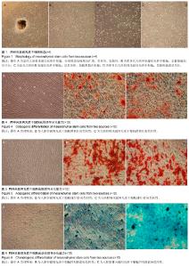
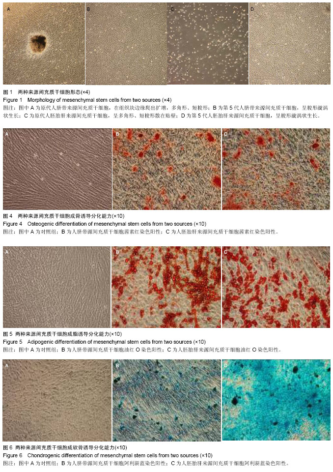
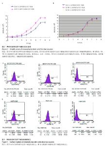
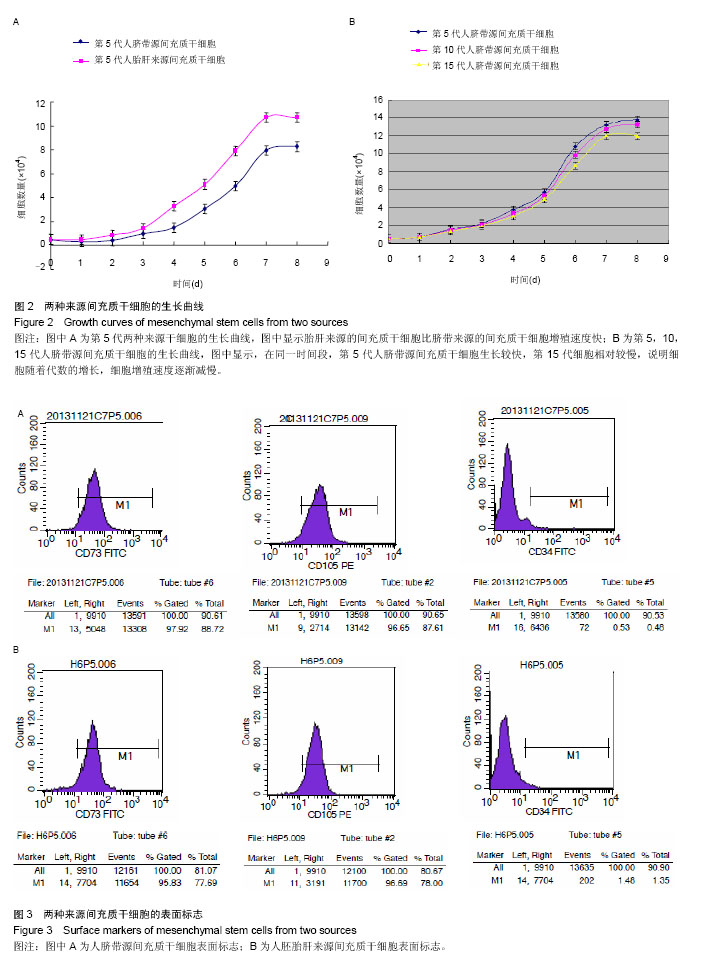
2.1 两种来源间充质干细胞形态 细胞培养过程中,随时观察照相。两种来源间充质干细胞生长形态基本相同。原代培养中均为多角形、短梭形,散在贴壁生长。传代培养过程中细胞增殖并逐渐变为长梭形,呈漩涡状生长(图1)。 2.2 两种来源间充质干细胞的生长曲线 第5代胎肝来源的间充质干细胞比脐带来源的间充质干细胞增殖速度快(图2A)。在同一时间段,第5代人脐带源间充质干细胞生长较快,第15代细胞相对较慢,说明细胞随着代数的增长,细胞增殖速度逐渐减慢(图2B)。 2.3 两种来源间充质干细胞的倍增时间 第5代人胚胎肝来源间充质干细胞的倍增时间为(34.37±0.31) h,第5代人脐带源间充质干细胞的倍增时间为(35.63±0.38) h,两者比较差异无显著性意义(P > 0.05)。 第5,10,15代人脐带源间充质干细胞的倍增时间分别为(35.63±0.38) h,(52.60±0.53) h,(53.27±0.92) h,第5代与第10代和第15代比较,差异有显著性意义(P < 0.05),第10代与第15代比较差异无显著性意义(P > 0.05)。 2.4 流式细胞术鉴定结果 流式细胞术检测两种来源第5代间充质干细胞表面CD73,CD105表达阳性,大于95%,而CD34表达阴性,小于2%(图3)。 2.5 细胞分化能力检测 成骨诱导:两种来源第5代间充质干细胞经成骨诱导14-21 d后,经茜素红染色,可见细胞爬片上出现红色钙结节(图4)。 成脂诱导:诱导14-21 d后,经油红O染色出现红色脂滴(图5)。 成软骨诱导:诱导第15天时,经阿莉新蓝染色可见软骨胶原基质被染成蓝色(图6)。"
"
| [1] | Chen Jianquan, Chen Maoshui, Lyu Zhouming, Chen Rongbin, Yu Zhaoyu, Liu Wanpeng, Lin Xinyuan, Lin Dingkun. Biomechanical properties of cortical bone trajectory combined with pedicle screw fixation on vertebral body motion unit: a finite element analysis [J]. Chinese Journal of Tissue Engineering Research, 2023, 27(22): 3457-3462. |
| [2] | Qiu Hao, Zhu Yun, Weng Zheng, Liu Dun, Chen Shimou, Jin Guorong, Chen Yu. Application of tranexamic acid in elderly patients with femoral intertrochanteric fracture [J]. Chinese Journal of Tissue Engineering Research, 2023, 27(22): 3550-3554. |
| [3] | Duan Jiahao, Tan Xuyi, Lu Min, Kuang Gaoyan, Fang Chuanlong, Xiao Man. Effects of Sanhua Jiegu San on Wnt/beta-catenin signaling pathway in osteoblasts [J]. Chinese Journal of Tissue Engineering Research, 2023, 27(20): 3230-3235. |
| [4] | Sun Fangyuan, Meng Jialei, Ma Yuhui, Geng Huan, Zhang Tao. Effects of ligustrazine on inflammation and oxidative stress in rat cardiomyocytes induced by lipopolysaccharide [J]. Chinese Journal of Tissue Engineering Research, 2023, 27(20): 3253-3258. |
| [5] | Xu Zhi, Li Yuwan, Jin Ying, Liu Yi. Effect of partial posterior root tear of medial meniscus on biomechanics of the knee joint during gait cycle [J]. Chinese Journal of Tissue Engineering Research, 2023, 27(18): 2824-2830. |
| [6] | Gao Xu, Xing Wenhua. Application of finite element analysis in spine surgery [J]. Chinese Journal of Tissue Engineering Research, 2023, 27(18): 2921-2927. |
| [7] | Gao Yue, Lin Jianwen, Li Di, Lan Xiaoyan, Li Shen, Chu Chengyan. Oligodendrocyte progenitor cells prolong the survival of glioblastoma-bearing rats after high-dose radiotherapy [J]. Chinese Journal of Tissue Engineering Research, 2023, 27(17): 2669-2674. |
| [8] | Li Jingna, Zeng Qingfeng, Yu Shuyin, Qin Yue, Ai Zizheng, Dong Xieping. Preparation and properties of porous hydroxyapatite scaffolds with biphase calcium phosphate coating [J]. Chinese Journal of Tissue Engineering Research, 2023, 27(16): 2493-2500. |
| [9] | Zhou Lanxi, Shao Lu, Dong Shiwu, Yu Zhengwen. Molecular mechanism of angiogenesis promoted by medical metal materials [J]. Chinese Journal of Tissue Engineering Research, 2023, 27(16): 2616-2624. |
| [10] | Ling Xuwei, Sun Jie, Liu Chang, Wang Yi, Shi Qin, Yang Huilin. Valproic acid promotes osteogenic differentiation of rat bone marrow mesenchymal stem cells [J]. Chinese Journal of Tissue Engineering Research, 2023, 27(15): 2304-2310. |
| [11] | Tang Jianhong, Zhang Nini, Huang Guilin, Long Yuanzhu, Cui Tianning, Luo Qinliang, Lang Jiachan, Dai Min, Zhang Ligang. Biological characteristics of human amniotic mesenchymal stem cells pretreated with different volume fractions of oxygen [J]. Chinese Journal of Tissue Engineering Research, 2023, 27(15): 2318-2324. |
| [12] | He Zike, Wang Shangzeng. Eucommia ulmoides Oliver aqueous extract promotes bone marrow mesenchymal stem cell proliferation and osteoblastic differentiation through upregulating Nur77 protein expression [J]. Chinese Journal of Tissue Engineering Research, 2023, 27(15): 2371-2378. |
| [13] | Li Ran, Jia Hongling, Zhang Chunxiao, Zhao Ruoxi, Dong Jingwen, Cheng Yixin, Cui Wenzhe, Zhang Jing. Role of exosomes and miRNAs in the mechanism, diagnosis and treatment of premature ovarian insufficiency [J]. Chinese Journal of Tissue Engineering Research, 2023, 27(15): 2453-2460. |
| [14] | Ye Haimin, Zou Huachun, Ding Linghua, You Murong. Finite element analysis of hollow screw fixation for sacroiliac dislocation [J]. Chinese Journal of Tissue Engineering Research, 2023, 27(13): 1993-1998. |
| [15] | Wang Shangzeng, Wang Zhen, You Mingcan, Ma Shang, Shen Jintao. Acetabular wear after femoral head replacement: cause and management [J]. Chinese Journal of Tissue Engineering Research, 2023, 27(13): 2028-2032. |
| Viewed | ||||||
|
Full text |
|
|||||
|
Abstract |
|
|||||