Chinese Journal of Tissue Engineering Research ›› 2020, Vol. 24 ›› Issue (30): 4841-4846.doi: 10.3969/j.issn.2095-4344.2846
Previous Articles Next Articles
Radiomics for diagnostic value of osteoporosis in female patients based on lumbar magnetic resonance imaging
He Li, Liu Zhai, Gao Zhimei, Liu Chunying, Zhao Junlu, Ren Qingyun
- Department of Radiology, First Hospital of Hebei Medical University, Shijiazhuang 050031, Hebei Province, China
-
Received:2020-01-10Revised:2020-01-16Accepted:2020-03-06Online:2020-10-28Published:2020-09-19 -
Contact:Ren Qingyun, Chief physician, Professor, Department of Radiology, First Hospital of Hebei Medical University, Shijiazhuang 050031, Hebei Province, China -
About author:He Li, Master, Associate chief physician, Associate professor, Department of Radiology, First Hospital of Hebei Medical University, Shijiazhuang 050031, Hebei Province, China -
Supported by:the Key Research Plan Project of Hebei Province, No. 182777126D
CLC Number:
Cite this article
He Li, Liu Zhai, Gao Zhimei, Liu Chunying, Zhao Junlu, Ren Qingyun. Radiomics for diagnostic value of osteoporosis in female patients based on lumbar magnetic resonance imaging[J]. Chinese Journal of Tissue Engineering Research, 2020, 24(30): 4841-4846.
share this article

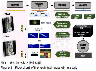
基于独立MRI序列及T1WI+T2WI联合序列组学特征建立的建立3个模型,分别为T1WI、T2WI、T1WI+T2WI模型。将年龄和性别临床因素纳入联合模型3(radiomics_female)而建立的临床影像组学模型4(radiomics+clinical_female)。纳入研究的50例样本均做为训练集来建立模型。每个模型采用5折交叉验证的平均值(mean Predicting accuracy)对模型的诊断性能进行评估,避免模型过拟合。模型评价指标为接受者操作特征曲线下面积、灵敏度、特异度和准确度,模型选择依据决策曲线的标准净收益的大小。研究的技术路线如图1所示。 "

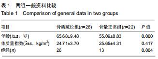
2.1 受试者一般情况 纳入的50例受试者中,骨质疏松28例,骨量正常22例。骨质疏松组与骨量正常组体质量指数比较差异无显著性意义(P > 0.05),两组年龄、绝经情况比较差异有显著性意义(P < 0.05),见表1。 "
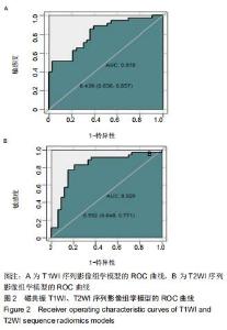
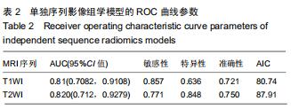
2.2 特征降维及单个序列诊断模型的建立 每个病例图像得到396×3个特征值,经过特征降维, T1WI序列筛选出4个特征,T2WI序列筛选出6个特征,T1WI+T2WI联合序列筛选出4个特征。lasso模型采用十折交叉验证降维。使用T1WI及T2WI序列筛选出来的特征建立逻辑回归模型,绘制2独立序列模型的ROC曲线,见图2。T1WI模型AUC值为0.810,T2WI模型AUC值为0.820,见表2。 "


2.2 特征降维及单个序列诊断模型的建立 每个病例图像得到396×3个特征值,经过特征降维, T1WI序列筛选出4个特征,T2WI序列筛选出6个特征,T1WI+T2WI联合序列筛选出4个特征。lasso模型采用十折交叉验证降维。使用T1WI及T2WI序列筛选出来的特征建立逻辑回归模型,绘制2独立序列模型的ROC曲线,见图2。T1WI模型AUC值为0.810,T2WI模型AUC值为0.820,见表2。 "
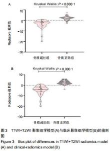
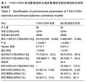
根据组学标签公式计算出的Radscore值在组间分布具有统计学差异(P < 0.000 1,Kruskal Wallis test),即Radscore值是进行骨质疏松症鉴别和预测的显著因素,根据Radscore的截断值(Optimal criterion)可将病患分为患骨质疏松的高风险组和低风险组(图3)。联合模型及临床影像组学联合模型的ROC曲线见图4,模型诊断性能参数见表3,可以看出在模型中加入临床特征能以显著提高模型的AUC值。此外加入临床特征后,模型的灵敏度和准确度也有提高,但是会降低模型的特异度。 "
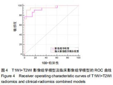
根据组学标签公式计算出的Radscore值在组间分布具有统计学差异(P < 0.000 1,Kruskal Wallis test),即Radscore值是进行骨质疏松症鉴别和预测的显著因素,根据Radscore的截断值(Optimal criterion)可将病患分为患骨质疏松的高风险组和低风险组(图3)。联合模型及临床影像组学联合模型的ROC曲线见图4,模型诊断性能参数见表3,可以看出在模型中加入临床特征能以显著提高模型的AUC值。此外加入临床特征后,模型的灵敏度和准确度也有提高,但是会降低模型的特异度。 "

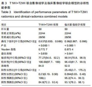
根据组学标签公式计算出的Radscore值在组间分布具有统计学差异(P < 0.000 1,Kruskal Wallis test),即Radscore值是进行骨质疏松症鉴别和预测的显著因素,根据Radscore的截断值(Optimal criterion)可将病患分为患骨质疏松的高风险组和低风险组(图3)。联合模型及临床影像组学联合模型的ROC曲线见图4,模型诊断性能参数见表3,可以看出在模型中加入临床特征能以显著提高模型的AUC值。此外加入临床特征后,模型的灵敏度和准确度也有提高,但是会降低模型的特异度。 "
|
[1] 马远征,王以朋,刘强,等.中国老年骨质疏松症诊疗指南(2018)[J].中华健康管理学杂志,2018,12(6):484-489.
[2] LINK TM. Osteoporosis imaging: state of the art and advanced imaging.Radiology. 2012;263(1):3-17.
[3] HOFBAUER LC, RACHNER TD. More DATA to guide sequential osteoporosis therapy.Lancet.2015;386:1116-1118.
[4] PISANI P, RENNA MD, CONVERSANO F, et al. Major osteoporotic fragility fractures: Risk factor updates and societal impact.World J Orthop.2016;7(3):171-181.
[5] 韩亚军,帖小佳,伊力哈木·托合提.中国中老年人骨质疏松症患病率的Meta分析[J].中国组织工程研究,2014,18(7): 1129-1134.
[6] MILLER PD. Underdiagnosis and Undertreatment of Osteoporosis: The Battle to Be Won. J Clin Endocrinol Metab. 2016;101(3):852-859.
[7] SAAD MM, AHMED TA, MOHAMED KE, et al. Role of lumbar spine signal intensity measurement by MRI in the diagnosis of osteoporosis in post-menopausal women. Egyptian Journal of Radiology and Nuclear Medicine.2019;50(1):35-42.
[8] BANDIRALI M, DI LEO G, PAPINI GD, et al.A new diagnostic score to detect osteoporosis in patients undergoing lumbar spine MRI.Eur Radiol.2015;25(10):2951-2959.
[9] KUMAR V, GU Y, BASU S, et al. Radiomics: the process and the challenges.MagnReson Imaging.2012;30(9):1234-1248.
[10] E L, LU L, LI L, et al. Radiomics for Classification of Lung Cancer Histological Subtypes Based on Nonenhanced Computed Tomography.Acad Radiol.2019;26(9):1245-1252.
[11] HORVAT N, VEERARAGHAVAN H, KHAN M, et al. MR Imaging of Rectal Cancer: Radiomics Analysis to Assess Treatment Response after Neoadjuvant Therapy.Radiology. 2018;287(3):833-843.
[12] 吴亚平,刘博,顾建钦,等.基于影像组学的脑胶质瘤分级方法[J].中华放射学杂志,2017,51(12):902-905.
[13] 袁清玉,江玉明,吕闻冰,等.基于18F-FDG PET/CT图像的影像组学列线图对胃癌术后的预后评估[J].中华核医学与分子影像杂志, 2019,39(1):2-5.
[14] 王超,刘侠,董迪,等.基于影像组学的非小细胞肺癌淋巴结转移预测[J].自动化学报,2019,45(6):1087-1093.
[15] MACKAY JW, MURRAY PJ, KASMAI B, et al. Subchondral bone in osteoarthritis: association between MRI texture analysis and histomorphometry.Osteoarthritis Cartilage. 2017;25(5):700-707.
[16] AREECKAL AS, JAYASHEELAN N, KAMATH J, et al. Early diagnosis of osteoporosis using radiogrammetry and texture analysis from hand and wrist radiographs in Indian population. Osteoporos Int.2018;29(3):665-673.
[17] TABARI A, TORRIANI M, MILLER KK, et al. Anorexia Nervosa: Analysis of Trabecular Texture with CT.Radiology. 2017;283(1):178-185.
[18] KANIS JA. Assessment of fracture risk and its application to screening for postmenopausal osteoporosis: synopsis of a WHO report. WHO Study Group.Osteoporos Int. 1994;4(6): 368-381.
[19] 王平,和建伟,黄刚,等.应用双能CT与定量CT对椎体骨密度测量的对照研究[J].中国骨质疏松杂志,2017,23(2):159-162.
[20] DEYO RA, WEINSTEIN JN. Low back pain. N Engl J Med. 2001;344(5):363-370. [21] MANENTI G, CAPUANI S, FUSCO A, et al.Osteoporosis detection by 3T diffusion tensor imaging and MRI spectroscopy in women older than 60 years.Aging Clin Exp Res.2013;25 Suppl 1:S31-S34.
[22] LI GW, XU Z, CHEN QW, et al. Quantitative evaluation of vertebral marrow adipose tissue in postmenopausal female using MRI chemical shift-based water-fat separation.Clin Radiol.2014;69(3):254-262.
[23] 何杰,方浩,李晓娜,等.腰椎MR扩散加权成像对骨质疏松的定量诊断价值[J].临床放射学杂志,2015(5):95-99.
[24] GRIFFITH JF, YEUNG DK, ANTONIO GE, et al. Vertebral bone mineral density, marrow perfusion, and fat content in healthy men and men with osteoporosis: dynamic contrast-enhanced MR imaging and MR spectroscopy. Radiology. 2005;236(3):945-951.
[25] 常荣,张红,刘正华,等.MRI m-Dixon-Quant技术快速评估中老年骨质疏松症的可行性研究[J].实用放射学杂志,2018,34(12): 1908-1911.
[26] 郭志,许崇永.IDEAL-IQ技术在2型糖尿病性骨质疏松症诊断中的应用价值[J].中国中西医结合影像学杂志,2019,17(5): 489-492.
[27] SCHWARTZ AV. Marrow fat and bone: review of clinical findings.Front Endocrinol (Lausanne).2015;6:40.
[28] QIU X, FU Y, CHEN J, et al.The Correlation between Osteoporosis and Blood Circulation Function Based on Magnetic Resonance Imaging. J Med Syst.2019;43(4):91.
[29] BURIAN E, SUBBURAJ K, MOOKIAH MRK, et al.Texture analysis of vertebral bone marrow using chemical shift encoding-based water-fat MRI: a feasibility study. Osteoporos Int.2019;30(6):1265-1274.
[30] 翁子敬,黄学菁,俞健力,等.常规MRI信号评价腰椎骨质疏松的意义[J].中国组织工程研究,2018,22(35):5667-5673.
[31] LECLER A, DURON L, BALVAY D, et al.Combining Multiple Magnetic Resonance Imaging Sequences Provides Independent Reproducible Radiomics Features.Sci Rep. 2019;9(1):2068.
[32] 刘伯亮,潘万敏.骨质疏松发生与年龄、性别的关系:5200例分析[J].中国临床康复,2004,8(33):139-140.
[33] BLACK DM, ROSEN CJ. Clinical Practice. Postmenopausal Osteoporosis.N Engl J Med. 2016;374(3):254-262. [34] FERIZI U, BESSER H, HYSI P, et al.Artificial Intelligence Applied to Osteoporosis: A Performance Comparison of Machine Learning Algorithms in Predicting Fragility Fractures From MRI Data.J MagnReson Imaging. 2019;49(4): 1029-1038. |
| [1] | Min Youjiang, Yao Haihua, Sun Jie, Zhou Xuan, Yu Hang, Sun Qianpu, Hong Ensi. Effect of “three-tong acupuncture” on brain function of patients with spinal cord injury based on magnetic resonance technology [J]. Chinese Journal of Tissue Engineering Research, 2021, 25(在线): 1-8. |
| [2] | Xu Feng, Kang Hui, Wei Tanjun, Xi Jintao. Biomechanical analysis of different fixation methods of pedicle screws for thoracolumbar fracture [J]. Chinese Journal of Tissue Engineering Research, 2021, 25(9): 1313-1317. |
| [3] | Jiang Yong, Luo Yi, Ding Yongli, Zhou Yong, Min Li, Tang Fan, Zhang Wenli, Duan Hong, Tu Chongqi. Von Mises stress on the influence of pelvic stability by precise sacral resection and clinical validation [J]. Chinese Journal of Tissue Engineering Research, 2021, 25(9): 1318-1323. |
| [4] | Zhang Tongtong, Wang Zhonghua, Wen Jie, Song Yuxin, Liu Lin. Application of three-dimensional printing model in surgical resection and reconstruction of cervical tumor [J]. Chinese Journal of Tissue Engineering Research, 2021, 25(9): 1335-1339. |
| [5] | Zhang Yu, Tian Shaoqi, Zeng Guobo, Hu Chuan. Risk factors for myocardial infarction following primary total joint arthroplasty [J]. Chinese Journal of Tissue Engineering Research, 2021, 25(9): 1340-1345. |
| [6] | Wei Wei, Li Jian, Huang Linhai, Lan Mindong, Lu Xianwei, Huang Shaodong. Factors affecting fall fear in the first movement of elderly patients after total knee or hip arthroplasty [J]. Chinese Journal of Tissue Engineering Research, 2021, 25(9): 1351-1355. |
| [7] | Wang Jinjun, Deng Zengfa, Liu Kang, He Zhiyong, Yu Xinping, Liang Jianji, Li Chen, Guo Zhouyang. Hemostatic effect and safety of intravenous drip of tranexamic acid combined with topical application of cocktail containing tranexamic acid in total knee arthroplasty [J]. Chinese Journal of Tissue Engineering Research, 2021, 25(9): 1356-1361. |
| [8] | Xiao Guoqing, Liu Xuanze, Yan Yuhao, Zhong Xihong. Influencing factors of knee flexion limitation after total knee arthroplasty with posterior stabilized prostheses [J]. Chinese Journal of Tissue Engineering Research, 2021, 25(9): 1362-1367. |
| [9] | Huang Zexiao, Yang Mei, Lin Shiwei, He Heyu. Correlation between the level of serum n-3 polyunsaturated fatty acids and quadriceps weakness in the early stage after total knee arthroplasty [J]. Chinese Journal of Tissue Engineering Research, 2021, 25(9): 1375-1380. |
| [10] | Zhang Chong, Liu Zhiang, Yao Shuaihui, Gao Junsheng, Jiang Yan, Zhang Lu. Safety and effectiveness of topical application of tranexamic acid to reduce drainage of elderly femoral neck fractures after total hip arthroplasty [J]. Chinese Journal of Tissue Engineering Research, 2021, 25(9): 1381-1386. |
| [11] | Yao Rubin, Wang Shiyong, Yang Kaishun. Minimally invasive transforaminal lumbar interbody fusion for treatment of single-segment lumbar spinal stenosis improves lumbar-pelvic balance [J]. Chinese Journal of Tissue Engineering Research, 2021, 25(9): 1387-1392. |
| [12] | Wang Haiying, Lü Bing, Li Hui, Wang Shunyi. Posterior lumbar interbody fusion for degenerative lumbar spondylolisthesis: prediction of functional prognosis of patients based on spinopelvic parameters [J]. Chinese Journal of Tissue Engineering Research, 2021, 25(9): 1393-1397. |
| [13] | Lü Zhen, Bai Jinzhu. A prospective study on the application of staged lumbar motion chain rehabilitation based on McKenzie’s technique after lumbar percutaneous transforaminal endoscopic discectomy [J]. Chinese Journal of Tissue Engineering Research, 2021, 25(9): 1398-1403. |
| [14] | Chen Xinmin, Li Wenbiao, Xiong Kaikai, Xiong Xiaoyan, Zheng Liqin, Li Musheng, Zheng Yongze, Lin Ziling. Type A3.3 femoral intertrochanteric fracture with augmented proximal femoral nail anti-rotation in the elderly: finite element analysis of the optimal amount of bone cement [J]. Chinese Journal of Tissue Engineering Research, 2021, 25(9): 1404-1409. |
| [15] | Du Xiupeng, Yang Zhaohui. Effect of degree of initial deformity of impacted femoral neck fractures under 65 years of age on femoral neck shortening [J]. Chinese Journal of Tissue Engineering Research, 2021, 25(9): 1410-1416. |
| Viewed | ||||||||||||||||||||||||||||||||||||||||||||||||||
|
Full text 147
|
|
|||||||||||||||||||||||||||||||||||||||||||||||||
|
Abstract |
|
|||||||||||||||||||||||||||||||||||||||||||||||||