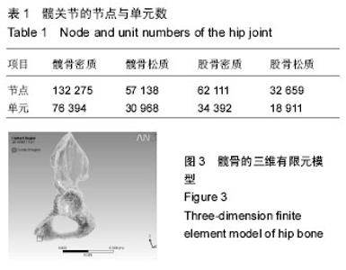
| [1] 张轩轩,王钢,赵辉,等.经皮内固定技术治疗骨盆前环骨折的现状[J].中华创伤骨科杂志,2012,14(5):444-446.
[2] 吴锦隆. 骨盆骨折的流行病学研究[J].中国急救医学, 2015,35(6):518-521.
[3] 赵岩,江建明,李筱贺,等.中下胸椎肋椎单元三维有限元模型的建立[J].中国组织工程研究,2012,16(22):3996-4000.
[4] 樊黎霞,丁光兴,费王华,等.基于CT图像的长管骨有限元材料属性研究及实验验证[J].医用生物力学, 2012,27 (12): 102-108.
[5] 张华,赵鹏,曾昭洋,等. 成人股骨中下段的解剖学与扁弧形长针的设计应用[J]. 解剖学杂志, 2009,32(6):806-808.
[6] 蔡兵,于沈敏,林文,等.人工肱骨头假体柄界面剪切应力的生物力学研究[J].创伤外科学杂志,2013,15(3):234-236.
[7] 刘耀升,陈其昕,廖胜辉,等. 腰椎L4-L5活动节段有限元模型的建立与验证[J]. 第二军医大学学报, 2006, 27(6): 665-669.
[8] 闵飞炎,杨明,王子才. 仿真模型的智能化验证方法[J]. 大连海事大学学报,2010, 36(2):59-64.
[9] 宋承龄.关于仿真模型验证[J]. 计算机仿真, 2000, 17(4): 8-11.
[10] 汤洋,章云童,张春才,等. 髋臼后壁解剖学测量及定量分析[J].中国骨伤,2014,27(12):1024-1028.
[11] 周海,王燎,王金武,等. 人工髓关节脱位失效的生物力学分析与推理[J]. 医用生物力学,2012,27(1):13-20.
[12] 马文辉,张学敏,王继芳. 髋臼缺损有限元模型的建立及力学分析[J]. 中国组织工程研究与临床康复,2010, 14(43):8004-8007.
[13] 邬培慧,傅明,康焱. 髋臼CE角及关节作用力方向的生物力学作用[J].第三军医大学学报,2011,33(11):1174-1177.
[14] 付朝江. 随机有限元分析的二级区域分解并行求解算法[J]. 应用力学学报, 2012,29(4):475-480.
[15] 袁平,王万春. 膝关节三维有限元模型的建立及生物力学分析[J].中南大学学报(医学版),2010, 35(1):85-89.
[16] 徐诗雄,洪顺红,孙文栋. 老年股骨粗隆间骨折术后髋关节功能恢复的影响因素[J].中国老年学杂志,2014, 34(12):3353-3355.
[17] 聂涌,马俊,胡钦胜,等. 髋臼假体放置角度对髋臼周围应力分布的影响[J]. 医用生物力学,2014,29(4):1-7.
[18] 汤洋,胡小鹏,陆雄伟,等. 髓臼后壁骨折固定的生物力学研究[J]. 中国修复重建外科杂志,2015,29(8):925-930.
[19] 陈海南. 骨性关节炎软骨下骨三维结构和力学性能改变及早期干预的实验研究[D].苏州大学,2013.
[20] 叶臻,李民,陈定家.骨关节炎软骨下骨的微结构改变[J]. 中国骨质疏松杂志,2016,22(5):624-627.
[21] 江岩,褚立希. 运动对膝骨关节炎的作用机制探讨[A]. 2015第十届全国体育科学大会论文摘要汇编(三) [C],2015.
[22] 冯宁. 不同负荷跑步运动对生长期大鼠股骨骨密度和生物力学性能的影响[A]. 2015第十届全国体育科学大会论文摘要汇编(二) [C],2015.
[23] 姜维浩,苏秀云,刘耀升,等. 三维有限元分析椎体联合后部结构损伤胸椎转移瘤的生物力学特性[J]. 中国组织工程研究,2016,20(13):1925-1931.
[24] 史强,李旭.儿童继发性骨质疏松症病因学研究进展[J]. 中国骨质疏松杂志,2014,20(5):584-588.
[25] Schlecht SH, Pinto DC, Agnew AM, et al. Brief Communication:the effects of disuse on the mechanical properties of bone: what unloading tells us about the adaptive nature of skeletal tissue. Am J Phys Anthropol. 2012;149:599-605.
[26] 李霞. 生物补片材料进展与运动性跟腱断裂的修复[J]. 中国组织工程研究与临床康复,2008,12(36):7159-7162.
[27] 王猛.改善老年人肺功能的日常简易锻炼方法[J]. 中国老年学杂志,2015,35(22):6605-6606.
[28] 邱长茂. 髋臼骨折术后股骨头内移对臼顶负重区应力影响的有限元分析[D].天津医科大学,2015.
[29] 李宁远,龚亚莉,刘煊文,等.不同材料人工髋关节假体对骨界面应力分布及生物力学的影响[J]. 中国组织工程研究, 2016,20(9):1268-1274.
[30] 聂涌,马俊,康鹏德,等. 正常步态周期中髋臼周围区域的应力分布及其在THA髋臼重建中的指导[J]. 医用生物力学,2014,29(1):31-37.
[31] 段金凤.实施FMEA模式管理预防髋关节脱位[J].临床医学工程,2013,20(8):1011-1012.
[32] 赖晓荣,朱欣娟.全程规范化护理在全髋关节置换术患者的应用[J]. 南昌大学学报(医学版),2013,53(6):59-61.
[33] 高泓一.K-L入路切开复位内固定治疗髋臼骨折伴股骨头脱位的临床疗效[J].广东微量元素科学,2016,23(3):60-63.
[34] 新苏雅拉图,吕福润,杨存虎,等. 手术治疗髋臼前柱合并前壁骨折的临床疗效[J]. 中国矫形外科杂志,2014, 22(6):548-551.
[35] 李力更,安志刚,郑煦光,等.198例复杂髋臼骨折的于术治疗[J].中国矫形外科杂志,2010,18(18):1570-1572.
[36] 邱少东,王拯,朱涛.影响髋臼骨折手术预后的因素分析[J]. 中国矫形外科杂志,2016,24(10):884-888.
[37] 倪建龙,王坤正,党晓谦.全髋关节置换术治疗髋臼骨折后创伤性关节炎的疗效观察[J].中华关节外科杂志(电子版), 2014,8(4):456-46.
[38] 李广彬,陈秀民,王在斌,等. 髋臼内壁截骨全髋关节置换术治疗髋关节发育不良[J].实用骨科杂志, 2016,22(4): 305-309.
[39] Hopley C, Stengel D, Ekkernkamp A, et al. Primary total hip arthroplasty versus hemiarthroplasty for displaced intracapsular hip fractures in older patients: systematic review. BMJ. 2010; 40:c2332. |